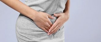
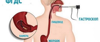
A gastric biopsy is a diagnostic procedure of taking a sample of tissue from the stomach lining and then examining it under a microscope. The tissue sample is called a biopsy. From a medical point of view, a biopsy sample is taken intravitally.
A biopsy is considered mandatory if cancer is suspected. Only under a microscope can you see the structure of a tissue sample and determine the presence and type of cancer cells. For this purpose, a special database of visual images of all studied cancer cells has been developed. They are simply compared with the sample taken.
However, in recent years, gastric biopsy has become increasingly used for the diagnosis of other gastrological diseases.
There is one subtle point in this procedure - the introduction of an endoscopic probe. Yes, the procedure is not dangerous and without pain. However, there are people who are not psychologically ready for it. The urge to vomit prevents the insertion of a probe. You just need a psychological attitude.
What is a gastric biopsy
Gastric biopsy is a diagnostic technique that allows intravital examination of stomach tissue.
When performing a gastric biopsy, organ tissue or individual cells are collected for subsequent histological, cytological or immunohistochemical examination.
To obtain the most reliable information, tissue is collected from several areas of the mucosa (the most suspicious from the point of view of the proposed diagnosis). That is, for one gastric biopsy, three or more samples (biopsy) are usually taken.
Currently, gastric biopsy is widely used in gastroenterological practice. This study is highly accurate, well tolerated by patients and rarely leads to complications.
Gastric biopsy is considered the “gold standard” for diagnosing malignant neoplasms of the stomach and precancerous pathologies of the gastric mucosa. Gastric biopsy is also used in the diagnosis of gastritis, polyps, ulcers, etc.
What does a gastric biopsy show?
Gastric biopsy analysis allows:
- assess the cellular composition of tissues;
- assess the secretory function of cells;
- assess the pH level;
- identify Helicobacter pylori;
- identify the presence of atypia (tumor atypical cells);
- carry out differential diagnosis between malignant and benign tumors;
- assess the activity of the inflammatory process;
- establish the type of gastritis (hyperacid, hypoacid, with normal secretion);
- identify rare forms of gastritis (lymphocytic, eosinophilic, granulomatous, autoimmune);
- identify and determine the type of dysplasia of the gastric mucosa;
- identify intestinal metaplasia of the gastric mucosa;
- assess the risk of malignancy of a stomach ulcer or polyp;
- assess the depth of the ulcer, etc.
The importance of a biopsy is confirmed by the fact that surgical treatment of gastric tumors is not performed without histological confirmation of cellular atypia.
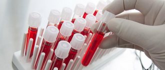
The material collected during the biopsy is sent to the laboratory, where it is stained and examined by histological, cytological or immunohistochemical methods.
Mode
Eradication rate (%)Bismuth subsalicylate 525 mg 4 r.s. + metronidazole 250 mg 4 r.s. + tetracycline 500 mg 4 r.s. 14 days + H 2 blocker for 28 days 77-82 Clarithromycin 500 mg 2 r.s. + amoxicillin 1 g 2 r.s. + lansoprazole 30 mg 2 r.s. 10-14 days 84-92 Clarithromycin 500 mg 2 r.s. + amoxicillin 1 g 2 r.s. + omeprazole 20 mg 2 r.s. 10 days 78-90 Bismuth subsalicylate 525 mg 4 r.s. + metronidazole 500 mg 3 r.s. or 250 mg 4 r.s. + tetracycline 500 mg 4 r.s. + proton pump inhibitor (omeprazole 20 mg or lansoprazole 30 mg) 2 r.s. 14 days97.6Clarithromycin 500 mg 2 r.s. + metronidazole 500 mg 2 r.s. + proton pump inhibitor (omeprazole 20 mg or lansoprazole 30 mg) 2 r.s. 14 days89-91Stomach biopsy - what is it taken for?
A gastric biopsy is performed when a patient has:
- stomach tumors of any etiology (as well as if there is a suspicion of a tumor or if it is necessary to perform a differential diagnosis between benign and malignant neoplasms);
- chronic gastritis (to clarify the types, stages and activity of inflammatory processes, as well as to assess the risk of transformation of gastritis into gastric ulcer);
- acute gastritis and stomach ulcers, difficult to treat (the study is carried out to clarify the cause of the development of the disease and assess the degree of activity of the inflammatory process);
- long-term non-regenerating ulcers of the stomach and duodenum;
- gastric polyps;
- Barrett's esophagus (stomach biopsy is performed in combination with esophageal biopsy);
- dysplasia of the mucous membranes of the stomach;
- pernicious anemia;
- dyspepsia, combined with anemia, fever, weight loss, jaundice, abdominal pain, etc.;
- dysphagia;
- chemical damage to the esophageal and gastric mucous membranes;
- intestinal metaplasia of the gastric mucosa;
- erosions of mucous membranes;
- space-occupying formations in the epigastrium.
A gastric biopsy is also indicated:
- when other diagnostic methods are not informative;
- after surgical interventions to remove tumors and polyps (to control the quality of treatment and timely detection of relapses);
- when conducting screening methods for diagnosing malignant neoplasms in patients at risk.
Indications
It must be emphasized that FGDS is prescribed strictly according to indications. Sometimes - in the absence of complaints from the patient. However, there are those who like preventive examination of the stomach using this method or who require a doctor to prescribe a study for any negative symptoms. This is fundamentally wrong and even dangerous. FGDS should only be performed by a very qualified doctor who knows the anatomy of the digestive tube well, since with any deviation from its natural course, complications are possible, even fatal, due to the risk of perforation of the organ wall. Therefore, it is unwise to do FGDS again. The doctor decides everything. Fibrogastroscopy is recommended for:
- abdominal pain of unknown etiology;
- discomfort in the esophagus;
- suspicion of a foreign object;
- prolonged, intractable heartburn;
- constant nausea for no apparent reason;
- uncontrollable vomiting;
- regurgitation after eating;
- dysphagia – inability to swallow;
- sudden, causeless weight loss;
- lack of appetite;
- anemia of unknown origin;
- pathologies of the liver, biliary system, pancreas;
- hereditary pathology such as stomach cancer or ulcers;
- preparation for complex abdominal interventions;
- medical examination;
- monitoring the effectiveness of treatment for diseases of the digestive tract;
- polyp removal;
- clinical follow-up after polypectomy.
Contraindications
Despite the fact that gastric biopsy is a minimally invasive and low-traumatic procedure, it
has a certain list of contraindications.
Absolute contraindications to performing a gastric biopsy include the presence of the following in the patient:
- ischemic or hemorrhagic strokes;
- myocardial infarction;
- stenotic lesions of the esophagus (with this disease it is not possible to pass a probe from the esophagus into the stomach);
- attacks of asthma (bronchial asthma).
Relative contraindications to performing a biopsy include the presence of the patient:
- fever;
- severe runny nose of any etiology (this is due to the fact that during endoscopy the patient must breathe exclusively through the nose);
- inflammatory processes in the pharynx;
- infections;
- epilepsy;
- bleeding disorders, hemorrhagic diathesis;
- hypertensive crisis;
- HF (heart failure);
- mental pathologies.
Treatment regimens
According to the definition, HP eradication is the absence of bacteria (both in vegetative and cocoid forms) 4 weeks after completion of drug treatment. There is a certain difference between the eradication regimens recommended in Europe and the USA. In the United States, the Food and Drug Administration (FDA) has approved 6 regimens for the treatment of HP infection in patients with duodenal ulcers; the recommendations given in the Maastricht Accords differ slightly. The choice of antimicrobial therapy regimen should be based on its effectiveness (degree of eradication), safety, drug interactions, microbial resistance, tolerability, patient comfort, and cost. There is no HP eradication regimen that would be 100% effective; regimens with 80% or higher eradication rates are eligible for use. The medication regimen should be convenient for the patient because skipping even one of the techniques significantly reduces the chances of successfully eradicating the infection.
Stomach biopsy - preparation
The gastric biopsy procedure does not require complex and lengthy preparation. Before the biopsy, the patient undergoes general tests (blood and urine tests, blood clotting tests, etc.).
The patient must warn the doctor about the presence of concomitant diseases (diabetes mellitus, cardiovascular pathologies, etc.) and about allergies to medications.
Eight to ten hours before the biopsy, you should not eat or drink anything. If the patient has a stenotic lesion of the pylorus, it is recommended to lavage the stomach before the biopsy. This is due to the fact that with pyloric stenosis there is often stagnation of undigested food.
Patients with an easily excitable psyche are premedicated with antispasmodics, tranquilizers and atropine.
To increase the accuracy of the study, the patient is simultaneously subjected to an FGDS and a gastric biopsy.
It should be noted that a gastric biopsy is not painful, but the procedure can cause significant discomfort in the patient (especially due to the gag reflex and difficulty breathing when the endoscope is inserted).
The entire procedure is carried out under local anesthesia (irrigation of the throat with a ten percent lidocaine solution). Irrigation with lidocaine is required to reduce the intensity of the gag reflex when inserting a fibrogastroscope. After passing the endoscope through the pharynx, there is practically no pain.
Minor trauma to the gastric mucosa when collecting tissue with a biopsy needle (or forceps) does not cause pain. Minimal damage to the mucosa heals quickly, so there are no scars left after the biopsy.
Discomfort in the throat and sore throat after the procedure usually disappear within 2-3 days.
Treatment regimens according to the Maastricht agreements
- First line therapy:
- Second line therapy:
- Special groups:
Proton pump inhibitor 2 times a day (or ranitidine-bismuth citrate 400 mg 2 times a day) + clarithromycin (500 mg 2 times a day) + amoxicillin (1000 mg 2 times a day) or
Proton pump inhibitor 2 times a day (or ranitidine-bismuth citrate 400 mg 2 times a day) + clarithromycin (500 mg 2 times a day) + metronidazole (500 mg 2 times a day)
The minimum duration of therapy is 7 days.
Proton pump inhibitor 2 times a day + Colloidal bismuth preparation (120 mg 4 times a day) + metronidazole (500 mg 3 times a day) + tetracycline (500 mg 4 times a day)
The minimum duration of therapy is 7 days.
In older adults, triple therapy regimens can be based on lower doses. For uncomplicated duodenal ulcers, proton pump blockers may not be taken after stopping antibacterial therapy.
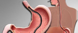
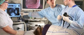
How is a gastric biopsy procedure performed?
The subject lies on his left side on a special table. A special mouthpiece is inserted into the patient's mouth, through which the endoscopic probe will be inserted.
The patient is asked to breathe calmly, measuredly and deeply, as this reduces the severity of the gag reflex and reduces spasm of the throat muscles, facilitating the advancement of the endoscope.
After inserting the endoscope to the required depth, the doctor examines the mucous membrane of the organ. When pathological formations are identified, biomaterial is collected using special biopsy forceps.
To increase the accuracy of the study, the doctor takes at least three (and if necessary, more) samples of the mucous membrane. For gastritis, a minimum of four samples is required, and for tumors and ulcers, a minimum of six fragments are required.
Mucosal samples are taken from more than one place. When taking a biopsy, attention is paid to suspicious areas identified during the preliminary examination.
The resulting samples are sent to the laboratory for further embedding in paraffin blocks, division into thin sections, staining and examination under a microscope (histological, cytological or immunohistochemical).
An immunohistochemical study is indicated when detecting malignant neoplasms. In this case, the biopsy material is treated with antitumor agents and the sensitivity of atypical cells to various drugs is assessed.
Progress of manipulation
Before the procedure, the doctor takes the patient’s written consent to perform it. Thus, the subject takes responsibility for possible flaws in the study, since he was warned about them in advance.
The oral cavity is treated with lidocaine (Falimint tablet under the tongue is an alternative). The patient takes a comfortable position on the left side: the cheek is pressed to the pillow, hands are on the chest (stomach). You can move your left hand over your stomach to your back, and your right hand to your stomach. After this, a special mouthpiece is placed in the mouth, which the patient clamps with his teeth. This is a concern for the safety of the gastroscope tube.
The doctor slowly inserts the endoscope probe into the mouth through the mouthpiece to the root of the tongue. The patient must take a deep breath and swallow saliva, at which time the fiberscope is placed in the esophagus. The doctor carefully moves the probe down the digestive tube, examining the walls of the esophagus, stomach and duodenum. All this time, the patient breathes deeply, which minimizes the urge to vomit. The person feels the advancement of the probe as itching. The procedure can take from 5 minutes to half an hour, depending on the severity of the pathological process. In some cases, a biopsy or removal of a small polyp or cauterization of the erosion is necessary. After all manipulations are completed, the fiberscope is slowly and carefully removed from the digestive tube.
After the procedure, you should not eat or drink for about an hour until the normal environment in the areas being examined is restored. If a biopsy has been taken, hot or cold food is limited until the evening (warm only), and for a day or two it is recommended to exclude fatty and coarse foods from consumption.
Since anesthetics are used during FGDS studies, you cannot drive a car immediately after the procedure: concentration of attention is reduced.
How to recover after a gastric biopsy
After the study, the patient should be under the supervision of a specialist for several hours. If there are no signs of bleeding, the patient is sent home.
Eating after a gastric biopsy is prohibited for 3-4 hours. Also, for three to four weeks you should stick to a light diet and avoid eating fatty, fried, spicy, etc. food.
Avoid drinking alcohol for at least two weeks.
After a gastric biopsy, it is not recommended to smoke (if possible for a day). You should also limit your intake of foods that increase gastric secretion (coffee, strong tea, marinade, pickles, sauerkraut, etc.) for several weeks.
Physical activity is excluded for a month.
It is necessary to limit emotional stress and avoid, if possible, stressful situations. If necessary, the doctor may prescribe mild sedatives.
Implications of the study
This type of biopsy is a low-traumatic examination, so most often, a gastric biopsy does not have serious consequences.
The main consequence of the procedure is pain and sore throat after insertion of the endoscope. This effect goes away on its own within a few days.
In rare cases, discomfort in the epigastric region, nausea, and slight bloating are noted. It is also possible to experience minor bleeding that stops on its own.
In isolated cases, severe bleeding, inflammation of the gastric mucosa, aspiration pneumonia, gastric perforation, etc. may develop.
If these symptoms appear, indicating the development of serious complications, you should immediately call an ambulance:
- development of brown vomit, reminiscent of coffee grounds;
- the appearance of severe nausea and abdominal pain;
- addition of febrile symptoms, chills;
- difficulty breathing;
- chest pain, persistent cough;
- the appearance of blood in the stool (tarry black stools - melena).
Diagnostics
Today, medicine uses several fairly effective methods for diagnosing BDS diseases. Let's take a closer look at some of them.
Endoscopic ultrasonography is a technique in which an optical device, an endoscope, is used to study the structure of the BDS. A photo of the papilla taken during a similar study is shown above.
Transabdominal ultrasonography is a screening method of examination using ultrasound, which allows one to very accurately identify structural changes in the gallbladder, liver, pancreas, and ducts. In addition, the technique determines the homogeneity of the gallbladder cavity and its contractility, the presence/absence of intracavitary inclusions.
The next method for diagnosing BDS pathologies is ultrasound cholecystography - a manipulation that examines the motor-evacuation function of the gallbladder within two hours from the moment of taking a choleretic breakfast.
Dynamic hepatobiliscintigraphy is a procedure based on the assessment of the absorption and excretion function of the liver. Fractional chromatic duodenal sounding allows you to determine the tone of the gallbladder; colloidal stability of the hepatic bile fraction and its bacteriological composition. During gastroduodenoscopy, the condition of the gastrointestinal tract is assessed, as well as monitoring the nature of bile flow. In addition to these methods, there are computed tomography and laboratory diagnostics.
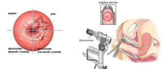
Stomach biopsy - interpretation of results
The interpretation of the tests should be carried out by the attending physician. Independent interpretation of the research results and subsequent self-medication are absolutely unacceptable.
In conclusion, the biopsy results indicate the following parameters:
- thickness of the gastric mucosa;
- cellular composition of the epithelium;
- level of secretory function of cells;
- the presence of atrophic changes, hypertrophy, etc.;
- presence of atypical cells;
- the presence of epithelial dysplasia and metaplasia;
- signs of the inflammatory process;
- Hh (the presence of HP on a gastric biopsy means the detection of Helicobacter pylori);
- degree of stomach contamination Hp (detection of Helicobacter during a gastric biopsy for an ulcer or gastritis indicates the Helicobacter nature of the mucosal lesion);
- the presence of lymphocytic cells, eosinophilic infiltrates, etc.
Results table
Also, with a gastric biopsy, a diagnosis is indicated based on morphological changes in the gastric mucosa.
In addition to the most common diagnoses such as Helicobacter pylori gastritis and ulcers, precancerous diseases and malignant neoplasms can be detected.
The table provides examples of histological precancerous diagnoses and their brief descriptions:
The price of the procedure depends on the region (in Moscow the study costs more than in the regions) and the number of biopsy samples. On average, the price for one biopsy ranges from 500 to 2000 rubles.