Today, a lot of people suffer from various diseases of the gastrointestinal tract (GIT). If pathological processes are suspected, the patient is often referred for examination of the abdominal cavity using fibrogastroscopy (FGS), esophagogastroduodenoscopy (EGDS), fibrogastroduodenoscopy (FGDS), fibroesophagogastroduodenoscopy (FEGDS) for diagnosis. What are the fundamental differences between the procedures? How to prepare for them?
What is gastroscopy?
This is a method for diagnosing diseases of the upper gastrointestinal tract:
- Esophagus.
- Stomach.
- Duodenum.
Since the test is carried out for the purpose of diagnosing pathologies of different parts of the gastrointestinal tract, the term “gastroscopy of the stomach” is not entirely correct. During the procedure, both the esophagus and part of the intestine are examined; this process is ensured by a special optical tube.
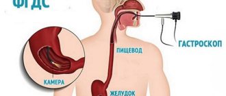
What is the name of the procedure in medicine? The abbreviation “FGDS” is most often used, which means fibrogastroduodenoscopy. There are also slightly modified abbreviations “EGDS” and “FGS”.
How is the procedure performed?
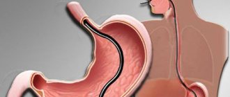
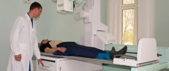
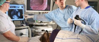
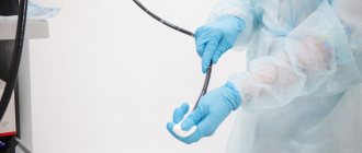
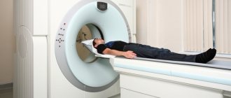
There are no difficulties in performing gastroscopy of both types, the biggest of which is to get yourself in the right psychological mood. The examination is carried out without the use of anesthesia. The patient is placed on the couch on his left side. After this, a flexible probe equipped with a mini-camera at the end - a gastroscope - is inserted into the stomach through the mouth and esophagus.
Features and stages of the procedure:
- To ensure that the probe slides well into the esophagus, the larynx can be treated with a local anesthetic - a spray containing lidocaine.
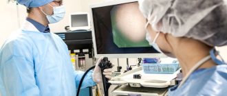
- The camera image on the tip of the probe is displayed on the monitor, the doctor assesses the condition of the digestive organs, takes appropriate photographs and notes.
- The procedure is painless - only uncontrolled salivation may occur; for this purpose, a tray is placed at the head of the couch.
The most unpleasant thing is the final stage, when you need to remove the probe. This action may provoke vomiting. But this is not a necessary phenomenon: if the patient sets himself up correctly, nothing negative or unexpected will happen.
FAQ (Frequently Asked Questions)
Fibrogastroscopy is often used in planned and emergency medical practice - and invariably terrifies patients. Although you have already learned from the previous section what FGS is, you need to clarify other common questions that may be of interest to patients and their relatives. Let's start.
When is gastroscopy needed?
This procedure is routinely prescribed in clinics and inpatient departments for complaints of symptoms such as:
- change in appetite;
- belching;
- heartburn;
- nausea;
- unpleasant taste in the mouth;
- vomit;
- heaviness and pain in the epigastrium;
- flatulence.
You should also be wary of weight loss (exhaustion), dark color of stool, changes in taste preferences (in particular, intolerance to meat dishes). In emergency cases, FGDS is used for bleeding from the gastrointestinal tract, acid burns, injuries, and foreign bodies entering the lumen of the digestive tube.
How is gastroscopy done?
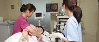
The diagnostic method involves the use of a special flexible tube with light, inserted through the mouth. Because of this, many patients refer to FGDS as a test during which you need to swallow a light bulb for the stomach. How is the research going? It includes the steps:
- anesthesia by irrigating the pharynx with a drug (usually “Lidocaine” in the form of a spray”);
- the patient adopts the desired position on the couch - lying on his left side with his right leg bent;
- biting the mouthpiece designed to protect the endoscope tube from the teeth;
- insertion of a probe;
- letting in air to “open” the lumen of the digestive canal;
- FGDS is performed - examination of the gastrointestinal tract, the study lasts from 3 to 5, less often up to 10-15 minutes - depending on the nature of the pathology;
- the tube is removed, the mouthpiece is removed, the patient is allowed to slowly get up from the couch - this is where the examination ends.
If you are allergic to local anesthetics, the anesthesia step is excluded. This will not make the study unbearable - modern tubes have a small diameter and easily pass into the lumen of the gastrointestinal tract .
Before the procedure, a nurse or doctor will definitely check the direction for FGDS. It is necessary to unbutton tight clothes (in general, you should not wear anything that prevents you from breathing freely), remove dentures, glasses and jewelry (bracelets, beads, etc.).
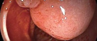
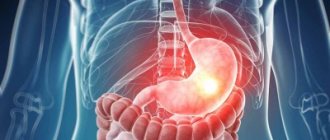
What does gastric FGS show?
The procedure is prescribed by gastroenterologists and therapists, and in emergency cases it is used by surgeons. Based on the results of the study, we can judge various pathological conditions:
- Inflammatory process.
- Suspicion of the presence of a foreign body.
- Defects of the mucous membrane (ulcers, erosions, scars).
- Narrowing of the lumen of the gastrointestinal tract.
- Neoplasms.
- Bleeding.
What does a gastrointestinal tract check show when using additional tests? Tests for acidity and Helicobacter pylori may be performed. This bacterium often causes gastritis. FGS of the stomach reveals changes that are interpreted unambiguously: after all, the doctor sees the pathology with his own eyes, and does not rely on indirect signs. A medical procedure done in a timely manner makes a valuable contribution to the early diagnosis and prevention of cancer.
Is the procedure painful?
In the vast majority of cases, the examination does not cause any painful sensations. Of course, the patient experiences discomfort - but this is quite reasonable, since the endoscope tube comes into contact with the mucous membrane, and a feeling of a “foreign body” arises. The device does not interfere with breathing. Soreness may be due to:
- narrowed lumen of the gastrointestinal tract;
- the presence of defects in the pharyngeal mucosa;
- contact of the device with the throat.
This pain is not severe and quickly disappears after the tube is removed. The use of anesthetics helps to avoid its occurrence. If several hours have passed after esophagogastroduodenoscopy and discomfort is still bothersome, you should consult a doctor for additional examination.
How often is EGD of the stomach performed?
In simple cases, a single procedure is enough; depending on the patient’s condition and the expected pathology, the doctor includes additional tests:
- determination of acidity, or pH-metry;
- analysis for Helicobacter pylori;
- biopsy (taking a piece of tissue from the affected area to send to a histology laboratory).
Sometimes gastroscopy of the stomach is repeated some time after the initial examination - for example, to monitor the eradication of Helicobacter pylori or in the event of an emergency condition (bleeding) or the need to remove previously detected small tumors.
FEGDS, EGDS
FEGDS/EGDS is (fibro-)esophagogastroduodenoscopy. This diagnosis is considered the most comprehensive, since it involves examination of the esophagus. Pathological changes are also common at this level:
- volumetric formations;
- varicose veins with bleeding;
- diverticula;
- stenosis;
- Barrett's esophagus (dilated esophagus with epithelial metaplasia);
- esophagitis;
- gastroesophageal reflux.
It should be noted that the difference in the listed terms is quite conditional. For example, if there is a suspicion of a gastric ulcer, and the endoscopist, when passing a gastroscope through the esophagus, notices pathological changes in it, then in any case he will record and describe them. Therefore, the use of one or another abbreviation by doctors is more a matter of convenience and habit.
Important Features
Most patients who underwent FGS say that they were much more worried than they should have been. Of course, the examination is unpleasant, but a qualified doctor and nurse will do everything possible to make it as comfortable as possible. The patient himself can influence the results. Let's find out what features of preparation exist and how the conclusion is deciphered.
Preparation
Since the gastrointestinal tract is being examined, you need to remember the following rules:
- Do not eat for 8-12 hours before the test.
- 48 hours before the procedure, avoid eating chocolate, spicy, fatty and smoked foods, nuts and seeds, and drinking alcohol.
- Be careful with coloring preparations, in particular those based on bismuth. They should not be drunk 24 hours before the test.
- On the eve of the diagnosis, no later than 22.00 there should be a light dinner (without salads from fresh vegetables and fruits).
- In the morning before inserting the probe, you should not smoke or take pills.
- To eliminate increased gas formation, the drug “Simethicone” (“Espumizan”) can be used in advance and during the test.
Patients with drug allergies should be warned about the sensitivity of the endoscopist.
Take a sheet with you (some clinics use disposable bed covers). You will also need a medium size towel. Wear loose clothing, you can brush your teeth, but do not swallow the water you use to rinse your mouth. It is better to come to the examination in advance, 10-15 minutes in advance.
How to behave during EGD of the stomach?
You need to lie quietly, establish a breathing rhythm that is comfortable for you - remember, the tube does not block the access of air to the lungs. Under no circumstances try to change position, much less pull out the probe, or touch the doctor’s hands - this can lead to dangerous injuries to the esophagus, stomach and intestines.
Be prepared for the following effects:
- copious secretion of saliva, which you cannot swallow (use a towel for absorption);
- uncontrollable belching (as air is blown in to improve vision);
- urge to vomit (associated with irritation of the throat with the probe, decreases with anesthesia, but in any case is not critical);
- a feeling of movement in the abdomen (as the tube has passed into the lumen of the gastrointestinal tract, and the doctor moves it to examine the necessary areas).
All these phenomena stop immediately after removing the probe, although belching may persist for some time - until the “extra” air comes out. If the examination was carried out without anesthesia, a feeling of rawness and soreness in the throat is likely; it disappears without medical help and special medications after a few hours.
Decoding the results
It is carried out by a doctor in the endoscopy room (he describes the picture seen during the procedure and issues a conclusion) and by the attending physician or gastroenterologist. In some clinics you can get a video recording, but in most cases the patient is given a printout indicating the detected changes. The description is based on an assessment of such characteristics as:
- The lumen of the gastrointestinal tract (normal, enlarged, stretched, expanded, narrowed).
- Sphincter area (no features, gaping, asymmetry, spasm, dyskinesia, occlusion, stenosis).
- Contents (mucus, bile, blood, saliva, digestive juice, pus, fecal matter, food, foreign body, parasites).
- Peristalsis (normal, decreased, increased, absent, reflux).
- Mucous membrane (unchanged, atrophy, hyperemia, stagnation, aphthae, ulcers, bleeding).
- Thrombi (parietal, fresh).
- Vascular picture (no features, indistinguishable, bright).
- Folds (normal, enlarged, stagnant, deformed).
- Neoplasms (polyp, granuloma, tumor).
- Pathological openings (diverticulum, fistula, stoma, perforation).
In inflammatory processes, the transcript of FGDS contains a mention of hyperemia; There may be local bleeding, the vascular picture is bright. Neoplasms are described by general characteristics, since it is possible to draw a conclusion about which category they belong to only after a histological examination.
A positive test for Helicobacter pylori indicates the infectious nature of the unfavorable process in the gastrointestinal tract. With pH-metry, normal, increased or decreased acidity is determined, which also has diagnostic value.
Rules for preparing for the FGS
Basic rules for preparing for research:
- The procedure is carried out strictly on an empty stomach. Within 10-12 hours before FGS, you need to refuse food and any liquid (except water). You should also refrain from drinking any liquid 2 hours before the test. Otherwise, excessive secretion of gastric juice and the presence of food in the stomach will distort the examination results and also provoke vomiting.
- It is necessary to follow a diet. Dinner on the eve of a gastroscopic examination should be light and quickly digestible, and in addition, difficult-to-digest foods should be avoided for 1-2 days before the examination.
- You should not smoke for 4-5 hours before the examination. This is due to an increase in the secretory function of the gastric glands when smoking even one cigarette.
- You should avoid brushing your teeth and chewing gum on the day of the examination. These actions also lead to an increase in the secretion of gastric juice.
- If possible, you should stop taking medications. If the subject is taking medications, you must first consult with a doctor about the possibility of temporarily canceling them.
The patient must take with him to the FGS:
- direction;
- outpatient card (if available);
- shoe covers or replacement shoes;
- disposable diaper (place on the couch);
- towel (place under your head).
Before FGS, it is necessary to remove glasses or contact lenses and remove dentures.
Contraindications
Fibrogastroscopy of the stomach is not performed in such conditions as:
- aortic aneurysm;
- acute myocardial infarction;
- severe cardiovascular and respiratory failure;
- resting angina.
A relative, that is, removable contraindication may be inflammation of the pharynx, tonsils and mediastinum, tuberculosis, acute respiratory viral or intestinal infection. In these cases, appropriate treatment is provided and the study is rescheduled to another date. For panic attacks and epilepsy, the procedure is performed under anesthesia.
When is fibrogastroduodenoscopy performed?
In terms of the set of indications, there are not many differences between FGDS and EGDS, because the main source of symptoms is, in one way or another, the stomach. Nevertheless, they exist, therefore, when prescribing gastroscopy, the doctor always chooses between FGDS and EGDS based on the clinical picture and patient complaints.
Gastroenterologists prescribe fibrogastroduodenoscopy in the presence of symptoms indicating diseases of the mucous membrane of the duodenum and stomach:
- nausea before or after eating;
- feeling of heaviness in the stomach on an empty stomach or after eating;
- pain in the epigastrium (upper abdomen in the cavity between the ribs);
- formation of gases in the intestines in the absence of visible reasons;
- loss of appetite or inability to eat the usual portion of food due to a feeling of fullness in the stomach;
- sudden weight loss or weight gain while maintaining your diet.
Similar changes are observed in the presence of an inflammatory process in the stomach (gastritis) or duodenum (duodenitis), diverticulosis of the duodenum, ulcerative lesions of the mucous membranes, duodenal reflux (when the contents of the duodenum are thrown into the stomach) and neoplasms in the stomach (polyps or cancerous tumor).
Important! Despite the differences in FGDS in terms of the area examined, the doctor can note in the results the condition of the mucous membranes of the esophageal tube.
Complications
Endoscopy must be performed responsibly and all preparation recommendations must be followed. The EGD procedure is considered safe and simple, but there is a risk of complications:
- allergy to anesthetic;
- damage to the esophagus;
- bleeding;
- entry of gastric contents into the respiratory tract.
Reasons for concern are weakness, fever, pain in the chest, back and/or neck and when swallowing, hoarseness, shortness of breath.
The likelihood of complications is higher in severe conditions, the presence of previous injuries (burns, mechanical damage) to the esophagus, diverticula and tumors in the stage of decay. A preliminary survey (information about allergic sensitivity), familiarization with the medical history or medical record, and careful manipulation (in which the patient’s calm and immobility plays a significant role) help reduce the risk.
Biopsy during FGDS
Fibrogastroduodenoscopy is a study that makes it possible to perform a biopsy. It is necessary for additional study of the tissues taken as a sample. With its help, you can make a more accurate diagnosis and even identify the cause of the pathology. This research method is highly informative. The procedure is performed during fibrogastroduodenoscopy.
The doctor, using an additional umbrella, collects cells from the mucous membranes of the stomach and duodenum . In this case, the patient does not experience additional discomfort or pain. More often than not, the patient does not notice how this process occurs. The biopsy requires abstinence from food for 5 hours after diagnosis.