X-ray is one of the main and mandatory research methods, which is necessary if pneumonia is suspected. X-rays confirm or exclude the diagnosis. This is the only way a doctor can see the full picture of the development of the disease and make the necessary conclusion.
Repeated radiography allows you to monitor the dynamics of the pathological process, analyze the effectiveness of treatment, and identify the development of complications. X-ray diagnostics are also performed after treatment to assess the condition of the lungs after the disease.
What are the purposes of radiography?
Pneumonia is an inflammatory disease caused by pathogenic microorganisms - viruses, bacteria, fungi. Symptoms include fever up to 38-39 degrees, cough, shortness of breath, wheezing in the chest, and deterioration in general health. The difficulty of diagnosing pneumonia lies in the fact that the above signs are characteristic not only of pneumonia, but also of other diseases - acute bronchitis, pleurisy, oncological processes, etc.
X-ray examination is based on scanning internal organs using a special device that emits electromagnetic waves of a certain length. Passing through the tissues of the human body, they leave traces on the surface of the film, from which an image of the corresponding organ is formed. It can be used to identify pathological changes and create a complete picture of various diseases, including inflammatory processes in the respiratory organs.
Indications for a chest x-ray include cough with copious sputum, high fever (38-39 degrees), chills, chest pain and other signs that may indicate pathological processes in the organs of the respiratory system.
IMPORTANT! The diagnosis of pneumonia should be based not only on x-rays, but also on the results of listening to the chest, blood and sputum tests, and other examination methods.
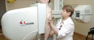
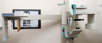
There are two main ways of carrying out the procedure - conventional (film) X-ray and digital, which allows you to obtain clearer and more informative images, and also reduces the X-ray load on the body. The choice of technique depends on the equipment of a particular medical institution - not all hospitals and clinics have devices for digital diagnostics.
What is pneumonia most often confused with?
X-ray examination allows you to distinguish pneumonia from the following diseases:
- Bronchitis. With this disease, there is no shading of the lung tissue in the image, but strands of connective tissue are visible in the roots of the lung. Because of this, it is often confused with hilar inflammation of the lung tissue, which on x-ray gives a picture of severe bronchitis.
- Presence of a foreign body. If it really got into the lungs, then its shadow in the picture will be in the lower parts of the lung, while pneumonia most often originates in the upper parts. However, a foreign body is often confused with infarction-pneumonia.
- Pleurisy. This pathology in most cases appears if a person did not treat pneumonia or treated it incorrectly.
- Hemothorax and pneumothorax. These pathological conditions in the pictures do not resemble pneumonia, but there is one common symptom - severe chest pain when breathing.
How is it different from fluorography?
X-ray and fluorographic examination have the same principle, but when diagnosing pneumonia, preference is given to x-ray .
Fluorography makes it possible to detect changes in lung tissue in the early stages, but does not provide a clear enough x-ray picture to make an accurate diagnosis.
At the same time, the radiation coefficient during the procedure is much less than during an x-ray examination, so fluorography is done for preventive purposes, and x-rays are done directly for diagnosing pathological processes in the presence of corresponding symptoms.
X-rays for pneumonia in children
In children, this disease looks a little different
than in adults. This is explained by the fact that the child’s body reacts to any inflammation much more acutely. Even the smallest infiltrate can cause a lobar form of the disease.
On an x-ray, pneumonia in a child looks like this:
- Foci of shading are localized in the lower parts of the lungs.
- The size of the infiltrates is very small. The higher their density, the more severe the disease.
- In the mediastinum there is an increase in lymph nodes. This is clearly visible on x-rays.
- The affected areas can blur the structure of the lung root and completely hide the pattern of the lung tissue.
Due to the fact that in children the lung tissue often swells without any inflammation
, diagnosing pneumonia in the initial stages of development using x-rays can be quite difficult. In addition, the correct diagnosis of pneumonia in children is hampered by the small volume of lung tissue.
Can it not show pneumonia
Pneumonia has many forms of clinical course, and foci of inflammation can be located in any segment of the lungs, and in some cases it can be difficult to determine the disease from an x-ray, especially if a small area of the organ is affected. If after the examination the doctor remains in doubt, the patient is prescribed additional diagnostic procedures - usually CT or MRI.
REFERENCE! It is most difficult to identify pneumonia in children suffering from immunodeficiency conditions, so in such cases it is better to immediately send the child to a CT or MRI.
Radiography for pneumonia in children
X-rays for pneumonia in children look somewhat different than in adults. Due to the increased reactivity of the immune system, any smallest infiltrate in a child can quickly lead to lobar inflammation (affecting both lungs). Obviously, it is very important to carry out timely diagnosis of the disease and identify focal inflammation.
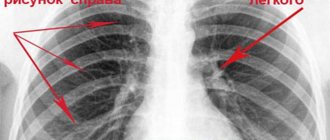
X-ray of the child’s chest organs: infiltration of the right root
What are the signs of pneumonia in children:
- Focal darkening in the lower parts of small sizes;
- Infiltrates do not exceed 1-2 mm in diameter;
- High density of spots is detected with advanced inflammation;
- Consolidation and enlargement of the lymph nodes of the mediastinum are difficult to discern in the image;
- After the disappearance of pathological shadows, the deformation of the pattern persists for another 7 days;
- With an unfavorable course, small focal pneumonia develops into pseudolobar pneumonia;
- The high density of the affected area overlaps the structure of the root and pulmonary pattern.
In children, swelling of the lung tissue is often observed, which complicates the diagnostic criteria of radiography for pneumonia in a child.
The inadequacy of the radiological and clinical picture of the disease can also be explained by the small volume of lung tissue and the large content of elements of the pulmonary pattern per unit area.
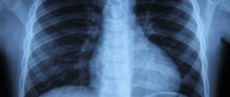
What does the disease look like in the picture?
After the x-ray, the images should be examined by a specialist , and based on the data obtained and the results of other studies, draw appropriate conclusions and make a diagnosis. Normally, a person’s lungs and bronchi look like this:
- the pulmonary lobes have the same, uniform black tint;
- a white lumen is observed in the area of the heart;
- the ribs and collarbones are gray, with the usual outlines;
- white diaphragm domes;
- The spinal column is located in the center.
Signs of pneumonia on an x-ray largely depend on the form of the disease and its stage, as well as on the localization of the pathological process. If the study shows signs of pneumonia, it is necessary to begin treatment as soon as possible - an advanced pathological process is dangerous not only for health, but also for human life.
Lobar pneumonia on x-ray
When individual foci of inflammation of the lung tissue merge, lobar pneumonia occurs. A photo of an x-ray taken with this form of the disease clearly shows the differences.
As a rule, with the lobar form of pneumonia, one or several lobes of the lungs are affected at once. This poses a serious threat to the patient's life.
On x-ray, the lobar form of the disease is manifested by the following signs:
- Pronounced large shadows affecting the entire lobes of the lung. Both lungs may be affected.
- The mediastinum shifts towards the most inflamed lung.
- Signs of deformation are clearly visible on the diaphragm domes.
- The pattern of lung tissue may not be visible.
Lobar pneumonia is most easily identified by X-ray examination. However, to make an accurate diagnosis, doctors prefer to take x-rays in two projections. This gives them the opportunity to determine the number of organ segments affected by inflammation and determine the condition of the mediastinum.
Main features, description
The first sign of pneumonia on an x-ray is the appearance of dark spots with uneven contours in different parts of the lung, which can have different sizes, from 3-4 to 12 mm.
Shadows are distinguished by appearance (round, oval ring-shaped) and color intensity - the darker the spot, the more pronounced the pathological process.
If the lymph nodes are affected and the blood supply to the organ is disrupted, changes in the roots of the lungs may be observed, and if the disease affects the pleura, there may be a disturbance in the pattern of the domes of the diaphragm. Otherwise, the manifestations of pneumonia depend on the stage, form and clinical features of the disease:
- Focal form. X-rays show small (1-1.5 cm) shadows with weak or moderate color intensity, heterogeneous structure and unclear boundaries. The lesions can be single or multiple, and in some cases they merge into one large spot. The roots of the lungs are expanded, and disturbances in the normal pattern of the organ may persist for several days after recovery.
- Lobar pneumonia. Changes in the normal pulmonary pattern, fluid in the pleural cavity, signs of infiltration of one of the lobes of the lung, and expansion of the roots are observed. As the inflammatory process develops, the severity of the changes and the intensity of the color of the shadows intensify.
- Interstitial form. The image shows compaction of the roots of the lungs and other changes that form a pronounced pattern reminiscent of tree branches.
- Abscess pneumonia. It manifests itself as extensive darkening of the affected area, signs of thickening of the pleura and the presence of cavities of different sizes filled with fluid.
- Aspiration form. X-ray is characterized by triangular spots with a uniform structure, light foci and a raised diaphragm.
IMPORTANT! Focal pneumonia is the most difficult to diagnose - in the first stages it manifests itself as small foci of infiltration, which are not always visible on x-ray.
Differentiation of pneumonia from other lung pathologies
An important stage in making a diagnosis remains to distinguish it from other lung diseases, coronavirus. So, with bronchitis there will be no darkening in the image, but instead an increase in the pulmonary pattern.
Strengthening the pulmonary pattern
In the presence of a foreign body, a darkening with clear edges is observed, localized in the lower lobe of the lung. It is difficult to confuse it with inflammation of a typical nature.
With pleurisy, the picture shows an accumulation of exudate in the affected area. Pleurisy acts as a complication of untreated pneumonia.
In pneumothorax, a characteristic level of fluid is present. The image shows a clearing; the pulmonary pattern is not visible.
An X-ray is the main examination, and if necessary, a computed tomography scan is prescribed.
Stages of the disease
The inflammatory process in pneumonia occurs in several stages, and each of them is characterized by certain changes on X-ray photographs.
- Tide stage. The first stage of the disease lasts from 12 to 72 hours and is manifested by increased blood circulation in the lungs, deterioration of their functions and the formation of fluid in the alveoli. The photographs show a clear pattern of the organ, which resembles a lattice, blurring of the extreme points and enlargement of the roots.
- Hepatization stage. The lung tissue thickens and becomes similar to liver tissue. The pattern of the lungs at this stage is not so intense; dark spots with light stripes form on it, the roots of the organ are expanded, and this is especially noticeable on the affected side. As the pathological process develops, the foci of inflammation darken, and the presence of fluid is clearly visible in the photographs.
- Resolution stage. The beginning of regeneration of lung tissue: a decrease in the intensity of the organ pattern and the color of the dark spots, the disappearance of large elements at the site of the lesion and signs of the presence of fluid.
REFERENCE! Changes in the pulmonary pattern may be present for several weeks after recovery.
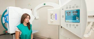
X-ray for pneumonia - indications and contraindications
An X-ray for pneumonia is then shown; the symptoms of pneumonia are characterized by cough, chills, sputum production, and laboratory tests show an increase in the number of leukocytes.
If a person is diagnosed with lobar or focal pneumonia, repeat radiographs are ordered to monitor changes in the “bad” shadows during treatment.
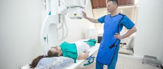
A specific indication for an X-ray of the lungs is a serious suspicion of an inflammatory process in the lung tissue or other dangerous disease. To take a picture of a person, you need to take into account the harm and benefits of the examination. Only if the benefits of x-ray exposure outweigh the harm, x-rays can be taken.
There are no contraindications for the study. The only restriction is pregnancy. However, if pneumonia is suspected in pregnant women, an x-ray of the lungs is performed. At the same time, the X-ray room staff does everything possible to protect the woman’s organs from radiation (lead aprons, reducing the time and number of procedures).
How often can you undergo x-ray diagnostics?
On average, it is recommended to undergo an X-ray examination no more than 1-2 times a year, but in case of pneumonia, the procedure is performed more often , since the risk of complications exceeds the danger from radiation exposure. As a rule, x-rays are performed at least 3 times - upon diagnosis of pneumonia, 3-5 days after the start of therapy to monitor its effectiveness, and also after the disappearance of clinical symptoms. Repeated photographs after the patient’s recovery should be taken to prevent complications and consequences of the disease - lung abscesses, proliferation of fibrous tissue, etc.
What does an x-ray photo show in focal pneumonia in adults?
An X-ray photo for pneumonia in adults shows the structure of the root and the deformation of the pulmonary pattern, not only in focal pneumonia in the initial stages. Similar symptoms are observed with bronchitis.
X-ray symptoms of focal and lobar pneumonia are described above. Let us describe the features of x-ray photography for aspiration pneumonia.
The disease occurs due to the entry of stomach contents into the bronchial tree. Against this background, blockage of the bronchi is observed, so foci of clearing and darkening appear.
In those places where the patency of the bronchial tree is impaired, atelectasis occurs. In the photo they look like triangular shadows with a homogeneous structure. If such changes occur, the diaphragm dome will rise upward. At the same time, the mediastinum shifts towards the lesion.
In adults, atelectasis is usually combined with the presence of dark spots of infiltrative origin.
What does interstitial pneumonia look like on an x-ray?
Interstitial pneumonia on a radiograph of an adult is manifested by the following changes:
- Peribronchial compactions;
- Perilobular shadows;
- Uneven expansion of the bronchovascular bundle;
- Radical infiltration looks like root expansion.
Staphylococcal inflammation of the lung tissue is unilateral. It appears on x-ray (or digital photo) as a limited compaction.
On days 2-5 of the disease, airy and dry bullae appear, which contain liquid and air. The size and number of infiltrates are determined on the image with a low degree of reliability, since the summation photo does not reveal changes in the thickness of the lung tissue.
As inflammation intensifies, fibrous tissue grows, resulting in fibrous thickening of the roots of the lungs.
Interstitial inflammation looks specific. The radiologist determines it just by looking at the radiograph. Intense “tree branch” in the lesion is a specific symptom of the disease.
Contraindications
Relative contraindications to the procedure are pregnancy (especially the first trimester) and the patient’s serious condition. However, if there are characteristic symptoms, the procedure is recommended for everyone without exception, including expectant mothers - to reduce the negative impact on the fetus, the pregnant woman’s belly is covered with a special apron that does not allow x-rays to pass through. It is almost impossible to accurately determine pneumonia without an x-ray - due to the lack of specific symptoms, the likelihood of error and improper treatment is too high.
Restriction on the procedure
X-ray diagnostics of the chest organs has one undeniable advantage - the procedure does not require special preparations, in addition to observing basic hygiene rules. However, this study has several contraindications:
- the patient's age is under 14 years,
- pregnancy,
- open bleeding.
Note! The listed contraindications are not strict, they are only advisory. This means that if an x-ray is needed to treat a pregnant woman or child and the patient’s life may depend on it, then diagnostics are possible .
Distinctive features of atypical forms of the disease
In some cases, inflammation may manifest itself with signs atypical for pneumonia. This is due to the origin of the disease, concomitant factors that influenced the development of inflammation.
Caseous pneumonia
This is a specific form of pneumonia with severe necrotic lesions of the parenchyma.
There are no symptoms characteristic of pneumonia. Pathology often creates a risk of death. The organ tissue melts, cavities of different sizes are formed. X-rays show a change in the topographic location of the mediastinal organs. They shift towards the pathological lung. Due to insufficient ventilation function, the diaphragm dome rises upward. The x-ray clearly shows a narrowing of the distance between the ribs. In photographs, cavities (cavities) appear as circles or ovals with darkened edges and a light center. On average, their diameter is 3 cm. With multiple small cavities, which are often formed on both sides, in the picture the top of the lung is dark, the lobes of the lower section have cavernous foci.
Atypical pneumonia
The disease is characterized by a sluggish course caused by microorganisms atypical for pneumonia (chlamydia, mycoplasma, legionellosis). More often it is diagnosed in children. Inflammation can be determined by x-ray, based on the following signs:
- intensity of the pattern;
- darkening in the area of parenchyma compaction (single or multiple);
- damage to a lobe or the entire organ;
- darkened stripes (a sign of pleural effusion).
With pneumonia caused by Legionella, a repeated x-ray shows a clear progression of the disease, individual shadows merge together, occupying a large area.
Pneumocystis pneumonia
This is a fungal infection of the lungs, which is characterized by a hidden (latent) course, without pronounced clinical manifestations. The lungs are affected symmetrically on both sides. The X-ray shows a pattern in the form of butterfly wings. The root sections lose their transparency and become cloudy and dull. In the picture with fungal pneumonia, the organ looks like a piece of cotton wool. In some cases, light areas are observed (consequences of air accumulation).
X-ray of the lungs for pneumonia is decisive in making a diagnosis and determining a treatment strategy. The images allow you to correctly determine the type and stage of pathology and the degree of damage to the organ. In atypical forms of pneumonia, the study makes it possible to differentiate the disease with relatively normal laboratory test results.
conclusions
Without a doubt, performing a plain X-ray of the lungs is a very important point in the process of diagnosing the disease. He will point out the characteristic signs, however, one should not lose sight of the fact that, one way or another, an x-ray of the lungs for pneumonia is an additional research method that helps in making a diagnosis. You cannot make a diagnosis based only on these additional research methods; you must also rely on the clinic. And treatment, it should also be noted, should be carried out based on the severity of the patient’s symptoms.
Even if the doctor does not see pneumonia in the image, but empirical antibiotic therapy with broad-spectrum drugs is started, then, by and large, this will not have any particular significance for the patient. In this case, the value of the image for pneumonia will be only retrospective - that is, it will confirm or refute the established diagnosis. After suffering from pneumonia, it is also worth performing a control X-ray in order to make sure that the treatment is effective and to look for signs that will confirm this.
Video: Elena Malysheva. Symptoms of pneumonia
Pneumonia or pneumonia is an infectious and inflammatory disease of lung tissue of various origins. Due to its widespread prevalence and high mortality rate, the issue of timely diagnosis of the disease is of great importance. In order to detect pneumonia in time and prescribe the correct treatment, it is necessary to conduct a comprehensive examination of the patient. The main diagnostic method is x-ray of the lungs, which has characteristic features for pneumonia of various origins.
Indications for x-rays
Main indications for primary radiology:
- increased temperature, fever, chills;
- cough;
- copious sputum;
- heavy breathing, shortness of breath;
- herpes on the cheeks, around the nose;
- high white blood cell count on a general blood test;
- possible inflammatory processes of the respiratory system.
In children, the indications are slightly different, since inflammation begins quickly and occurs in an acute form. This is due to the small size of the lungs and weak immunity. There may be no cough or fever - in such cases it is recommended to pay attention to the child’s breathing, the presence of whistling and shortness of breath.
Indications for repeated x-rays:
- when diagnosing lobar and focal pneumonia, x-rays are re-administered to determine the rate of development of questionable darkening of the lungs;
- to ensure that there are no residual effects after the disease.