Radiography is the study of internal structures that are projected onto a special film or paper using x-rays. Most often, X-rays are used in traumatology, but they are no less often used in pulmonology. Chest X-rays are used to diagnose and monitor treatment.
Patients undergo chest X-rays less often, but fluorography should be performed every year for preventive purposes. The classical examination has been replaced by digital radiography. Modern technologies provide clear images, eliminating the possibility of distortion. And the main advantages of modern X-ray equipment are a lower radiation exposure to the patient.
X-ray vs fluorograph: main differences
X-ray examination is often confused with fluorography, however, these are two different types of human examination. They have fundamental differences among themselves.
X-ray of the lungs is considered a more informative study, so it is used to:
- confirm the suspected diagnosis after FLG;
- clarify the results of treatment;
- track pathology over time.
A beam of X-rays penetrates the organ being examined in two projections, and is then transferred to a CCD matrix, or film (old version). The study produces a picture with 256 shades of white and black.
Dense structures are indicated in a light color, and hollow ones are indicated in a dark color. As a result, the doctor receives an image and diagnoses normal or pathological conditions.
Fluorographic research is a technique in which a shadow image of organs is photographed in one projection from an optical screen or x-ray screen onto 70 or 100 mm film.
To obtain a fluorogram, an X-ray fluorography apparatus is used. It consists of an X-ray tube (X-ray tube), a diaphragm, and a CCD matrix.
This technique is often used to diagnose tuberculosis, cancer and pathology of the bronchopulmonary system. The advantage of fluorography is that it is a less harmful research method, which is why it is used in mass screening of the population.
It is recommended to undergo fluorography no more than once every two years, and for some categories of the population - once a year.
The disadvantages of fluorography are considered significant in the diagnosis of many pathologies:
- The fluorography image is not as sharp and contrasty as an x-ray;
- the size of the chest in the image is reduced;
- It is more difficult to distinguish lung pathologies.
Therefore, doctors try to prescribe x-rays if necessary, and in order to prevent tuberculosis, fluorography will be sufficient.
Fluorography and x-rays are different methods for studying the human body. Both of these methods are used to confirm a particular diagnosis or identify a specific pathology.
To take an X-ray, a dose of 0.1-0.3 mSV is used, and for fluorography - 0.03-0.05 mSV, which is ten times less than with an X-ray.
FAQ
What is the difference between X-ray and fluorography?
The essence of fluorography is as follows:
Using a special installation, the shadows of the area under study are photographed from a fluorescent monitor onto film. The procedure is widely used in screening for tuberculosis and pneumonia.
Radiography is a modern, improved and highly accurate alternative to fluorographic examination, since organs are recorded on film or a digital matrix in real scale. If during fluorography it is possible to obtain shadows measuring 5 mm, then during X-ray diagnostics shadows of 2 mm are visible.
Today, fluorographic diagnostics are more of a preventive nature and give only a general idea of the state of the body.
To clarify the diagnosis, the patient must undergo a chest x-ray.
How often can I have the procedure?
The latest X-ray equipment has made it possible to make radiography as fast and safe as possible for the patient’s health. The condition of the chest organs can change significantly even over a short period of time.
Relatively healthy people can have radiation examination once a year. More frequent examinations are recommended only if indicated.
Is x-ray dangerous for a child?
When examining a child, doctors often resort to this procedure. An x-ray allows you to diagnose various diseases of the chest organs with minimal loss of time and money. Of course, an x-ray is prescribed only after an examination by a pediatrician and if the child has strict indications for it.
It is important to remember that children's bodies are more susceptible to x-ray radiation. Therefore, the maximum permissible dose of radiation during medical research for a child will be less than for an adult. During the year, a total radiation dose may be received not exceeding 1 mSv. If this rule is violated, the risk of developing various oncological pathologies increases.
The duration of diagnosis and the amount of x-ray exposure are determining factors when choosing equipment for examination. With old X-ray equipment, which is still used by municipal health institutions, the body receives a radiation dose of 0.3 mSv. The duration of contact of the chest with the device is one second.
Innovative digital equipment emits 10 times less radiation, and the procedure lasts no more than 0.02 seconds. The examination itself takes only 10–15 minutes. To identify diseases in children, preference is given to digital radiography. This choice is especially relevant when there is a need to conduct multiple studies.
Performing the procedure for children under 12 years of age has its own characteristics. During the study, a parent or any other adult must be in the room with the child. It is important to ensure that the subject does not move and that metal jewelry is removed from the body.
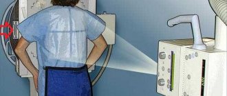
Medical staff puts a special lead apron on the child. This protects the body from unnecessary radiation. The area of the body to be examined remains open.
A timely diagnosed disease is more easily amenable to therapeutic correction, which means that various negative consequences for the body of a small patient are prevented. The study helps to identify pathologies of the musculoskeletal system, heart, lungs and bronchial tree. These may be congenital anomalies, consequences of traumatic injuries, infectious-inflammatory or tumor processes. Using an x-ray examination, the doctor evaluates the results of the treatment and the dynamics of the disease.
How quickly are research results released?
After 15–30 minutes, the patient receives pictures and a conclusion about the diagnostic results. Based on the results of X-ray diagnostics, additional laboratory or instrumental examinations, as well as consultations with relevant specialists, may be prescribed.
What are the benefits of x-rays?
Today, radiography is carried out using modern installations, which makes it possible to obtain detailed images with high resolution and detect dangerous diseases in the early stages of development. The radiation dose is minimal, 5 times lower compared to the film fluorography method. However, given that the study still involves radiation exposure, it is performed exclusively on the direction of the attending physician.
In the Doctor Nearby network of medical clinics, X-ray diagnostics are carried out by qualified specialists. The radiation dose is selected taking into account the constitutional characteristics of the patients.
Indications for the study
There are clear indications for performing a chest x-ray. If a pulmonary pathology is suspected, the doctor will prescribe an x-ray if the patient has complaints about:
- cough lasting at least a week;
- increased temperature and heat;
- sputum discharge;
- chest pain;
- wheezing in the lungs;
- shortness of breath;
- spitting up blood.
These signs primarily illustrate pulmonary problems. After a visual examination, the doctor will make a preliminary diagnosis, but can only confirm it with an x-ray.
X-ray examination helps not only to make diagnoses, but also to carry out differential diagnostics and separate one disease from another. This is extremely important, because many pulmonary pathologies have similar symptoms and it can be difficult to determine a specific diagnosis.
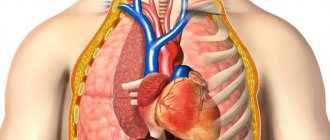
In addition to respiratory diseases, chest x-rays also visualize heart pathologies. Typically, diagnostics for heart diseases are performed along with electrocardiography, which will also illustrate abnormalities in the functioning of this organ.
X-ray of the chest is indicated for shortness of breath, chest pain, and rapid fatigue from the slightest physical exertion. These signs may be symptoms of chronic heart failure.
Using a chest x-ray, doctors determine the following diseases:
- heart attack and post-infarction changes in the heart;
- pulmonary embolism;
- heart defects, both congenital and acquired;
- chronic heart failure;
- cardiomyopathy;
- aortic aneurysm.
Changes in the aorta.
Aortic aneurysm. The procedure is performed for diseases of the skeletal system and spinal column. First of all, X-rays are taken if injuries are suspected, and 100% of patients who have already sustained injuries to the sternum are subject to examination.
The picture will show bruises and fractures. Most often, these can be injuries in the area of the ribs, spine and collarbone. In the image, the doctor sees not only the bone fragments themselves, but also the presence of foreign bodies and displacement of the bones in relation to each other.
If the victim has a pneumothorax and air gets into the chest cavity, this can also be seen using an x-ray.
Processing the results obtained
Based on the visual picture presented on the monitor, the doctor makes a conclusion about the condition of the chest organs. Data obtained in different planes may indicate, for example, the presence of air in the pleural cavity, which is manifested by light areas. The presence of fluid in the pleural cavity is expressed by a kind of line on the screen. In the process of monitoring the dynamic change in the volume of organs, the specialist monitors changes in fluid levels, paying special attention to signs characteristic of purulent processes.
In addition, in the process of studying fluoroscopy data, the doctor assesses the location and size of detected changes (if any). The roots of the lungs, in which large vessels pass, are carefully studied. If the density of any area is increased, then there will be a shadow in the image in this place. Then the doctor describes in the conclusion their structure, shape, quantity and other parameters.
How is an X-ray performed?
Diagnostics of OGK is often prescribed - every person has done this test at least once in his life. A referral for the procedure can be issued by a therapist, traumatologist, cardiologist, pulmonologist, surgeon, oncologist and doctors of other specialties who believe that the cause of the disease is in the chest organs.
The procedure is carried out in a specially designated room where the installation is located. The research will not take much time.
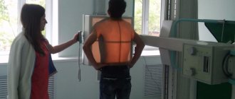
Before taking an x-ray, the x-ray technician will instruct the patient how a chest x-ray is done and what needs to be done. The patient is then placed in the required position in front of the projection screen.
The picture is taken in different projections. This is done to avoid images layering on top of each other. Sometimes pathologies can be invisible on a direct projection, but perfectly visualized on a lateral projection.
During the procedure, the patient is required to remove clothes to the waist and metal jewelry. During the AP X-ray, the radiologist will give the command to hold air in the lungs for about 10-15 seconds.
In the lateral projection, the same actions occur, only the patient’s hand is placed behind the head and the patient is turned sideways to the screen. No special preparation is required before examining the chest.
Compaction syndrome. Low to medium density lesion. Tuberculosis.
Frequency
How long an X-ray is valid is a rather relative concept. The picture in the chest organs can change in a fairly short period of time. The generally accepted opinion is that x-rays/fluorography should be performed once a year. But how often x-rays can be taken varies greatly among different patients.
The following groups are distinguished:
- Patients who are considered relatively healthy should not undergo x-rays more than once a year.
- Patients who work in a hazardous enterprise, live in an unfavorable ecological zone, are long-time smokers, can have X-rays no more often than every six months.
- Persons who work in public catering or are constantly in contact with children should be exposed to x-rays once every 6 months.
- Patients suffering from severe pneumonia have to undergo x-rays 2-3 times a week.
X-ray is a radiation diagnostic method and if possible, it is better to avoid it. However, there are situations when it is urgently necessary to do it. X-ray of the chest and thoracic spine has not lost its relevance over the years and remains an important diagnostic method for making accurate diagnoses.
Projection diagnostics of the lungs
When performing an X-ray of the lungs, it is possible to do the study in two projections. Naturally, the harm from radiation is higher than with one photo.
However, with the help of a plain chest x-ray in two projections, a person’s life can be saved, because not all diseases are visible with a direct projection.
X-ray of the OGK in two projections is performed for diagnosis:
- pneumonia;
- pulmonary tuberculosis;
- cancerous tumors;
- pleurisy;
- the presence of abscesses, cysts;
- airiness of the lung;
- pneumothorax;
- heart sizes.
Lateral projection
Diagnosis of the chest in two projections is carried out in a direct and lateral view. The direct view is also called the anteroposterior view, a name based on the way the X-rays pass through the patient's chest cavity. When examining the lateral position, it makes no difference whether the patient is placed on the right or left side of the screen.
The image in the lateral projection is secondary - it helps to better see those organs that were closer to the screen.
Targeted lateral projection is extremely important for determining the volume of pneumonia and localizing the source of inflammation, as well as for determining the location of tumors in the lungs.
X-ray for a newborn
It is almost impossible to fully predict how the delivery process will proceed. Often, a child may experience various types of injuries while passing through the birth canal. This is the most common reason why a newborn is referred for an X-ray examination of the head and body.
X-rays of an infant may be indicated for the following reasons:
- diagnosis of injuries received during childbirth, as well as after a fall from a height;
- suspicion of the presence of congenital skeletal pathologies - disorders of mineral metabolism and bone formation, bone fragility, congenital inferiority of the hip joint;
- entry of foreign objects into the respiratory tract or gastrointestinal tract;
- intestinal obstruction;
- diagnosis of intrauterine pneumonia, which develops as a result of infection of the lungs in the womb;
- examination for cancer pathologies.
During radiography, special attention is paid to protecting the reproductive organs of both girls and boys from radiation.
It has been proven that the effect of X-ray radiation on a child’s body is twice as high as on an adult’s body.
Interpretation of diagnostic results
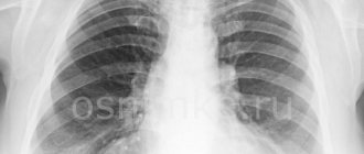
Healthy lungs.
Survey X-ray of the chest. The image is interpreted by a radiologist. More often, doctors have to work with images in two projections. Normally, the lungs are unchanged, the roots are well visualized and have no pathological extensions.
The diaphragmatic contour and sinuses have no pathological signs. The vessels provide a shadow of the usual configuration. Joints, bones and soft tissues are normally unchanged.
The above decoding data is typical for patients under fifty years of age. As you age, changes occur that appear differently on x-rays.
Elderly people have an enlarged heart shadow, their lungs are more transparent, and due to the loss of elasticity of the parenchyma, the vascular pattern is deformed.
For a qualitative assessment of the results, it is necessary to contact doctors with experience, since the description of the results is often subjective.
Chest X-ray: indications and contraindications
Indications for prescribing an x-ray of the lungs:
- Suspicion of pneumonia.
- Monitoring treatment for pneumonia.
- Assessment of the dynamics of the pathological process in tuberculosis.
- Elimination of exacerbation of inflammatory diseases of the lung tissue.
- Early diagnosis of cancer tumors.
X-ray of the lungs in an elderly patient: pronounced thickening and deformation of the pulmonary pattern, ring-shaped shadows in the roots due to chronic bronchitis, unfolded aortic arch.
X-ray of the chest cavity is prescribed by doctors in the presence of the slightest inflammatory changes in the lung tissue in adults. Children under 16 years of age are not even prescribed fluorography, which creates a low radiation dose.
The danger of radioactive radiation is that it can cause damage to the genomic apparatus of the cell. This effect is most active when exposed to rapidly dividing cells. Remember, chest x-rays are performed for children only for emergency indications.
Other contraindications to chest x-ray:
- preventive fluorography - no more than 1-2 times a year;
- High radiation exposure per person throughout the year is a relative contraindication.
There are no absolute contraindications to chest x-ray diagnostics. It is necessary to understand that high radiation exposure throughout the year negatively affects the health of children and adults. They are recorded on the “individual dose load” sheet, as well as the patient’s x-ray fluorographic passport. As a rule, attending physicians simply write down the individual doses received during irradiation there.
Advice to readers! Independently calculate the doses received during X-ray diagnostics for the year. For example, if you took a photograph of the spine in frontal and lateral projections, you received a dose of 2.9 mSv. A subsequent chest x-ray will increase the total radiation dose to 3.08 mSv (2.9 +0.18 mSv). If there is no urgent need, do not take an x-ray of the lungs so as not to be exposed to excessive radiation exposure.
Chest X-rays are contraindicated in pregnant women and children to prevent ionizing radiation from affecting rapidly dividing cells. If necessary, to reduce the harmful effects, targeted X-rays of the lungs can be performed.
X-ray of the chest cavity in a 55-year-old patient with myopathy (expansion of the cardiac shadow to the left)
What do inflammatory foci indicate?
In some cases, x-rays of the lungs reveal foci of pathology and inflammatory foci. They indicate not so much inflammation as the presence of pathology. Lesions may be a sign of:
- oncology;
- pulmonary cysts;
- tuberculosis;
- benign neoplasms;
- fungal infection of organs;
- arteriovenous malformations.
If abnormalities are detected, the doctor needs to differentiate the pathology and determine what disease gave such an image.
For example, a small nodular formation may be a sign of thromboembolism, while a larger one may indicate a cyst or cancer metastasis.
To conduct a detailed study and clarify the diagnosis, doctors recommend performing a computed tomography or MRI.
Radiation exposure during the procedure
Compared to film radiography and fluorography, digital fluoroscopy provides less radiation exposure per unit of time. Thus, when conducting an examination using a digital fluoroscopic method, the patient is exposed to a radiation dose of 0.02-0.03 mSv. During a fluorography study, this figure is 0.15-0.25 mSv, and with film radiography it reaches 0.4 mSv.
Digital X-ray units can significantly reduce patient radiation doses during examination
However, the decisive factor is not a single dose of radiation, but the total dose over the entire period of time of the procedure.
And in this regard, fluoroscopy, of course, is inferior to its counterparts, since its duration can reach up to 15 minutes. In this case, the patient receives radiation in an amount of almost 3.5 mSv.
Roots of the lungs and diaphragm on x-ray
In the image, the diaphragm appears below the pulmonary field and forms a dome. The diaphragm stands high in the central part, and lowers towards the periphery, forming angles - sinuses. Normally, the dome of the diaphragm is located at the level of the fifth or sixth rib. When you take a deep breath, it flattens.
It is difficult to see the roots of the lungs during X-ray diagnostics, since they are covered by the shadow of the mediastinum. In the picture, the visible part is divided into upper, middle and lower parts.
The main shadow is given by the pulmonary artery and the smaller one by the veins, and the bronchi give contrast to the image. Externally, the root of the lung is a whole plexus of vessels and bronchi, which give the shadow in the picture.
Interpretation of results
To make a correct diagnosis, the quality of the image obtained after the procedure is important. If the image is blurry or poorly taken, the doctor may send the patient for a repeat x-ray.
X-ray is assessed taking into account:
- pulmonary pattern;
- lung size;
- lung shapes;
- condition of the bronchi;
- diaphragm conditions;
- heart conditions;
- location of organs in the chest;
- tissue structures;
- state of airiness;
- shape and location of lymph nodes;
- number and position of shadows;
- condition of blood and lymphatic vessels.
Two doctors may give different interpretations of the same image.
Norm
Normal X-ray values for people under 50 years of age:
- no visible focal shadows;
- the structure of the roots is unchanged;
- the contours of the diaphragm are smooth, without changes;
- free costophrenic sinuses;
- lack of gas under the diaphragm domes;
- standard lung transparency;
- no change in bone structure.
The doctor may consider some deviations in radiography to be normal when studying images of people over 50 years of age.
Elderly patients may experience:
- expanded heart shadow;
- increased transparency of the lung fields;
- deformation of the pulmonary pattern;
- loss of tissue elasticity.
A radiologist talks about the norms when assessing a chest x-ray.
Anatomical damage
Chest injuries may include:
- closed;
- open.
| Type of damage | External manifestations, classification |
| Concussions | There are no deviations from the norm in appearance. |
| Bruises | Bruises fall into this category:
The following are diagnosed as bruises:
|
| Compression | The patient complains of suffocation. |
Inflammatory lesions in the picture
Darkened areas on an x-ray are a sign of inflammatory processes in the body.
They are distinguished by size:
- up to 3 mm – small-focal;
- from 3 to 7 mm – medium focal;
- from 8 to 12 mm – large-focal.
Pneumonia
Pneumonia in the image is defined as:
- pronounced shadows;
- additional fabrics;
- venous stagnation, “Butterfly wings”;
- swelling of the lung tissue.
Tuberculosis
When studying an x-ray, tuberculosis can be identified at an early stage by symptoms:
- primary focus (affect) in the upper division of the pulmonary field;
- “path” to the root of the lung;
- enlarged lymph nodes.
On an x-ray, the tuberculosis process is visible as:
- darkening with a clear lower border and blurred upper border;
- an annular cavity with different external and internal contours.
Tumor formations
A benign or malignant tumor is visible on x-ray in cases where:
- the size of the inflammation focus is more than 2 mm;
- the pathological formation does not overlap with other tissues.
Signs of a tumor in the image:
- "plus-shadow" syndrome;
- darkening;
- malignant tumors are denser than lung tissue; in photographs it looks whiter;
- excessively clean lungs;
- displacement of the mediastinum at the entrance;
- pulling up the dome of the diaphragm;
- presence of air in soft tissues.
Tietze syndrome
Tietze syndrome is characterized by aseptic inflammation of one or more upper costal cartilages in the area of their articulation with the sternum. Most often it occurs in women 20–40 years old. It manifests itself as local pain at the site of the lesion.
With Tietze syndrome, pain increases with:
- increased loads;
- operations on organs located in the chest;
- palpation;
- deep breathing.
The appearance of the syndrome is not life-threatening, but radiography may be prescribed to exclude more serious pathologies. Changes will be visible on the pictures 2-3 months after the onset of pain.
Chest X-ray for children
MRI is the safest research method.
Diagnostics for children is recommended only according to indications. If a child has a questionable Mantoux reaction, as well as signs of tuberculosis, then doctors refer the patient for an x-ray. An X-ray of a child is also required in case of a chest injury, since the bones in children are fragile and any blow can lead to a fracture or crack of a rib, collarbone, etc.
Using X-rays, congenital pathologies of organs are visible, so doctors will, without a doubt, send the baby for an X-ray. Many mothers worry about the harm of the study - this is in vain, because it is much more important to diagnose the pathology in a timely manner and cure it.
Advantages and disadvantages of the method
The advantages of chest x-ray include the following:
- high information content;
- efficiency of identifying pathological foci;
- the ability to conduct research in any position of the patient;
- observing changes in dynamics, body movement or breathing;
- allows you to obtain information about the state of the mediastinum (including during breathing);
- makes it possible to examine the diaphragm.
X-ray of the lungs, like any other research method, has its pros and cons
However, along with the advantages, the classical method also has disadvantages. The main one is that the procedure should be carried out in a darkened room (this condition is necessary). Therefore, in order to correctly interpret the resulting image, the doctor must first get used to the darkness. However, in the case of a more modern version, using equipment that displays the image on the screen, this drawback is gradually becoming a thing of the past.
Another disadvantage is that such an examination gives a large radiation dose (as discussed above). Modern digital devices, of course, differ significantly from older models, but the duration of the procedure is still alarming. However, this is not a reason to refuse to carry it out, since it allows you to timely clarify the diagnosis, and therefore prescribe the correct treatment.
Alternative to X-ray
The most harmful method of examination
An X-ray is not the only examination of the chest that helps to obtain an image of the internal organs. Computed tomography and magnetic resonance imaging are no less informative, and sometimes doctors can make a diagnosis after receiving the results of fluorography.
In this case, the patient will even receive much less radiation compared to the classical one. Therefore, when prescribing an x-ray, you should not despair - a conventional examination can be replaced with a digital one, and sometimes even use an alternative diagnostic method.
An X-ray of the OGK is an informative study of organs that cannot be seen otherwise. Therefore, when ordering an x-ray, it must be done correctly, following the doctor’s recommendations. Then you can get a reliable result and, if necessary, begin timely treatment.
Where can I get tested?
Chest X-ray can be performed at any medical or diagnostic institution (center) that has special X-ray equipment. Typically, large centers, public clinics, and hospitals are equipped with such equipment. Since the examination involves a large radiation exposure, the office must be equipped with all the necessary protective devices.
X-ray room
How is diagnosis carried out for infants and older children?
X-rays for children are performed with the lowest dose of radiation. If it is possible to perform an X-ray on a digital device in the clinic, but if not, then parents can go to a diagnostic center with modern equipment, take an X-ray photograph there, and provide the result and transcript to the attending physician.
The procedure is carried out quickly. Before the procedure, the child is undressed to the waist, metal objects and crosses are removed. The body that is not subject to irradiation is covered with lead aprons - this will avoid unnecessary exposure. Attention is focused on ensuring that the baby does not move.
When taking an x-ray, it is fixed, otherwise the x-ray image will be blurry and unclear, which will lead to the need for a second pass.
If the child is under one year old, the picture is taken in a lying position. Parents in protective clothing are nearby to calm the baby and secure him during the procedure. The examination takes a few minutes, after which the baby is allowed to get dressed.
Indications for x-rays of the chest cavity in children
The procedure is prescribed according to indications. The doctor prescribes a chest x-ray for children in the following situations:
- Injury.
- X-rays for bronchitis in children under one year of age suffering from asthma are performed for diagnostic purposes. Assess the condition of the breathing tubes.
- If the cough continues for weeks, the image will show whether the child has developed bronchopneumonia.
- Suspicion of an abscess in the lungs due to pneumonia in children.
- Positive response to a diagnostic test for tuberculosis.
- Preparation for surgery.
On X-ray, signs of pneumonia appear on the images as rounded areas of clearing, covering small areas or a significant part of the organ.