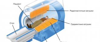
Fluorography (one projection) is a screening (mass) method for diagnosing primary pathologies of the chest organs, and is a type of x-ray examination.
X-rays can adversely affect human health, provoking pathological processes at the cellular level. Large doses can even lead to the development of cancer. Today, doctors use digital fluorography - a safe type of examination . To determine radiation exposure, use the Dosimeter.
Film fluorography: features and differences from x-rays
Film apparatus (There is a cassette on the left side of the CCD matrix)
Film fluorographic examination is not a safe procedure. Like X-rays, it uses rays passing through the human body. They provide a film image of the heart and lungs, from which the doctor makes a diagnosis.
With film fluorography, the image is small in size, and is shown by whole reels of film used per shift. That is why the results of fluorography must wait 1-1.5 days. Since the film size is small, the image quality is unsatisfactory - signs of tuberculosis, pneumonia or oncology can be seen in the image, but a more detailed picture can be seen on an x-ray of the lungs (two projections).
Radiography (lung x-ray) is rarely used. If fluorography is done annually, then radiography is performed only if the signs of pathology described above are confirmed in the primary image.
An X-ray image is a kind of clarifying image, specifying the patient’s condition. How often you can do this will be determined by your doctor based on the permitted annual radiation load. The data can be viewed on large format film, i.e. life-size. Therefore, the doctor prescribes an x-ray only for pre-existing reasons.
It’s quite difficult to find a film machine these days, but such clinics exist!
ANALYSIS OF IMAGES IN LATERAL PROJECTION
The criterion for correct placement for lateral photographs of the lumbar spine is the uniformity of the posterior surface of the bodies, presented as a clear line (Fig. 1). If the laying is not entirely accurate, this line bifurcates (Fig. 16).
The physiological curvature of the lumbar spine is lordosis. Deviations from this condition on lateral radiographs should be correctly assessed, distinguishing true violations of relationships from apparent ones that depend on the projection.
Rice. 1. Scheme of a normal lateral photograph of the lumbar spine of a 14-year-old boy with the torso in an upright position. The line drawn along the posterior surfaces of the vertebral bodies is straight.
Rice. 2. Scheme from a normal lateral photograph of the lumbar spine of the same boy with the torso tilted backward. The line drawn along the posterior surfaces of the vertebral bodies is arcuate.
In Fig. 1, 2 and 3, diagrams with 3 lateral photographs show the normal relationships between the lumbar vertebrae in a normal position, during flexion and extension, i.e., possible excursions of the lumbar spine under normal conditions are presented. As can be seen from the above diagrams, normally there are no angular bends; lines drawn along the posterior surfaces of the vertebral bodies can be straight, convex and concave. Thus, under normal conditions, a physiological mild round kyphosis can be observed during flexion (Fig. 3), but angular kyphosis is always an expression of pathology, just like axial displacement. The shape of normal vertebral discs on lateral radiographs of the lumbar vertebrae depends on the degree of flexion and extension of this part of the spinal column at the time of the image.
The wedge-shaped shape of the intervertebral discs of the lumbar spine, expressed in the greater height of the intervertebral spaces in the ventral section, is captured only when the spine is in a straight position and when tilted back (Fig. 2). When flexed anteriorly, the endplates of the vertebral bodies become parallel to each other.
Rice. 3. Diagram from a normal lateral photograph of the lumbar spine of the same boy with the torso tilted anteriorly.
Rice. 4. Diagram from a lateral photograph of the lumbar and sacral spine of a 4-year-old boy.
Normal relationships between the bodies of the lumbar vertebrae on lateral photographs are characterized by a uniform increase in the height of the intervertebral spaces in the caudal direction. It should be emphasized that the highest disc is between Lv and SI, and that its shape always remains wedge-shaped, even with sharp anterior flexion of the lumbar spinal column. With the destruction of intervertebral discs, the indicated relationships are violated, the height of the discs is reduced and the contours of the vertebral bodies are disrupted. At the appropriate level, physiological lordosis disappears and the entire overlying part of the spinal column is often straightened. Physiological lordosis of the lumbar spine under normal conditions is most pronounced in the lower section, on the border with the sacrum, and the degree of severity of lordosis varies individually. This depends, as indicated above, on the degree of anterior tilt of the Lv body, the severity of the wedge-shaped shape of its body and the wedge-shaped shape of the underlying disc, as well as on the position of the sacrum (Fig. 5, 6, 7). Under normal conditions, regardless of the degree of lordosis, the lines drawn along the posterior surface of the bodies of the lumbar and sacral vertebrae are always arched.
Rice. 5. Different positions of the sacrum and different angles of pelvic inclination with spondylolisthesis (I), with an “arched” sacrum (II) and with an “acute” sacrum (III). Increased lordosis of the lumbar spine is present only with an “acute” sacrum; with spondylolisthesis, a stepped curvature is observed. With spondylolisthesis, due to a slight tilt of the pelvis, the spina iliaca anterior superior (marked with an asterisk) approaches the spine.
It is possible to evaluate the features of the relationships in the lumbosacral region on lateral photographs only on the posterior surfaces of the bodies due to the fact that the phenomena of deforming spondylosis, determined on the anterior surface, can obscure the existing violations of the relationships. For example, with Lv spondylolisthesis, as a rule, cornice-like calcifications of the anterior longitudinal ligament are formed on the sacrum, which smooth out the geniculate bend of the lines drawn along the anterior surfaces of the bodies. Meanwhile, curves along the back surface can always be traced without difficulty. When analyzing lateral images of the spine, it should be remembered that the vertebral bodies can shift not only anteriorly, but also posteriorly due to diseases and injuries. In Fig. Figure 5 shows diagrams from lateral radiographs of the pelvis, which clearly show various individual features of the position of the sacrum and pelvis with an “arched” sacrum, sacrum arcuatum (//) and with an “acute” sacrum, sacrum acutum (III) - in Sherb’s terminology, as well as pathological ratios for spondylolisthesis (I). In the presence of an arched sacrum, when lordosis in the lumbar region is small and the sacrum approaches a vertical position, there is a large lumbosacral angle; with an acute sacrum, when the sacrum is located almost horizontally and there is a pronounced lordosis, a small lumbosacral angle is observed. In Fig. 6 with slight lordosis, with an arched sacrum, a rather large lumbosacral angle is presented. In Fig. 7 with significant lordosis, with an acute sacrum, there is a small lumbosacral angle. One must be able to distinguish spondylolisthesis (anterior sliding of the body and anterior part of the arch of the fifth lumbar vertebra) from increased lordosis. There is a misconception that spondylolisthesis is a consequence of increased lordosis (Whiteman). In fact, this is not true at all.
Rice. 6. Diagram of a lateral view of the normal lumbosacral spine of a 42-year-old man.
Rice. 7. Diagram of a lateral view of the normal lumbosacral spine of a 41-year-old man. The position of the sacrum is approaching horizontal, there is a small lumbosacral angle (marked with a dotted line) - the so-called “acute” sacrum.
Analysis of the position of the sacrum with significant spondylolisthesis (Fig. 5, /) leads to the conclusion that with spondylolisthesis, the sacrum approaches a vertical position (which in the rear image causes excellent visibility of the sacral foramina) and the tilt of the pelvis decreases (as the symphysis rises upward). In addition, it is clearly visible that with spondylolisthesis there is not increased lordosis, as with an acute sacrum, but a cranked bend of the line drawn along the posterior surfaces of the bodies of the lumbar and sacral vertebrae; this bending is due to the exposure of the upper surface of the sacrum due to the anterior displacement of the body. From the analysis shown in Fig. 5, 6, 7 diagrams show that with spondylolisthesis the sacrum occupies a position opposite to the position with an acute sacrum, when the lumbosacral angle is very small. As a rule, Rokhlin, Shakov and Schmorl indicate that with spondylolisthesis, the lumbosacral angle does not decrease, but increases. Thus, it is certainly incorrect to consider increased lordosis in acute sacrum as a predisposition to spondylolisthesis, like prespondylolisthesis according to Whiteman.
Rice. 8. Scheme of lateral (/) and posterior (//) images of the lumbosacral spine of a 53-year-old man.
With small spondylolisthesis, when, nevertheless, on the lateral image (Fig. 8, I) there are all the radiological signs of sliding of the Lv body anteriorly, namely: the phenomenon of osteochondrosis of the disc between Lv and SI, an increase in the lumbosacral angle, a gap in the interarticular area of the arch and the crankshaft the bend of the line passing along the posterior surfaces of the Lv and SI bodies of the sacrum tends to be in a vertical position. This is confirmed by the posterior image (Fig. 8, II), where the sacral foramina are clearly visible in the sacrum adjacent to the film, but the spondylolysis gap is not visible due to the inclined position of Lv relative to the sacrum (the spinous process of Lv is displaced upward). In addition to the anterior tilt of the Lv body, in this case there are also phenomena of listetic scoliosis (i.e., anterior sliding is accompanied by scoliosis and torsion). These features are considered by Bik, Glorier, Movsesyan, Rokhlin to be due to the asymmetrical arrangement of the spondylolysis fissures. There are two theories about the origin of spondylolisthesis. Most authors following Neugebauer consider the cause of spondylolisthesis to be spondylolysis, i.e., an individual feature expressed in non-fusion of the arch in its interarticular area. Others, in particular Meyer-Burgdorf, regard spondylolysis not as an anomaly of ossification, but as a secondary change - the result of a single injury or the summation of microtraumas. It must be assumed that in most cases, spondylolisthesis is a consequence of congenital non-fusion of the Lv arch in its interarticular area and subsequent osteochondrosis of the underlying disc, which bears the maximum load of the entire body.
Rice. 8a. Diagram from a lateral image of the lumbosacral spine of a 54-year-old woman.
Spondylolysis occurs in any part of the spinal column, but, however, most often in the Lv arch. In Lv, spondylolysis is detected on posterior images only when the physiological lordosis is straightened, and then not always, which is explained by the unique location of the spondylolysis gap. Anatomical and x-ray analyzes of the corresponding preparations show that the spondylolysis gap is visible only when its plane coincides with the direction of the central ray bundle. Meanwhile, in this way the spondylolysis gap is located quite rarely. In contrast to spondylolisthesis, with pseudospondylolisthesis, due to deforming arthrosis in the intervertebral joint (art. inter-vertebralis), a change in the relationship of the articular facets in this joint occurs, in particular, an anterior tilt of the upper articular facet, lengthening of the arch of the overlying vertebra and an anterior displacement of the body of this vertebra ( Fig. 8a). The intervertebral disc between the bodies of these lumbar vertebrae usually undergoes degenerative changes. In these cases, a decrease in the height of the corresponding intervertebral space is observed (Fig. 8a). Having a clear idea that spondylolisthesis entails a vertical position of the sacrum, with increased lordosis of the lumbar spine, when the sacrum in the rear image is projectively shortened, there is no need to look for a spondylolysis gap. The gaps identified in some cases against the background of the Lv body (Fig. 9) cannot be interpreted as spondylolysis, as is often incorrectly done; these clearings are due to normal anatomical details, namely, the X-ray joint spaces of the intervertebral joints between Lv and SI, which have a usual frontal location on the left (/), and a rarer sagittal location on the right (II). It is possible to look for a gap caused by spondylolysis in the posterior image only when there are x-ray anatomical signs of straightening the physiological lordosis, i.e., the visibility of the end plates in the body of Lv and the visibility of the sacral foramina (Fig. 10).
Rice. 9. Diagram from posterior images of the lumbosacral region with increased lordosis (/ and II).
In such cases, if the spondylolysis gap can be detected, it is also visible against the background of the Lv body, but it has a completely different location than the gaps of the intervertebral joints. The spondylolysis gap is directed from the median plane obliquely downward laterally; it is located in the area of the interarticular section of the arch. The spondylolysis gap is also very clearly visible on oblique photographs, where it, together with the x-ray joint spaces of adjacent intervertebral joints, causes a zigzag line, called by Rokhlin the “lightning symptom”. It is necessary to clearly differentiate spondylolisthesis from pseudospondylolisthesis on radiographs. Clinically, this is possible only in advanced degrees of spondylolisthesis, which alone can lead to complete sliding of the Lv body (and the entire overlying part of the spinal column) onto the anterior surface of the sacrum. With pseudospondylolisthesis, anterior slippage of the fifth or fourth lumbar vertebra is also observed. However, the genesis of this slippage is different than in spondylolisthesis. With spondylolisthesis, the cause is most often congenital spondylolysis, i.e., a gap in the interarticular part of the arch, and with pseudospondylolisthesis, apparently, also a congenital oblique arrangement of the corresponding intervertebral joints, but the interarticular part of the arch remains undisturbed.
Rice. 10. Diagram from a posterior image of the lumbosacral region of the spine of a 34-year-old man.
In both cases, with constant load, degenerative damage to the corresponding disc gradually develops, resulting in anterior slippage of the overlying part of the spinal column. With pseudospondylolisthesis, the slippage extends only a few millimeters, even in very pronounced cases, but with spondylolisthesis it tends to increase. Osteochondrosis with pseudospondylolisthesis is much less pronounced.
On lateral photographs with pseudospondylolisthesis, both a decrease in the height of the intervertebral disc and the phenomena of deforming arthrosis in the intervertebral joints, which have an oblique location, are clearly visible (Fig. 8a). Thus, consistent, correct X-ray analysis of posterior images of the lumbosacral region, taking into account the relationships between the lumbar and sacral vertebrae, makes it possible to clarify the X-ray diagnosis of spondylolysis, spondylolisthesis and pseudospondylolisthesis. The shape of the bodies of normal lumbar vertebrae in lateral projection resembles rectangles with slightly concave anterior sides. The height of normal vertebral bodies varies individually, but increases evenly in the caudal direction. The body of the fifth lumbar vertebra, as a rule, has a wedge shape, since in the ventral section it is significantly higher than in the dorsal section (Fig. 7). It is unacceptable to consider this normal feature, which is present in every person and at all ages, as a compression fracture. The anterior and posterior surfaces of the lumbar vertebral bodies in all age periods in strictly lateral photographs are limited by one contour, the anterior one being very thin and the posterior one more sclerotic (Fig. 12, 14, 15). If installed incorrectly, the contour of the rear surface bifurcates (Fig. 16). The surfaces of the lumbar vertebral bodies adjacent to the discs are single-circuit during the growth period, but double-circuit in an adult.
Rice. 11. Schemes from photographs of the lumbar and sacral vertebrae of a 9-year-old girl in two projections. Significant destruction of the bodies and intervertebral discs of several lumbar vertebrae, leading to their fusion into a single wedge-shaped formation, in which there are many caseous masses. It is possible to count the number of vertebrae involved in the tuberculosis process only by the arches, more precisely by the intervertebral foramina.
If there are deviations from the average norm in the shape and contours of the vertebral bodies and adjacent discs, it is necessary to resolve the issue of the reasons that caused them. Wedge-shaped deformation of the lumbar vertebral bodies on lateral photographs (smaller height of the ventral part of the body compared to the dorsal part) with visibility of the end plates and preservation of the discs is most often observed with compression fractures. In contrast, during inflammatory processes, the end plates appear to be destroyed, and with congenital fusions between the vertebrae, despite a decrease in the height of the corresponding intervertebral space, the end plates are preserved throughout life, if not throughout, then at some point. With the severity of destructive changes, the bodies of two or more destroyed vertebrae, collapsing in the anterior sections, together produce wedge-shaped formations. In such cases, it is possible to determine the number of vertebral bodies that form them only by the number of arches (the easiest way is by the intervertebral foramina), which are involved in the process much less frequently (Fig. 11). Peculiar changes in the shape of the vertebral bodies and intervertebral discs occur with the “fish vertebrae” symptom, which is observed in various diseases leading to osteoporosis of the spine (Fig. 12). With this deformation, the vertebral bodies are biconcave and the discs are biconvex (Schmorl and Junghans, Rokhlin, Shimanovskaya). The homogeneity of the structure of the bodies of normal lumbar vertebrae on lateral photographs in the upper part is disrupted by the projection overlap of the ribs adjacent to the film (usually XI and XII), and in the lower part by “fusion” with the crest of the iliac bone adjacent to the film.
Rice. 12. Diagram from a lateral photograph of the lumbar vertebrae of a 40-year-old man. Fish-like deformation of the vertebral bodies.
Depending on the standing height of Liv and Lv, either one Lv or also Liv is projectively covered (Fig. 5). The structure of the lumbar vertebral bodies (as well as the vertebrae of other parts) changes differently during various pathological processes. Thus, with tuberculous spondylitis, in the context of general osteoporosis, central cavities are sometimes identified (Fig. 16), and with osteomyelitis of the spine in a certain phase, the process is characterized by reactive transient sclerosis (Fig. 13). The presented series is very instructive both from the point of view of the course of the pathological process and in terms of age-related characteristics of the spine. Very interesting is the reflex spasm of the posterior part of the disc at the beginning of the disease and the disappearance of this spasm during recovery. The normal relationships between the bodies and arches in the lumbar spine on a lateral photograph are such that only the lower articular processes are located below the vertebral bodies. The spinous processes are visible behind the articular processes at the level of the bodies and have the shape of rectangles with rounded corners. The width of the three upper spinous processes is the same, on the fourth it decreases, the spinous process Lv is narrower than all (Fig. 6, 7, 8). All spinous processes are visible against the background of soft tissues, the spinous process of Lv is visible against the background of the iliac crests.
Rice. 13. Schemes from 3 lateral serial radiographs of the spine of the same girl suffering from spinal osteomyelitis: at 11 years of age (/), at 13 years of age (II) and at 16 years of age (III). On the left is a picture 4 months after the onset of the disease.
Rice. 14. Scheme of lateral and posterior images of the lumbar and sacral spine of a 28-year-old man.
Sometimes the individual characteristics of the spinous processes of the lumbar vertebrae are such that their apices are located very close to each other; this leads in some cases, with increasing load, to the formation of secondary articular joints in these areas, in which typical radiological signs of arthrosis and deforming arthrosis can develop and be detected. These features sometimes accompany lumbar pain syndrome (Rokhlin, Sviridov, Tager, Baastrup, Mayer, our observations). A compression fracture of the lumbar vertebral bodies, characterized by a wedge-shaped deformity on the lateral image, can sometimes be accompanied by a fracture of the corresponding arches. In such cases, due to the traction force of the yellow ligaments, fragments of the arch along with the spinous processes are displaced in the caudal direction. This is captured in both lateral and rear projections.
Rice. 15. Diagram from a correct lateral photograph of the lumbosacral region of a 19-year-old man. Single arrows indicate the apophyses of the spinous processes, double arrows indicate the apophyses of the iliac wing, and triple arrows indicate the bullet.
Rice. 16. Diagram from a not quite correct lateral photograph of the lumbar vertebrae of a 35-year-old man.
Fracture of the spinous process of Z.jjj due to a tangential wound (indicated by a single arrow). Traces of physiological sclerosis in the apophyses of the spinous processes of other vertebrae (marked with double arrows).
Due to the anatomical uniqueness of the intervertebral joints, there are no pure dislocations in the lumbar spine, as in the cervical spine, but only fracture-dislocations. This is reflected in the X-ray image (Fig. 14). The presence of apophyses at the apices of the spinous processes, which go around them in the form of scales, should not be interpreted as traumatic changes in lateral photographs (Fig. 15). In Fig. Figure 16 in the lateral projection shows the fragmentation of the spinous process of the spinous process due to a tangential wound and the phase of physiological sclerosis in the apophyses of the adjacent spinous processes. In this case, on the incorrect posterior image (Fig. 17, I), the damaged spinous process of the bm was projectively superimposed on the left lower articular process of the same vertebra, simulating damage to the latter. Meanwhile, on a repeated correct posterior image (Fig. 17, //) damage to the spinous process was established without difficulty.
Rice. 17. Scheme from the posterior, not quite correct (/) and from repeated correct (//) posterior images of the lumbar vertebrae of the same patient (see Fig. 16).
Rice. 18. Scheme of incorrect (/) and correct (//) posterior images of the spine of a 21-year-old man with scoliosis accompanied by torsion. In image I, the anatomical details are located asymmetrically: the spinous processes are deviated from the median plane to the convex side, and the X-ray joint spaces of the intervertebral joints and the roots of the arches in the form of circles are visible only on one opposite side. In image II, the location of these anatomical details appears to be symmetrical.
In addition, in this case, in the correct posterior image, the X-ray joint spaces of the intervertebral joints are captured on both sides between all vertebrae. The exception is the joint between Lv and Si where the gap is visible only on the left, while on the right it is not detected due to its frontal location. The soft tissues in the lumbar spine are homogeneous on lateral photographs from behind, but inhomogeneous from the front due to the presence of gas in the intestinal loops. Sometimes it is possible to catch the shadows of septic abscesses, which are especially well expressed when they are calcified.
What are the differences between digital fluorography?
Digital fluorography is a progressive technique for diagnosing the chest organs. The main feature of such research is the transfer of images into digital format. At the same time, the quality of the image does not deteriorate and becomes better, and the radiation dose to the patient is less.
The image during digital fluorography becomes comfortable primarily for the doctor. If a film image could be unclear, for example, due to development errors, and the doctor needed to carefully peer into the picture and look for pathology, then there are no such problems with a digital image.
Digital scanning fluorograph (the safest and most modern diagnostic method)
The image is of the highest quality; using the program you can enlarge individual areas, add or decrease brightness, and adjust contrast. This provides new opportunities for research.
The procedure time is minimal, it takes about one minute. The rest of the time is spent on paperwork and dressing the patient. The image is ready 5-10 minutes after the examination, and after another couple of minutes it is saved on any storage medium - disk, external memory.
The undeniable privilege of digital images is that the data is stored in DICOM or HL7 format. If necessary, an image of the lungs is printed on a DICOM printer or sent by e-mail.
The fluorography room sees up to one and a half hundred patients per day. These features make digital fluorography in demand.
Digital fluorography (matrix type)
X-ray and fluorography of the lungs are a reliable way to diagnose organ pathologies. Based on the results of the study, it is possible to make a timely diagnosis and begin treatment of the patient.
Projections in X-ray diagnostics
A radiographic projection is an image on a plane obtained when X-rays pass through a given body onto a plane (for example, onto a film in a cassette).
There are many different projections in X-ray diagnostics, but the following standard and additional projections remain the main and frequently used projections. In this case, standard projections are mainly performed in a lying position.
Standard projections:
- Right lateral projection.
- Left lateral projection.
- Ventrodorsal projection.
- Dorso-ventral projection.
Additional projections:
- Oblique projections.
- With a horizontal course of rays - lateral, straight.
Now let's take a closer look.
The right lateral projection (RLP) is performed with the animal lying on its right side, and if the area of study is the thoracic region, abdominal cavity or survey image, then the thoracic limbs should be retracted strictly forward so that they do not block the chest and spine and the sternum should lie parallel to the table, and the ribs should be in superposition.
The left lateral projection (LLP) is performed similarly to the right lateral one, but with this projection the animal lies on its left side.
The position of the animal when performing the lateral projection.
Ventro-dorsal projection (VDP) is the position of the animal lying on its back, while the sternum and spine should be in superposition throughout, and the pelvic limbs should be retracted evenly back.
The position of the animal when performing the ventro-dorsal projection.
The dorsoventral projection (DVP) is similar to the ventrodorsal one, but the animal lies on its stomach.
The position of the animal when performing a dorsoventral projection.
Oblique projections are projections when the animal lies on its side at an angle of 45º to the table surface (see figure). Such projections can be performed both on the right side and on the left. And other variations are also possible depending on the area being studied.
The position of the animal when performing oblique projections.
With horizontal ray path - with this projection, X-rays pass horizontally (from the side). These projections can be performed both in a lying and standing position, and can be lateral and straight.
The position of the animal when performing direct projections with horizontal rays.
The position of the animal when performing lateral projections with horizontal rays.
The position of the animal during lateral projections with horizontal rays.
Projections in X-ray diagnostics are selected based on the area of study and preliminary diagnosis.
It should be borne in mind that if the area of interest is the chest, then only the chest is imaged. From a survey image, it is impossible to evaluate any indicators for examining the chest due to the fact that, firstly, the image is distorted due to the inclination of the X-rays passing along the periphery (the further the object is from the center to the edge, the greater the distortion). Secondly, we will not see small details that should be taken into account when describing the chest and making a correct diagnosis. The same situation occurs when examining the abdominal cavity, head, etc. Therefore, survey photographs should be used very rarely and only in emergency cases.
Therefore, before the owner and the animal go for X-ray diagnostics, it is necessary that the doctor, at a minimum, conduct a full clinical examination, make a preliminary diagnosis and, only on this basis, determine the area of study and the necessary projections.
To identify pulmonary pathology, the following are necessarily used: right lateral and ventro-dorsal projections.
To identify metastases: right lateral, left lateral and ventro-dorsal projections.
For cardiology: right lateral and dorsoventral projections.
To examine the trachea (including to identify collapse): lateral during inhalation and exhalation, straight, oblique cranio-caudal tangentially.
To examine the abdominal cavity: right lateral and ventro-dorsal.
However, do not think that for the same pathology there will be a standard number of images, this is not the case. For each specific case there will be a different number of projections due to the pathophysiology of the ongoing process and the anatomical structure of the animal. In some cases, in order to identify and correctly make an x-ray diagnosis, 2 projections are enough, but in some cases, 4 projections are completely insufficient. Therefore, the main standard projections are initially performed, and then additional projections are made as necessary.
It should also be taken into account that in order for the image to be obtained qualitatively, a well-positioned and fixed animal is important; for this it is necessary that at least 2 people come with the animal for X-ray diagnostics.
I would also like to note that when you come for X-ray diagnostics, you must have a referral for X-ray diagnostics with you, which you can read and print on our website or in the VKontakte group.
You can take an x-ray in Tver at our veterinary clinic “Doctor Ay and Oy” at the address: Tver, T. Ilyina St. 1 B. If you have any questions, please call; (4822)737-077
Source of the VK article “Doctor Ay and Oy” - 2015
radiologist Alexandrova Maria Sergeevna
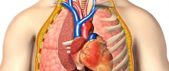
What will an x-ray show?
An X-ray of the lungs helps visualize the organs located in the chest. In the image you can visualize both soft structures and dense ones, but the rays will give different shades. Diagnostics makes it possible to look at the heart, bronchi, lungs, lymph nodes and trachea. The description of these organs is enough to suspect abnormalities in one of them.
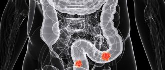
Tuberculosis
As for lung pathologies diagnosed by x-ray, these include:
- sarcoidosis;
- pneumonia;
- pleurisy;
- emphysema;
- oncology;
- swelling of the lungs;
- asthma;
- pulmonary tuberculosis.
X-ray copes with many diagnostic tasks successfully. Therefore, when sending for an x-ray, you should not worry about radiation exposure - it is much more important to see damage to organs.
Digital diagnostics of pulmonary diseases
Digital diagnostics has great capabilities, and therefore the image is more informative. For example, with a digital X-ray you can even find out who the picture belongs to - a man or a woman, since the results show shadows of the mammary glands in the form of contours.
Doctors receive information about the correctness of the study - how deep the breath was and whether the patient was motionless at the time of filming.
Photo of healthy lungs
When assessing the results of digital fluorography, doctors consider the following positions using x-ray irradiation:
- soft tissues;
- integrity of bone elements;
- location of the trachea;
- the presence of calcifications;
- contours of the heart;
- pulmonary fields.
The study is carried out in an anteroposterior projection. The images are informative and make it possible to assess or identify the presence of pathology in the lungs.
Purpose and preparation for radiography in two projections
An X-ray of the lungs in the right or left lateral projection is prescribed in the following cases:
- for the purpose of identifying heart diseases and pathological changes in the pulmonary fields;
- control of catheter placement in the heart, pulmonary artery, as well as for the purpose of assessing pacemaker electrodes;
- in the diagnosis of pneumonia, inflammatory changes in the bronchi, bronchiectasis.
X-ray of the lungs in two projections does not require special preparation, but a person will have to perform some manipulations:
- Remove clothing and foreign objects that cover the examination area.
- Leave your mobile phone and keys on the table, as well as other items that can accumulate radioactive radiation.
In the process of performing an X-ray of the lungs, it is necessary to follow all the recommendations of the x-ray technician. It is important to hold your breath while taking a photo to avoid dynamic blur.
Direct (posterior-anterior) projection during radiography of the lungs
Direct (posterior-anterior) projection during chest x-ray is performed as often as possible if pneumonia or tuberculosis is suspected. There are some technical subtleties when implementing it:
- the ideal focal length between the X-ray tube and the human chest should be on average 2 meters;
- when placing the patient on the stand, the x-ray technician ensures that the chin is positioned on a special holder;
- The height of the clamp is adjusted so that the cervical spine is straightened. When installed, a person leans his hands against the screen, and his chest is projected in the central part of the cassette;
- When exposing the photo, you must hold your breath.
This is how a posteroanterior (direct) projection is performed when diagnosing respiratory diseases.
Lower lobe pneumonia on x-ray of the lungs in direct projection
Anteroposterior view of the lungs
An anteroposterior photograph of the lungs in combination with left or right lateral projections is performed in the supine position. How to take a direct shot:
- the patient is placed on the couch;
- the head end rises up;
- the cassette is placed under the patient's back, and the distance between the X-ray tube and the object of study is selected as directed by the doctor. It should be taken into account that there should be no foreign objects in the path of X-ray penetration;
- exposure is carried out with a deep breath.
Performing right and left lateral chest films
To perform lateral images of the lungs (left and right), special positioning is required:
- hands are placed behind the head;
- the left side leans against the cassette;
- When exposed, you hold your breath or take a deep breath.
The patient is placed next to the cassette on the side that needs to be x-rayed.
Lower lobe pneumonia on an x-ray of the lungs in the left lateral projection
Precautionary measures
Chest X-ray is contraindicated in pregnant women. Radiation exposure to the fetus due to ionizing radiation involves the appearance of genetic mutations, which can lead to developmental abnormalities.
When performing the study, it is necessary to protect the human pelvic and abdominal area with a special lead apron.
In an outpatient setting (in a clinic), when a doctor prescribes radiography in two projections, postero-anterior images should be taken rather than anteroposterior, which is due to the greater reliability of the former.
When choosing lateral images (left or right), you must rely on the doctor’s prescription with a description.
X-ray and fluorography for pneumonia - application features
Pneumonia, or pneumonia, is a dangerous disease with vivid symptoms. It needs to be diagnosed as early as possible, since the pathology has a high mortality rate with late diagnosis. In this case, doctors are faced with a choice - which is better, fluorography or x-ray for diagnosing the disease.
Pneumonia can also be detected by fluorography. The x-ray will show lesions larger than five millimeters in diameter, which can be seen on healthy lungs. At the same time, such spots are not detected during fluorography.
It is impossible to judge the presence of pneumonia by the spots - the fluorography data were obtained at low resolution, and therefore are an assumption. X-ray gives a clear picture and the results will be decisive in making a diagnosis. This is a basic test for suspected pneumonia. With its help it is good to control pathology.
X-ray of the lungs in two projections – indications and contraindications
In two projections, x-rays of the lungs are performed according to absolute indications when the benefits of x-ray examination outweigh the harm. When the pulmonary parenchyma becomes inflamed, life-threatening conditions are formed that will lead to respiratory failure. X-ray of the lungs in two positions involves taking pictures in a straight and lateral position.
Chest X-ray in 2 projections – indications: 1. Inflammation of the alveoli of the lungs (pneumonia); 2. Tuberculosis of the pulmonary fields; 3. Peripheral and central cancer; 4. Diseases of the pleural cavity (pleurisy); 5. Cysts and abscesses; 6. Determination of heart size; 7. Airiness assessment; 8. Detection of pneumothorax (air of the pleural cavity).
The list goes on and on, but the diseases described above are studied using x-ray examination as often as possible.
X-ray signs of pneumonia
It is possible to detect pneumonia on an x-ray. It will look like a single darkening against the background of healthy lungs. However, differential diagnosis may also be required, since the same darkening is detected in the presence of metastases, tuberculoma, and cystic formations.
Therefore, with pneumonia, the difference is implicit and diagnosing a single focus of darkening does not indicate the disease. This can be either inflammation or a sign of tuberculosis or oncology. For a final diagnosis, it is recommended to do additional studies.
Doctors should not miss the possibility of the appearance of a cancerous tumor, so in parallel with the research they add diagnostics for tumor markers.
Is it possible to detect pneumonia?
It is quite simple to identify pneumonia in an image of the lungs. Doctors take into account the number of spots that appear, compactions in the lung segments, and the intensity of light and dark areas. The radiologist will definitely pay attention to the root of the lung and changes in it. They also indicate pneumonia.
If you have pneumonia, the following signs will be visible on the image:
- focal lesions;
- unclear outline of the infiltrate;
- strengthening of the pulmonary pattern;
- expansion or thickening of the root of the lung.
The spots are localized in the lower sections, the pattern around them is deformed, and shadows of the bronchi will be visualized nearby.
Radiography in frontal and lateral projections
Chest X-ray in two projections consists of frontal and lateral images. A direct radiograph is also called anteroposterior, since the X-rays pass through the object under study (the patient's chest cavity) in the anteroposterior direction.
For any examination of the lungs, an AP X-ray is always performed. A lateral view is taken at the request of the radiologist.
What shadows does a direct photograph of the chest organs show:
– Increased airiness of the pulmonary fields with emphysema; – Intense darkening with pneumonia or tuberculosis; – Violation of the innervation of the diaphragm (relaxation of the dome); – Deformation, strengthening or thickening of the pulmonary pattern; – Expansion of the heart; – Collapse of lung tissue – atelectasis; – Pathology of bone and soft tissue.
The list of radiological syndromes associated with the disease is more extensive. A qualified radiologist knows about them. The authors described the most common symptoms of pulmonary pathology.
Where are diagnostics done?
Fluorography and x-rays of the lungs are carried out in any medical institution. The test can also be done in a private diagnostic center, where the patient receives the result in hand and gives it to the doctor who sent it for an x-ray.
Digital fluorography for lung pathologies is a necessary study that identifies such dangerous diseases as tuberculosis, pneumonia and malignant neoplasms. X-rays are used not only for diagnostics. For example, they can treat retinoblastoma, a malignant pathology of the organs of vision. When conducting a digital examination, the radiation dose is minimal, which means that the diagnosis will not harm the patient’s health.
When is Fluorography in Two Projections Prescribed, Diagnostics
Various examination methods are used to detect lung pathology.
One of the most famous is fluorography. The method is used for mass screening and helps to identify many diseases of the respiratory system in the initial stage. Fluorography in two projections (direct and lateral) is used in diagnostically difficult cases.
Features of the examination
Fluorography is a study in which an image is photographed on a special fluorescent screen, which is obtained when X-rays pass through the human body. The end result is a thumbnail image.
Depending on the frame size, the following techniques are distinguished:
- small frame (35/35 mm or 24/24 mm);
- large-frame (100/100 mm or 70/70 mm) - can replace radiography.
Various devices are used to carry out the examination.
Options:
| Stationary | The device is installed in hospitals for continuous operation. |
| Mobile installation | Designed for repeated disassembly and reassembly. It is used for examining people living in remote regions, as well as during preventive examinations in educational institutions and large enterprises. The unit is equipped with a mobile power station. The equipment is transported to the desired location in disassembled form. |
| Mobile station | The device is installed in the interior of a van or other vehicle. A photo laboratory and a mobile power station are also located here. There are also workplaces for personnel. A more convenient option, since it does not require constant assembly and disassembly of equipment. |
Currently, digital fluorography is most often used, in which the image can be displayed on a monitor screen, printed or saved in a database.
Advantages of digital devices:
- minimum radiation dose (30 times less than with the film method);
- high clarity and information content of the image;
- convenience of storing examination results;
- no chemicals or x-ray film;
- low cost of research;
- good throughput.
Basic techniques:
| To save the image, a CCD matrix (a special chip made of light-sensitive photodiodes) is used. | |
| Layer-by-layer scanning using a fan-shaped X-ray beam. The operating principle of the device resembles a scanner. This method is more gentle and allows the use of low doses of radiation. | The result will have to wait |
The resulting image is stored on a computer disk. It can be retrieved, viewed, and, if necessary, sent by email at any time. With the help of special programs, additional studies are possible; there is no need to re-irradiate the patient.
Basic projections for fluorography
The examination of the lungs includes two images: direct (antero-posterior) and lateral. For this purpose, both film and digital fluorography can be used.
A direct projection photograph will help reveal:
- increased airiness of the lung tissue with emphysema;
- focal inflammation (with pneumonia or tuberculosis);
- relaxation of the dome of the diaphragm (due to impaired innervation);
- change in pulmonary pattern;
- atelectasis;
- expansion of the heart;
- pathology of the mediastinum and skeletal system.
Examination in lateral projection is additional. It helps to establish the exact location of the pathological focus, carefully examine the chest and examine nearby organs.
This study is especially informative for tuberculosis, since infiltration, invisible on the anteroposterior image, is clearly visible on the lateral one.
Fluorography in two projections is performed if the following pathologies are suspected:
Necessary information
No special measures are required to prepare for the procedure.
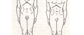
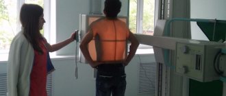
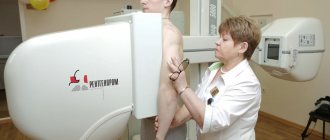
The chest should be pressed against the screen, elbows pulled back
Basic principles of the examination:
- the procedure can be carried out at any time;
- It is recommended to refrain from smoking for 2-3 hours before fluorography (this will make it possible to obtain a clearer image);
- Before the examination, expose the upper part of the body, remove all jewelry (necklaces and chains);
- The picture requires a vertical position, so this method is not suitable for bedridden patients;
- it is necessary to follow the doctor’s recommendations;
- The image is taken during a deep breath (during this phase of the respiratory cycle the lungs expand, which helps to obtain a clearer image).
If pathological manifestations were detected during fluorography, additional studies (computed tomography, x-ray) are necessary.
The examination is not prescribed:
- pregnant women (especially those less than 25 weeks);
- bedridden patients;
- children under 15 years of age;
- for claustrophobia (fear of closed spaces);
- patients with severe respiratory failure.
For timely detection of latent forms of pulmonary diseases, patients over 18 years of age should undergo fluorography at least once every two years. In the event of an epidemiological situation, more frequent examinations (1-2 times a year) of citizens from a high-risk group are necessary.
These include:
- patients with chronic lung diseases;
- military personnel;
- refugees;
- patients registered in a psychiatric or drug treatment clinic;
- HIV-infected;
- convicts from correctional institutions;
- patients receiving treatment for tuberculosis;
- persons who do not have a fixed place of residence.
Once a year, a mandatory examination is required for workers in child care institutions, social services and public catering.
Signs of the disease
Fluorography allows you to identify various pathological changes in the lung tissue.
Examples:
| Strengthening the pulmonary pattern | The pulmonary pattern is a plexus of blood vessels. With various hemodynamic disorders, it becomes deformed and becomes denser. Sometimes areas of increased transparency are identified. Pathological changes are detected with the following problems:
| You can't do without a visit to the doctor |
| Focal shadows | Characteristic for:
They can be either focal or multiple. The size of the pathological formation is of great importance. | Characteristic of tuberculosis |
| Calcifications | A sign of an infectious process. | |
| Pathological changes in the root of the lung | The following violations are possible:
| |
| Fibrosis | Replacement of pulmonary connective tissue is a consequence of inflammation. | Fibrous-cavernous tuberculosis |
| Accumulation of fluid in the pleural cavity | A sign of inflammation of the pleura. | |
| Displacement of the chest organs | The position is assessed:
Displacement is observed with tumors of the lungs and mediastinum, cardiac pathology and fluid accumulation in the pleural cavity. | Hydrothorax with mediastinal shift to the right |
| Changes in aperture position | Often observed after injuries and surgical interventions. | High position of the diaphragm dome |
Regular preventive examination helps to detect tuberculosis and many cancers at an early stage. Timely initiation of treatment will give hope for recovery.
Why do you need two photos?
Good afternoon My son was given a referral for fluorography in two projections. He is a student and lives in a dormitory. Of course, he doesn't eat very well. Recently, the boy who lives in the same room with him was diagnosed with tuberculosis. They said that all contacts need to be tested. But this means being irradiated twice?
Hello! In the initial stage of tuberculosis development, some changes can be seen only on a lateral image.
Examination during pregnancy
Hello! I get a job and go through a medical examination. They gave me a referral for fluorography. The problem is that I'm 8 weeks pregnant. Everywhere they write that “in an interesting position” x-rays cannot be taken. What should I do? What if they won’t hire me?
Good afternoon Present a certificate from a gynecologist stating that you are registered as pregnant, and you will be exempt from this examination.
Source: https://uPulmanologa.ru/diagnoz/instrumentalnaya/flyuorografiya-v-dvuh-proekciyah-25