Trigeminal nerve - structure, features and functions
The trigeminal nerve is a pair of the largest nerves connected to the brain, located symmetrically in the craniofacial area on both sides of the face.
The nerve consists of three branches emanating from the nerve ganglion in the temple area - the ophthalmic branch, the maxillary and mandibular, which stretch to the forehead, eyelids, cheeks, nasopharynx, jaw.
The branches control the motor activity of the muscles in the area of their action (chewing, swallowing), the functioning of the salivary glands, and provide sensitivity to the face, eyelids, tissues of the nasal cavity, mouth, pharynx, dura mater of the brain, soft tissues of the skull, teeth and jaws.
Brainstem stroke (pathology in the pons of the brain)
The picture of the disease, in this case, affects not only the root of the facial nerve, but also the nucleus of the abducens nerve, thereby negatively affecting the external muscle of the left or right eye. Ultimately, in addition to numbness of areas of the face and degradation of the extrinsic eye muscle, convergent strabismus may occur. This complex of deviations requires immediate consultation with a doctor to resolve all existing complications. The latter can also lead to partial blindness.
If hearing deterioration is also observed in parallel, then the brainstem stroke is further aggravated by damage to the nerve at the exit from the brainstem.
Diagnosis of MRI for damage to the trigeminal nerve
Magnetic resonance imaging is a safe, informative hardware diagnostic examination, which is based on a layer-by-layer study of the tissue structure and identifying the causes of nerve damage using high-frequency electromagnetic pulses.
The advantages of MRI of the trigeminal nerve include:
- safety, since the study is carried out without penetration into tissue and the use of x-rays;
- painlessness;
- obtaining images of precise detailed “slices” of tissue in the affected area;
- the ability to detect the slightest deviations from the norm.
Thanks to high-quality images, the technique helps to accurately understand the cause of nerve damage, distinguish pathologies with similar signs, and exclude the development of malignant lesions.
Autotransplantation as an option for returning to normal life
The essence of this procedure is plastic surgery of the facial nerve. The material itself (graft) is taken from the patient’s leg. The latter is used as a binding material without rejection, to which the facial nerve of the healthy half of the face is sewn at two points.
Ultimately, the nerve impulse of the healthy half of the face is the only one for all facial muscles, providing real symmetrical movements. Subsequently, after a few years, the patient can undergo laser removal of scar tissue.
In what cases is research carried out?
Damage to the trigeminal nerve due to hypothermia, infection, inflammation, traumatic influences and other damaging factors leads to a disorder of sensitivity and muscle movement in the corresponding areas, and often to acute, unbearably severe pain.
The patient is indicated for MRI diagnostics in the following abnormal conditions:
- the appearance of nagging or sudden intense pain in the temples, jaws, forehead, upper and lower lips on one side of the face (or both);
- pain when moving the muscles of the face and oropharynx - facial expressions, chewing, swallowing;
- pain when touching the skin of the face, head, mouth (applying makeup, shaving, brushing teeth);
- partial or complete loss of sensation (numbness) on one or both sides;
- the occurrence of sensations of heat or cold, goosebumps, tingling;
- paralysis, spasm or convulsive contractions of the muscles of the face, masticatory muscles, difficulty swallowing, eyelid tic, drooping corner of the mouth or eyelid;
- the appearance of asymmetry and swelling on the face;
- hearing disorders, pain inside the ear.
Similar manifestations occur when the trigeminal nerve is damaged, associated with its inflammation (neuritis), damage or spasm of the network of vessels supplying the nerve fiber (neuropathy).
The concept of neuralgia indicates acute pain along the branches. It is often combined with inflammation, but also occurs with mechanical damage - pinching, hypothermia, bruise.
Neuralgia is not characterized by changes in the structure of the affected nerve, motor disturbances and weakening of sensitivity.
Surgery
For the most part, it is a last resort measure that is recommended for use in cases of congenital neuritis of the facial nerve or its complete rupture due to mechanical damage. There are two options for this treatment. This can be either suturing the nerve or performing neurolysis. If there is no improvement 10 months after surgery and electrophysiological data still indicate significant impairment, repeat surgery may be required.
The operation is effective only in the first 12 months of the disease. This is evidenced by hundreds of clinical cases in which the result had varying degrees of success, but most often minimal. This statement is justified, since a year after the development and worsening of neuritis of the facial nerve, atrophy of the facial muscles begins. The latter in this condition are not amenable to any treatment.
Contraindications
Before prescribing a diagnostic procedure, the specialist must be sure that there are no contraindications to the examination.
MRI is not performed if the patient’s body contains:
- a pacemaker due to possible cardiac arrest under the influence of electromagnetic fields;
- electronic prostheses or neurostimulators (malfunction);
- clips installed on vessels (risk of displacement);
- metal implants, prostheses, braces, crowns and pins made of ferromagnetic materials (alloys of steel, gold), intrauterine device, fragments from bullet and other wounds (distortion of images, risk of overheating and displacement).
The procedure is contraindicated:
- if the patient is diagnosed with epilepsy, a tendency to seizures;
- for severe psychoneurological diseases, claustrophobia;
- during gestation (risk of negative effects of a high-intensity electromagnetic field on the fetus);
- with too high body weight (most tomographs are designed to study patients weighing up to 120-150 kg).
Diagnosis of this type is not recommended for patients with permanent eyeliner. Restrictions are possible with tattoos in the head area due to the risk of skin overheating, irritation and swelling.
Contraindications to contrast MRI
In order to increase the information content of diagnostic data, assess the condition of blood vessels and blood flow, improve the visibility of inflammatory, malignant foci in the image, about 20% of magnetic resonance studies are carried out with intravenous administration of a gadolinium-based contrast agent (Dotarem, Gadodiamide, Omniscan, "Gadovist")
The need for a contrast-enhanced study is determined only by a doctor.
Although contrast agents are harmless, there are additional contraindications:
- severe allergy to gadolinium or another substance;
- severe impairment of the excretory function of the kidneys, since the contrast agent in this case is removed from the blood slowly and can poison the body;
- feeding a baby with mother's milk - even small doses of gadolinium can cause allergies in the child and overload the kidneys.
It is not advisable to conduct contrast tomography in case of anemia and blood diseases, bronchial and cardiac asthma (due to the risk of an attack due to allergies).
Treatment of the disease
Treatment, first of all, must be comprehensive. If we are talking about a reliable traumatic injury, whether it is an iatrogenic cause or just trauma, it makes sense to think about microsurgical intervention. The only downside is that microsurgeons do not always take on work of such complexity.
Of the medications, an important role is played by:
- B vitamin;
- doctive acid;
- drugs that improve neuromuscular transmission (prozerin);
- drugs whose action is aimed at increasing the level of microcirculation;
- general strengthening drugs.
Non-drug treatment methods play an important role, namely:
massage;
- physiotherapeutic procedures;
- Plaster traction (the therapist helps to develop the affected part of the face);
- gymnastics (recommended for a year after the onset of symptoms and their removal with therapeutic drugs).
The meaning of gymnastics is to work and stimulate the nerve, stimulate the work of muscles while simultaneously restoring facial symmetry. A very important point in performing gymnastics is fixing the healthy side of the face. You can do this with your palm. Otherwise, all efforts will initially go to the more effective side, thereby provoking the development of symmetry with the affected area.
Facial nerve neuropathy is quite treatable, especially if you consult a doctor at the onset of the lesion. It is very rare that the full range of symptoms will remain for life.
The disease, caused by inflammatory processes, does not pose any threat to human life, with the exception of significant discomfort. Correct and timely treatment gives the most favorable prognosis for getting rid of facial neuropathy.
Taking estrogen drugs should be under the strict supervision of the attending physician so that the situation does not worsen due to other complications and inflammatory processes. Moreover, the cost of such a medicine is quite high.
Preparing for hardware diagnostics
No special preparation is required for magnetic resonance examination of the trigeminal nerve.
General recommendations:
- It is required to wear clothes without metal fasteners, remove metal chains, pendants, earrings, hair clips, and piercings.
- It is not advisable to apply makeup before the session.
- Before a procedure with contrast, you should not load your stomach with food.
If you are prone to any type of allergy, the patient must notify the radiologist about this.
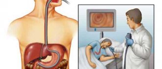
Diagnosis methods
Determining the presence of pathology is not difficult for a specialist - a neurologist during the initial examination. Palpation of the indicated points, causing discomfort and pain, indicates the presence of a disorder. Next, it is necessary to differentiate the anomaly and determine the cause. For this, the following diagnostic methods are used:
- Computer scan of the skull. Aimed at studying bone fibers, deformations of hard tissues, and narrowing of intracranial tracts.
- Magnetic resonance imaging of the head brain. Necessary to exclude developing tumor, cystic objects that can provoke intracerebral nerve compression.
- Magnetic resonance angiography. Looks for a connection between pathology and vascular changes.
Consultation with specialized specialists (dentist, ophthalmologist, otolaryngologist) may be required. This will exclude or confirm the presence of underlying diseases affecting the trigeminal branch.
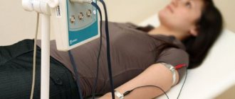
How is an MRI done?
The patient lies face up on a special table with soft straps to prevent involuntary movements.
During the contrast procedure, a dropper is installed for intravenous administration of the drug. The table is pulled inside the illuminated capsule of the tomograph and the scanner, emitting “clicks”, begins to work around the patient’s head. The procedure lasts about half an hour, with contrast enhancement the session lasts up to 60 minutes.
During an MRI, the patient may experience:
- dizziness due to decreased blood pressure;
- nausea due to exposure to electromagnetic radiation;
- warmth, tingling, coldness along the vessel into which gadolinium was injected, itching sensation at the injection site;
- metallic taste in mouth.
If during the diagnosis the patient is scared or feels unwell, you should immediately inform the radiologist about this using the button.
Experts' forecast
In 80% of cases, the client completes the full course of treatment and makes a full recovery. Such a positive picture is only possible if there are no associated complications, which include:
- otitis;
- mumps;
- herpes.
Full rehabilitation can take up to several years, which significantly complicates the quality of life of some patients. Cases of complete recovery after 3 months are much less common. A similar scenario is possible only when damage to the facial nerve occurs at its exit from the skull.
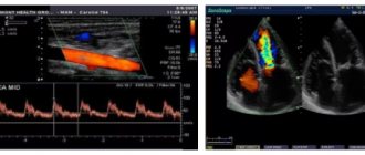
What do diagnostic data indicate?
Energy changes in the survey area that occur under the influence of a magnetic field are processed by a program that converts the data into a series of high-quality 3D images.
When deciphering the results, the doctor receives information about the causes of nerve damage, including:
- inflammatory processes, injuries to nerve fibers or nearby tissues;
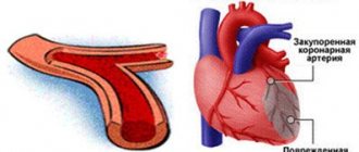
- blood supply disorders;
- necrotic changes in nervous tissue (cell death);
- infections in the nasopharynx and air sinuses (sinusitis, frontal sinusitis, sinusitis), diseases of the oropharynx in the chronic stage;
- bite defects, injuries in the face, jaw;
- abnormally active bone formation;
- tumors compressing the branches of the trigeminal nerve;
- pinching of the nerve fiber in the bone canal by veins or arteries due to a vascular anomaly;
- vascular aneurysm;
- multiple sclerosis, with damage to the protective myelin sheath of the nerve fiber.
A diagnostic procedure for trigeminal neuralgia gives the doctor the opportunity to analyze MRI scans, examine the structure and condition of the nerve fiber, blood vessels and surrounding tissues in section.
Thanks to the high accuracy of the images, the doctor identifies foci of inflammation, early pathological changes, areas of damage, pinching and compression of the nerve.
Facial neuritis (facial nerve neuropathy)
Paralysis of the facial muscles on one side of the face (prosopoplegia) as a result of damage to the facial nerve is a common disease that requires urgent treatment.
In a large number of cases, damage to the facial nerve (FN) occurs in the bony canal (pyramid of the temporal bone), before exiting the skull through the stylomastoid foramen. Bone canal L.N. is quite narrow, which contributes to compression of the nerve in it during the formation of edema. Edema is usually caused by impaired blood supply to the nerve as a result of hypothermia or a viral infection. In the first hours after the onset of paresis of the facial muscles, therapeutic measures should be aimed at relieving swelling of the facial nerve. Otherwise, irreversible death of nerve fibers may occur. On the first day of the disease, it is important to establish the location, nature and extent of nerve damage. In the following days, the diagnosis aims to accurately establish the etiological factor - infection, ischemia, etc.
In accordance with these standards, our clinic primarily performs brain tomography (MRI, CT) and electromyographic study of L.N.
Localization of facial nerve damage
First of all, it is important to differentiate between the intracranial localization of the lesion, the lesion in the bony canal of the temporal bone and after the exit of the nerve on the face.
1. If paralysis of the facial muscles occurs simultaneously with the appearance of hemiplegia (weakness) in the limbs of the ipsilateral (same) half of the body, then we are talking about a focal lesion of the opposite hemisphere of the brain. The most likely cause is a stroke. Brain tomography in this case makes it possible to clarify the cause of hemisphere damage (tumor, multiple sclerosis?). In mild cases, tomography does not reveal lesions. In this case, EEG (electroencephalographic study) allows one to differentiate between damage to the cerebral cortex and subcortical (lacunar) strokes. We do not perform EMG in cases of hemispheric lesions.
In rare cases, limited central damage (damage to the cerebral hemispheres) appears only on the face. In this case, a simple forehead wrinkling test can exclude a central lesion. The forehead muscles receive innervation from both hemispheres. Therefore, if one of the hemispheres is damaged, the forehead muscles do not suffer. At the same time, when the facial nerve itself or its nuclei are damaged, paresis of the facial muscles of the entire half of the face, including the muscles of the forehead, is observed.
*It must be remembered that the masticatory muscles receive innervation from the trigeminal nerve system. Therefore, their function is preserved. The oculomotor muscles, innervated by the 3rd, 4th and 6th pairs of cranial nerves, are also preserved. Ptosis (drooping eyelid) is not a symptom of facial nerve damage. On the contrary, damage to the facial nerve is characterized by the inability to close the eye.
2. Damage to the nuclei of L.N. in the brainstem is usually accompanied by paralysis or paresis of the limbs of the opposite side (Millard-Hubler syndrome) and/or paresis of the abducens nerve on the same side, due to the involvement of the nucleus n. Abducens (Fauville syndrome). The latter is manifested by convergent strabismus: the inability to move the eye of the affected side to the side.
*The upward movement of the eyeball when the eyes are closed (Bell's phenomenon) is not a symptom of damage to the oculomotor nerves.
We definitely do an MRI, since MRI is better at visualizing the deep structures of the brain than CT. Brain tomography visualizes structural abnormalities. We obtain additional information about the localization of functional disorders by conducting EMG studies of the blink reflex (R1 and R2 components of the blink reflex are generated in different parts of the brainstem) and acoustic brainstem evoked potentials (Components IV are generated in different parts of the brainstem).
Most often, disorders at this level are caused by demyelinating disease, tumors, vascular malformations, syringomyelia, etc. With the sudden appearance and development of symptoms (within hours) of damage to the hemispheres or brain stem, an acute cerebrovascular accident is assumed. The patient is admitted to the intensive care unit.
3. The third variant of intracranial lesion is the lesion of L.N. along the path from the brain stem to the entrance to the bony canal of the temporal bone (porus acusticus internus) in the so-called cerebellopontine angle. Here the facial nerve follows next to the auditory nerve and the intermediate (regulates tear and salivation, carries taste fibers from the anterior two-thirds of the tongue) nerve. Therefore, with pathology in the area of the cerebellopontine angle, in addition to paresis of the facial muscles, there is deafness on the same side, loss of taste on the same side of the tongue, dry mouth and decreased tear production may be felt.
The most common causes are acoustic neuroma, vascular malformations, basal gliomas, etc. MRI is performed to verify the diagnosis. If necessary, contrast-enhanced MR angiography is performed. Throughout the entire length from the cerebellopontine angle to the exit of the facial nerve to the face, the symptoms of its damage may indicate the presence of a serious ENT pathology: purulent otitis, with the formation of a fistula, mastoiditis (inflammation of the mastoid process), etc. Therefore, in case of the specified localization of the lesion, a consultation with an ENT specialist is required in our clinic.
4. Upon entering the bony canal, the facial and intermediate nerves diverge from the auditory nerve. Therefore, with damage to the canal, deafness (if it is not associated with ENT pathology) is not observed. On the contrary, so-called hyperacusis is detected - increased sensitivity of the ear to sounds, especially high tones. This phenomenon is associated with dysfunction of the nerve fibers that run as part of the L.N. to muscle m. Stapedius of the inner ear, which regulates the adjustment of the mechanical part of the sound-receiving apparatus.
5. Symptoms of damage to the facial nerve in the bone canal of the temporal bone. As the facial nerve moves from inside to outside in the canal, the following nerve branches are successively separated from it: n. Petrosus major (lacrimation), n. Stapedius (to the muscle m. Stapedius), Chorda timpani (salivation and taste fibers). Therefore, when the nerve lesion is localized before the origin of n. Petrosus major, no lacrimation observed. Hyperacusis can be observed only when the nerve is damaged before the n. Stapedius. Dry mouth and taste disturbances occur if the nerve is affected before the Chorda timpani leaves. The latter departs from the facial nerve close to its exit from the bone canal to the outside.
Bell's palsy - paralysis of the facial muscles of half the face with the addition of these symptoms, resulting from swelling and compression of the nerve in the bone canal - the most typical case of L.N. neuropathy.
Hunt's neuralgia (Ramsey Hunt syndrome) is Bell's palsy + pain and the presence of characteristic bubbles in the area of the external auditory canal, pinna and behind the ear. Hunt's neuralgia is a sign of herpetic nerve damage. In this case, we carry out a serological blood test for Herpes Zoster.
6. After the release of L.N. outward from the stylomastoid foramen, it branches on the face. Here it is available for direct study using electromyographic methods. Typically, M-responses of the nasal muscles, orbicularis oculi and oris muscles are studied when the nerve at the exit from the stylomastoid foramen is stimulated. Identification of signs of neuropathy in the peripheral part of the facial nerve on both sides indicates the presence of polyneuropathy. In this case, an EMG study of the nerves of the limbs is performed to verify the presence of polyneuropathy.
After branching, some branches pass through the parotid gland. Tumors of the parotid gland can cause their damage.
If an EMG study is performed in the first 4 days (preferably in the first two days) after the onset of paralysis of the facial muscles, then these studies allow for differential diagnosis of lesions on the face and inside the bone canal (when topical diagnosis based on the clinical symptom complex is impossible). After 4-7 days, Wallerian degeneration of distal nerve fibers (on the face) may occur if they are damaged proximally (in the canal).
NB: Intracranial lesion indicates the presence of serious diseases that threaten to lead to damage to other parts of the brain if not diagnosed and treated in a timely manner. The first examination of the patient is brain tomography.
Neuritis of the facial nerve in the classic form, i.e. in case of damage in the bone canal and in the facial area, it also requires urgent hospitalization of the patient in a neurological hospital with immediate anti-edematous therapy, the basis of which is corticosteroids. It is important to carry out timely measures to restore nutrition and blood supply to the nerve.
*Facial nerve neuritis is a common but outdated name. More correctly, neuropathy of the facial nerve, since this concept includes not only inflammatory (neuritis) diseases, but also nerve lesions of other etiologies.
Objective criteria for the severity of facial nerve damage.
Nature of the lesion and prognosis.
Please note that slight facial asymmetry without facial muscle weakness is not a consequence of facial neuritis. Close your eyes, stretch your lips in a wide smile, whistle, wrinkle your forehead and frown - make sure that the muscles are really paralyzed.
1. The first EMG study for neuropathy of the facial nerve is recommended to be carried out in the first 4 days after paralysis. The study consists of two parts: EMG of the facial nerve and study of the blink reflex on both sides. With EMG L.N. recording is made from the facial muscles innervated by direct stimulation of the nerve in the area where it exits the bone canal. The blink reflex is recorded from both orbicularis oculi muscles when the trigeminal nerve is stimulated. The impulse along the trigeminal nerve enters the brain stem, where it switches and enters the nuclei of the L.N. on both sides. After which, from the core of L.N. the impulse travels along the entire nerve (including the bone canal) to the facial muscles.
Three typical types of lesions with neuritis of the facial nerve and their interpretation:
— deviation from the norm in EMG L.N.: lesion on the face
- normal EMG results of L.N., but the amplitude of the R1 component of the blink reflex is reduced: lesion in the bone canal - complete destruction of axons or axonotmesis (incomplete damage to the axon with the formation of a persistent block of conduction along it). Complete absence of the reflex is an unfavorable prognosis.
- normal EMG results of L.N., but the latency of the R1 component of the blink reflex is increased: demyelination of the nerve (damage to the myelin sheath). Favorable prognosis.
2. The second EMG study is recommended to be carried out 10-15 days after paralysis. The following EMG signs allow you to verify the diagnosis:
— decrease in amplitude (%) of the M-response of the facial muscles during EMG L.N. compared with the first study in proportion to (%) irreversible degeneration of nerve fibers. If the amplitude has not decreased, the prognosis for complete recovery is favorable.
— the amplitude of the M-response is preserved, but the amplitude of the reflex response is significantly reduced with normal latency: axonotmesis, restoration of nerve function may take several months (with adequate therapy).
— the amplitude of the M-response remains at the same level, but the latency of the first component of the blink reflex is significantly increased. Compared to the first study, a clear correction of the deviation of the reflex component from the norm is noted. Recovery occurs due to remyelination (restoration of the myelin sheath of the nerve). The prognosis is favorable. Recovery within several weeks with adequate therapy.
— The M-response of the facial muscles has disappeared: an extremely unfavorable prognosis. Formation of facial muscle contractures.
- The M-response has sharply decreased, the reflex response is absent in the first and second studies of the blink reflex. The prognosis is unfavorable. Recovery is possible by the sprouting of new fibers into denervated muscles with the formation of aberrant conduction (hemifacial spasm, tics).
If it is not possible to compare with the first study (late hospitalization), from 2-3 weeks from the onset of the disease, it is possible to conduct needle EMG of the facial muscles to verify axonal damage.
3. The third study is recommended to be carried out 1.5-2 months from the onset of paralysis. In addition, during the treatment process there is often a need to evaluate the effectiveness of the therapy. Then additional research is carried out on an individual basis. In addition, if the restoration of the nerve as a result of axonotmesis lasts for several months, we repeat the neurophysiological study after 3-4 and after 5-6 months.
There is no need to dramatize the situation if you notice sudden paralysis of the muscles of half the face. As a result of systematic tactics of therapeutic and diagnostic measures, timely therapy aimed at limiting the spread of the pathological process, and a pathogenetically determined approach to identifying the etiology of the disease, it is possible to achieve complete restoration of facial muscle function in the vast majority of patients. But remember: if you have neuritis of the facial nerve, you must immediately consult a doctor. Treatment and examination should be started within the first hours of the onset of the disease.
For information about making an appointment with specialists, please contact us by phone: 8;, or by email. The email address is being protected from spambots. Javascript must be enabled in your browser to view the address. / The email address is protected from spambots. Javascript must be enabled in your browser to view the address.
Forecast and preventive recommendations
The disease does not pose a threat to life, but it greatly affects the patient’s quality of life and well-being. If you seek medical help in a timely manner, the prognosis for recovery is favorable. With instrumental treatment, relapses occur only in 3-15% of cases.
Preventive measures are:
- complete relief from diseases of the jaw area, ear diseases, intracranial sinuses;
- regular monitoring by a neurologist;
- avoidance of negative external factors;
- prevention with anticonvulsants during acute respiratory viral infections and influenza.
Remedial measures
Therapeutic assistance consists of long-term use of anticonvulsants. The dosage of such drugs is prescribed individually. Reception begins with small doses and gradually increases until the effective amount of the substance is determined. Treatment is carried out for several months, then a gradual reduction in dosage is prescribed.
As an accompanying, symptomatic therapy, antiallergic drugs are prescribed, indicated to reduce swelling. At the same time, antispasmodics are used to relieve pain. For intense attacks, therapeutic blockades with local administration of appropriate solutions are indicated. A good effect is achieved using physiotherapeutic methods.
If growing tissue objects compromising the trigeminal branches are detected, a decision is made to surgically remove the formations. The treatment is carried out by a neurosurgeon.