Esophagoscopy is an endoscopic examination of the esophagus, which is carried out using a bronchoesophagoscope or esophagoscope. The method allows you to diagnose diseases of the organ, carry out some therapeutic manipulations to remove small growths, coagulate bleeding, bougienage the narrowing of the esophageal tube, and remove foreign objects. This technique is similar to gastroscopy, but, unlike it, during esophagoscopy only the esophagus is examined. Next, we will consider the features of the procedure in more detail.
Equipment used
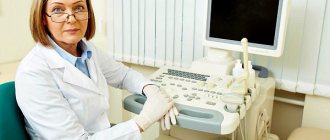
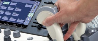
To carry out the examination, instruments called an esophagoscope or bronchoesophagoscope are used, as well as various small instruments, auxiliary equipment, medications, washing liquids, and an electric suction.
Bronchoesophagoscope is a device consisting of:
- handles;
- illuminator;
- a set of tubes of different lengths, diameters, cross-sections, materials, performing various functions during the examination;
- holders, probes, hooks.
It is intended for examination of the bronchi, trachea, and esophagus. The esophagoscope is designed similarly, but the tubes for examining the esophagus have an oval cross-section, in contrast to the round tubes for bronchoscopy, and there are fewer accessories for the device.
There are different designs of devices, for example, the Brunings system, where the illuminator is built into the handle; the light, reflected in a mirror installed at an angle of 450° to the tube, enters it and illuminates the walls of the organ being examined. The Friedel device is distinguished by solid tubes of various diameters and an illuminator on the handle, which simultaneously serves as a chamber for inhaled and exhaled air.
Before use, removable tubes, forceps, probes, holders are boiled or treated with a disinfectant solution. The handle with the illuminator is not boiled, but thoroughly wiped with alcohol. The rest of the instruments are laid out near the examination site in an order convenient for performing manipulations, that is, according to the order of use.
Special training
If the procedure is carried out for emergency reasons, then preparation for the study consists only of preliminary anesthesia, usually of a general nature.
All patients undergoing routine esophagoscopy should adhere to the following recommendations:
- on the eve of the study, the last meal is taken no later than 16.00-17.00, in the morning before the procedure it is forbidden to have breakfast, it is better not to drink liquid an hour before the procedure;
- if the patient is afraid of the examination, then mild sedatives or tranquilizers are prescribed, and sleeping pills are taken at night;
- Before the examination, it is better not to take any medications, smoke or drink caffeine-containing drinks, as they lead to increased salivation, and this complicates the visualization process;
- If the patient has removable dentures, they are removed during the procedure.
Shaykhnurova Lyubov Anatolyevna
Indications for esophagoscopy
Esophagoscopy is performed in the following cases:
- inflammatory processes in the esophagus;
- the presence of tumors and neoplasms;
- achalasia;
- presence of diverticula;
- foreign body entry;
- fused scars;
- varicose veins;
- reflux esophagitis;
- burns of the esophagus of various origins;
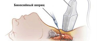
- the need to take a biopsy of the organ for cytological examination.
In some of the listed cases, esophagoscopy from examination flows into treatment. Neoplasms are removed, a foreign object is removed, medications, for example, are delivered to the burn site, and bougienage is performed.
Description of service
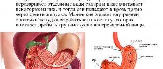
Endoscopy is an examination of the gastrointestinal tract from the inside by inserting a long flexible cable with a miniature camera that transmits an image to a monitor. Allows you to assess the condition of the mucous membrane of the organs being examined, take tissue samples for laboratory analysis, and also carry out micro-operations to remove certain formations, stones, cauterize damaged areas, and so on. Various specialists consult endoscopy: pulmonologists, urologists, gynecologists, gastroenterologists. This method allows you to assess the condition of the esophagus, pharynx, stomach, duodenum, colon, rectum, and bile ducts. Since the endoscope allows for minimally invasive treatment measures, endosurgery has gained momentum in its development. The endoscopic examination method is painless and does not require rehabilitation, but is very effective.
Diagnostic doctor Work experience - 7 years
Sign up for a consultation by phone
Preparing for a patient examination
The patient must come to the examination prepared as was explained to him during the conversation with the doctor. The preparation is as follows:
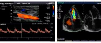
- undergo examination for pathology of the heart and blood vessels;
- do not eat for 6-8 hours before the procedure;
- eat light food in small portions for two to three days;
- do not drink alcohol 2-3 days before the examination;
- do not smoke on this day;
- take an x-ray of organs;
- take blood and urine tests;
- remove removable dentures (if any), glasses, lenses, jewelry;
- rinse your mouth with an antiseptic;
- perform hygiene procedures before the examination;
- Notify your doctor in advance about your medications and chronic diseases.
This preparation is explained by the fact that during esophagoscopy the doctor strives to minimize the likelihood of vomiting, the mass of which can cause the patient to choke. In addition, food that remains in the esophagus interferes with examination, changing the picture of the condition of the organ. The results of tests and X-ray examinations will help the doctor make a decision about the possibility of performing esophagoscopy of the esophagus.
On the day of the procedure, the patient comes to the appointment with a towel, sheet, water, and a change of clothes.
Where can I undergo the procedure and is it possible to undergo it for free, cost
Esophagoscopy of the esophagus does not require expensive equipment and is therefore available to all segments of the population. The study can be performed in an inpatient setting at a regular public hospital completely free of charge if the patient went there as prescribed by a doctor.
The procedure is offered by a wide network of private clinics, where the cost of this service is relatively low. Thus, in Moscow, endoscopic diagnosis of the esophagus in 2020 cost from 1,800 to 3,000 rubles. In small towns - about 900 rubles.
The price depends on the authority of the clinic, the equipment used, the name and qualifications of the esophagoscopist.
Features of the procedure
The difference with esophagogastroduodenoscopy and fibroesophagoscopy in esophagoscopy is that in the first case the esophagus, stomach and duodenum are examined at once, in the second - only the stomach cavity.
Before starting the procedure for examining or treating the esophagus, local anesthesia is administered - Lidocaine or Xylocaine. The purpose of local anesthesia is to reduce the patient’s discomfort during insertion of the probe into the esophagus, to reduce the sensitivity of the palate and other organs that react to the touch of the tube by gagging and coughing.
Under general anesthesia, esophagoscopy is performed in the following exceptional cases:
- the patient's mental illness;
- large foreign object in the tube;
- bleeding, injury;
- severe spasm or inflammation;
- deaf-mute;
- problems with the heart and blood vessels.
The procedure time usually ranges from 2 to 20 minutes. Esophagoscopy of the esophagus is performed with the patient lying on his left side or sitting in a special chair. In a sitting position, the assistant should hold the patient's head and shoulders, assisting the doctor during manipulations with the probe.
The study begins with the administration of relaxing and sedative drugs to the patient in order to achieve complete relaxation of the esophagus. A few minutes after treating the uvula and palate with an anesthetic, a special ring is inserted into the patient’s mouth so that he does not close his mouth, and the device begins to be inserted along the midline of the oral cavity into the larynx, then into the esophagus. To open the upper opening of the esophagus, the patient is asked to make a swallowing movement.
It depends on the skill and experience of the doctor how successfully the probe will be inserted without unnecessary problems and damage. If there is a deviation from the correct insertion of the tube, its direction is corrected by tilting the patient’s torso slightly forward. As it progresses, a thorough examination of the condition of the walls of the organ and its mucous membrane is carried out. For a diagnostic examination, a flexible probe is used, and for a therapeutic examination, a rigid probe is used.
Depending on the patient’s age, the diameter of the inserted tubes is selected. The picture of the folding of the walls, their severity, and the condition of the veins also varies depending on age. A normal healthy lining of the esophagus is pink in color without redness. The number of folds becomes greater closer to the stomach. In the presence of pathology, the structure of the surface and its color change. Erosion, ulcers, scars, and bleeding may occur. The condition of the esophagus during examination is described in a special protocol. Peristalsis and diameter of the esophagus are measured. Based on the protocol, further treatment is prescribed.
At the end of the examination, the esophagoscope is carefully removed. The manipulation is carried out with great care and attention due to the presence of other vital human organs around the esophagus.
Therapeutic esophagoscopy is divided into urgent and planned. Urgent is usually urgent and is associated with emergency care, for example, when a foreign body enters the esophageal cavity, a child swallows parts of toys, or food debris. A planned procedure can be carried out after an examination to install a bougie, remove tumors, and stop bleeding.
General idea of the methodology
Esophagoscopy (from the Greek oesophagus - esophagus and skopeo - examine, examine) means a visual examination of the walls and mucous membrane of the esophagus. The specialist examines the hollow organ or performs a series of manipulations on its internal surface using an esophagoscope. The optical device is an elastic rubber tube; rigid esophagoscopes are used to perform biopsies, remove foreign bodies, and also when it is necessary to expand the esophagus during stenosis.
For esophagoscopy you will need a fairly extensive set of instruments:
- Brunings bronchoscope.
- Friedel bronchoscope.
- Forceps.
- Fiber optics.
- Washing liquids and electric suction.
Means of infiltration anesthesia and resuscitation should be at hand. Endotracheal tubes are selected strictly according to size, taking into account the biological age of the patient. For example, for children under 3 years of age, esophagoscopy is performed using tubes of the smallest diameter - 5-6 mm, 35 cm long, while the diameter of the insertion tube for adults is from 10 to 14 mm. Local anesthesia (“freezing”) is indicated in the absence of aggravating reasons.
There are urgent and planned esophagoscopy. The first is often done without a preliminary examination and familiarization with the patient’s medical history, as it is carried out in emergency cases. Before a planned esophagoscopy, the patient undergoes an X-ray of the esophagus, laryngopharynx, spine, as well as a contrast examination of the stomach. Thus, the procedure is used for diagnostic and therapeutic purposes.
Contraindications
Contraindications to esophagoscopy of the esophagus are:
- the patient is in serious condition;
- acute stage of an infectious or chronic disease;
- pancreatitis;
- severe narrowing or congenital pathology of the esophagus;
- appendicitis;
- suspected or established aortic aneurysm;
- angina;
- cirrhosis of the liver;
- preeclampsia;
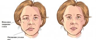
- neurological diagnoses of a special nature;
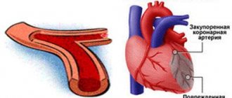
- severe damage to the cardiovascular system - stroke, heart attack;
- pulmonary edema;
- intestinal obstruction;
- severe traumatic brain injury, spinal lesions.
In these cases, the examination is postponed until the patient recovers or another diagnostic method is selected.
Possible complications
During the procedure for introducing an esophagoscope and manipulating it, some complications may arise:
- breathing problems during the procedure;
- allergic reaction, up to anaphylactic shock;
- small wounds as the tubes move into the esophagus;
- bleeding;
- perforation of the esophageal wall.
To avoid such troubles, the doctor is obliged to familiarize himself with the condition of the patient’s internal organs and the peculiarities of the location of the gastrointestinal tract, as well as the tendency to allergies before manipulating the esophagoscope.
We recommend: What does the sensation of a lump in the sternum or esophagus indicate?
Decoding the results
During the examination of the esophagus, its following characteristics are assessed:
- color of the mucous membrane of the organ;
- its folding, integrity, mobility;
- peristalsis and wall elasticity;
- changes in them depending on respiratory movements and heartbeat;
- the ability of folds to straighten due to the introduction of air.
The folded mucous membrane of a healthy esophagus is extremely thin, has a uniform pinkish color and a shiny surface.
There are no stains, defects or ulcers on it. There should be no compaction or growth of the mucous membrane normally. If they are visualized, then this is a sure sign of the disease.
A transverse fold, slightly protruding towards the spine, covers the gap - the so-called mouth of the esophagus, located at the entrance to the organ. The lumen of the thoracic part of the tubular organ gapes and looks almost round. When you inhale, it expands, and when you exhale, it contracts. Simultaneously with the increase in the lumen of the esophagus, the transverse folding, clearly visible in this segment of the organ, is smoothed out. There are practically no longitudinal folds here. However, they become more pronounced as the esophagoscope moves down.
The lumen of the esophagus at the junction with the stomach has the appearance of a rosette. This shape is formed by numerous longitudinal folds of the lower segment of the tubular organ and the loose tissue surrounding it. Peristalsis of a healthy esophagus is expressed by successive contractions of the muscles of its walls in the direction from top to bottom - from the mouth to the stomach. Strong pulsation, synchronous with heartbeats, becomes noticeable at the level of the aortic arch. After passing the esophagoscope through this place, a narrowing of the esophageal canal is observed, caused by pressure on it from the left bronchus.
A sheet of the results of the esophagoscopy performed is given to the patient for further interpretation by the attending physician.
The characteristics of the mucous membrane and the structure of the esophagus are described, and if deviations from the norm are detected, the nature of the pathological process, its localization and severity.
Using esophagoscopy you can diagnose:
- acute and chronic esophagitis (inflammation of the esophagus);
- bleeding;
- diverticulum;
- various leukoplakia, erosions and ulcers of the esophagus;
- strictures (scar narrowings) of the organ;
- varicose veins of the esophagus;
- burns;
- tumors (including cancer);
- hiatal hernia.