In the modern world, of all diseases, heart pathology occupies a leading position.
This fact creates an urgent need for accurate and timely detection of the disease for the purpose of further effective treatment.
Among the numerous methods of heart research, one of the most informative is transesophageal echocardiography (TEE), which allows examination of the organ through the esophagus.
The technique of transesophageal echocardiography has gained popularity since the end of the twentieth century. Its detailed description can be found in the book by Alekhine M.N. "Transesophageal echocardiography", which is a practical guide for doctors.
Advantages
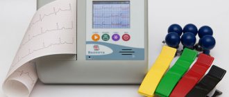
The most common method for identifying cardiac problems is an electrocardiogram (ECG), but it only shows a graphical record of heart activity, while the use of transesophageal echocardiography allows you to study the structure of the organ.
Systematic electrocardiogram examination is recommended even for healthy people
The use of transthoracic echocardiography (Echo CG) also has good information content and is widely used in medical practice to determine most heart diseases. The examination is safe for the body and does not cause discomfort to the patient due to the location of the ultrasound sensor on the surface of the body during the examination.
However, the occurrence of obstacles to ultrasound waves in the form of subcutaneous fat, ribs, muscle tissue, and lungs can significantly distort the results of transthoracic echocardiography and lead to errors.
Studying the heart through the esophagus has some advantages compared to Echo CG:
- the location of the sensor in close proximity to the heart during transesophageal echocardiological examination leads to obtaining a much better image than with EchoCG and, as a result, to greater information content;
- the presence of anatomical features of the patient’s body (close proximity of the ribs, excess weight, etc.) does not cause obstacles to his examination;
- the use of transesophageal echocardiography allows you to monitor the work of the heart during surgical interventions without interfering with the actions of doctors.
Cardiological studies also include transesophageal pacing (TEPS), which allows specialists to record the biocurrents of the heart.
When is TEE used?
Transesophageal echocardiography is used in the following cases:
- When examining a patient before surgery in order to clarify an existing diagnosis. As a rule, this is due to the condition of the heart valves, the likelihood of embolism, and the presence of space-occupying formations.
- To clarify not only the diagnosis, but also the surgeon’s action plan, the initial assessment of various indicators of the heart’s condition, monitoring the implementation of medical intervention and air removal during open-heart surgery, assessing the condition of shunts.
- For monitoring the state of the heart during transplantation and intensive care.
Equipment used
The examination equipment contains a standard echocardiograph, an endoscope (no light optics) and an ultrasound sensor. The endoscope, including the sensor, has a thickness of 9 to 11 mm, a length of 60 to 100 cm. The sensor, containing crystalline elements, is capable of creating ultrasonic waves with a frequency of 5 to 7.5 MHz.
The development of the first transesophageal sensor was first carried out in 1976. Currently, the equipment for the procedure has been significantly improved
Applying special marks on the endoscope allows you to adjust the depth of the sensor into the esophagus. In addition, with the help of special handles, it can bend and unbend, as well as rotate to the right and left, which significantly improves the visual inspection of the areas being inspected.
The presence of temperature-sensitive sensors in the endoscope leads to automatic shutdown of the device if the temperature of the esophagus exceeds 42ºC.
Contraindications for emergency echocardiography
The examination can be performed on patients of any age and pregnant women. However, during the procedure, damage to the esophagus of varying severity may occur. Therefore, the method has the following contraindications:
- The presence of malignant or benign neoplasms in the esophagus or stomach, strictures and scars, fistulas;
- Anomalies in the development of the esophagus (violation of the normal length of the organ, the appearance of a diverticulum);
- Bleeding from the upper digestive tract (esophagus and stomach);
- Varicose veins of the esophagus against the background of liver cirrhosis;
- Conducting radiation therapy on the chest area;
- Erosive inflammation of the esophagus;
- Diseases of the pharynx that complicate the process of inserting the sensor.
Indications
Transesophageal echocardiography is mandatory for:
- the need to study the aorta in cases of aneurysm and dissection;
- infective endocarditis, its diagnosis and identification of consequences;
- examining a patient before surgery to replace a heart valve with a prosthesis;
- the need to diagnose problems with the prosthesis.
The study is recommended in the following cases:
- diagnosis of congenital heart pathologies;
- detection of tumor formations and blood clots in the heart;
- the need to assess the condition of the coronary sinus (venous canal);
- the ineffectiveness of using transthoracic echocardiography in cases of large patient weight, pulmonary emphysema, etc., as well as clarifying its results;
- suspicion of severe cardiac pathologies.
A significant advantage of the study is the ability to diagnose certain diseases in the early stages.
Transesophageal echocardiography is a semi-invasive method and, as a rule, is prescribed after a transthoracic examination.
The essence and purpose of echocardiography
Echocardiography or echocardiography is a non-invasive method of examining the heart using ultrasound. The echocardiograph sensor emits a special high-frequency sound that passes through the tissues of the heart, is reflected from them, and is then recorded by the same sensor. The information is transmitted to a computer, which processes the received data and displays it on the monitor in the form of an image.
Content:
- The essence and purpose of echocardiography
- Benefits of EchoCG
- Indications and contraindications for Echo-CG
- Types of echocardiography
- Preparation for echo-CG
- Research methodology
- Decoding the results
- Where to get an echocardiogram
- Echocardiography in children
- Preparing and performing the procedure for children
- Fetal echocardiography
Echocardiography is considered a highly informative research method, since it makes it possible to assess the morphological and functional state of the heart. Using this procedure, it is possible to determine the size of the heart and the thickness of the myocardium, check their integrity and structure, determine the size of the cavities of the ventricles and atria, find out whether the contractility of the heart muscle is normal, find out about the condition of the valvular apparatus of the heart, examine the aorta and pulmonary artery. This procedure also allows you to check the level of pressure in the structures of the heart, find out the direction and speed of blood movement in the heart chambers and find out the condition of the outer lining of the heart muscle.
This cardiological examination allows you to diagnose both congenital and acquired heart defects, find out about the presence of free fluid in the heart sac, identify blood clots, changes in the size of the chambers, thickening or thinning of their walls, detect tumors and any disturbances in the direction and speed of blood flow.
Contraindications
The procedure is not prescribed for any factor that can lead to a serious threat to a person’s health, as well as:
- pathological changes in the esophagus, oral cavity and pharynx, dysphagia;
- difficult sensor insertion;
- bleeding in the upper gastrointestinal tract, as well as recent surgical interventions on them.
It should be noted that the patient has the right in writing to refuse any medical examination, including transesophageal echocardiography. In this case, it is the doctor’s responsibility to explain the possible consequences of this decision.
Pregnancy and childhood of the patient are not contraindications for the examination. In children under 7 years of age, the procedure is performed using special endoscopes with a diameter of 7 mm.
To whom and when is it prescribed?
Transesophageal echocardiography is prescribed in the process of diagnosing the following diseases and pathological conditions:
- Atherosclerosis . A pathological process occurring in the arteries, which lasts for many years and leads to a decrease in the diameter of their lumen.
- Cardiomyopathy . A disease of the heart muscle in which it thickens and contractility is lost.
Cardiomyopathy
- Congenital heart defects . Anatomical defects of this organ, its valves, vessels that arose during the period of intrauterine development. This includes a ventricular septal defect (a hole in the wall that separates the ventricles).
- Heart failure . A condition in which the heart muscle (myocardium) becomes weak due to various reasons and loses its pumping efficiency. This leads to so-called cardiac edema. They appear first on the legs (around the ankles) and then in other parts of the body.
- Aneurysm . Thinning of a section of the myocardium or aorta, followed by the formation of a protrusion in this area.
Left ventricular aneurysm
- Acquired heart valve defects . As a result of any diseases (for example, rheumatism, atherosclerosis), stenosis and valve insufficiency occur.
- Heart tumor.
- Pericarditis . Inflammation (both infectious and other etiologies) of the pericardial sac.
- Infectious endocarditis . Inflammation of the inner layer of the heart wall. As a rule, the valves are also affected.
Infective endocarditis
In general, there are three indications for transesophageal echocardiography (possible situations) in which a doctor may recommend this procedure instead of a standard transthoracic one.
First , TEE is prescribed when an “adequate” image to help make a diagnosis cannot be obtained using standard sonography (through the chest wall). This problem occurs in patients with emphysema or who have undergone chest surgery. Even for no apparent reason, sometimes the echo image obtained using the standard method is not clear enough. Transesophageal echocardiography comes to the rescue in such a situation.
Secondly , TEE allows you to perform an echo examination of the heart during surgery. It is especially useful when prosthetics or other manipulations with the valves of this organ are performed. For example, during surgical treatment of mitral valve insufficiency.
Thirdly , TEE helps to identify blood clots in the left atrium. The absence of blood clots in it is one of the conditions that allows cardioversion for atrial fibrillation.
To learn about what TEE is and its indications, watch this video:
Preparing for the examination
Read more: Transesophageal echocardiography
Transesophageal echocardiography is prescribed after the patient has undergone preliminary procedures:
- general blood and urine tests, as well as biochemical blood tests;
- ECG;
- transthoracic ultrasound diagnostics, esophagogastroduodenoscopy to exclude pathologies of the esophagus (tumors, venous dilatation, esophagitis, etc.).
Next, to reduce discomfort and obtain reliable information during the study, the patient needs to:
- Avoid eating 8 hours before the examination and drinking 2 hours before;
- do not smoke or drink alcohol in advance;
- do not take any medications without a doctor’s permission;
- immediately before the procedure, remove dentures.
During transnutritive echocardiography, the patient is subjected to local or general anesthesia. It is advisable that there is a person next to him who will help him get home after the procedure.
Carrying out the procedure
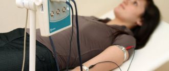
The procedure is carried out in a hospital setting if the specialist has a resuscitation kit, an oxygen inhaler, a saliva ejector, and equipment for determining blood pressure. During the entire examination, the patient is given a cardiogram.
Examination of the heart through the esophagus lasts no more than 15–25 minutes
During drug preparation, measures are taken to reduce:
- gag reflex during the procedure using local anesthesia of the oral cavity and pharynx (10% lidocaine solution in the form of a spray);
- excitement (Relanium 2 ml intravenously a few minutes before echocardiography);
- salivation and secretion of gastric juice (intravenously 0.1% atropine 1 ml).
Bacterial endocarditis in patients with valve implants is prevented by oral antibiotics.
The patient is placed on his left side, and an endoscope pre-lubricated with gel is inserted into his throat without excessive effort. After the subject makes swallowing movements, the device reaches the esophagus. The resulting feeling of discomfort felt at the beginning of the manipulation gradually disappears. All research data must be recorded on videotape.
After the procedure, the patient remains in a medical facility for several more hours under the supervision of doctors, and then can go home.
Minor injuries to the esophagus obtained during transesophageal echocardiography necessitate some restrictions at the end of the manipulation:
- the patient’s refusal to eat and eat for 2 hours;
- excluding tea and coffee drinks from the diet for several days, as well as eating food at room temperature.
Further consultation with a doctor is necessary if:
- sore throat that does not subside or gets worse;
- the appearance of pain in the sternum;
- difficulty breathing.
The results of the study are interpreted by qualified specialists, taking into account the Age, gender and physical parameters of the person. Based on the diagnostic conclusion, appropriate treatment is prescribed.
Transesophageal echocardiography – what is it?
Echocardiography is one of the main methods for diagnosing diseases of the cardiovascular system. It can be performed at any age, as it is not accompanied by radiation exposure to the body. Thanks to this instrumental study, it is possible to visualize the size and thickness of the heart chambers and assess the condition of the valves. Transesophageal echocardiography (TEE) differs in that it is performed from the inside rather than from the outside (the chest wall). This improves the quality of visualization. This examination is not prescribed to everyone, but only for special indications. To perform echocardiography through the esophageal cavity, it must first be performed transthoracically. This diagnostic method is carried out by a specially trained specialist in a hospital setting.
Risks of the procedure
The risk of complications from transesophageal echocardiography is minimal. Thus, a violation of the integrity of the esophagus is observed in less than 1 case in 3000, which is associated with a contraindication for examination in case of organ pathologies.
The procedure can also lead to:
- atrial and ventricular disturbances of heart rhythms;
- vagal vascular reactions;
- hypocemia;
- symptoms inherent in individual intolerance to certain drugs.
Transesophageal echocardiography has greater prognostic value than many diagnostic methods. This type of examination should not be neglected when prescribed by the attending physician.
Where to get an echocardiogram
Standard echocardiography is carried out both in public medical institutions (clinics and hospitals) and in private medical centers. To register for an examination, you must provide a referral from your attending physician or cardiologist.
More specific types of echocardiography - transesophageal examination or stress echocardiography - can only be performed in specialized medical institutions, since they require special equipment and personnel who have undergone special training.
Reviews
Elena Borisovna, Belgorod, 52 years old, housewife For a long time she suffered from heart pain, shortness of breath, and dizziness. The cardiogram showed normal results. The doctors doubted the effectiveness of a regular ultrasound of the heart because of my excess weight, so they prescribed transesophageal echocardiography. The examination took place in the hospital under local anesthesia. I was very worried. But the procedure turned out to be not so scary. There was discomfort, of course. But the main thing is that no serious diseases were discovered.
Kalutsky Valery Sergeevich, cardiologist surgeon, Minsk Transedible echocardiography provides an indispensable service for heart diseases. It is carried out before surgery, during surgery and allows you to identify the slightest disturbances in the operation of implants at the earliest stages. Any cardiac operation requires maximum precision from the surgeon, and examination of the heart through the esophagus contributes to this in every possible way. The doctor is not only able to correct his actions if necessary, but also monitor the functioning of the organ.
Svetlana Vasilyevna Babenko, 45 years old, engineer During a preventive examination, I was diagnosed with atrial fibrillation and was sent for transesophageal echocardiography. Of course, I had some worries, but the feedback from other patients calmed me down a little. The main thing is to listen to the doctor’s recommendations and be patient a little if any unpleasant sensations arise. I don’t regret that I took my husband with me, who helped me get home after the procedure.
Echocardiography in children
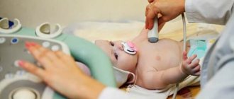
As noted above, the undeniable advantages of EchoCG are the non-invasiveness, painlessness and complete safety of this method of cardiac research. The manipulation is not associated with radiation exposure and does not provoke any complications. Therefore, if there are appropriate indications, the study can be recommended not only for adults, but also for children.
Diagnostics will help to timely detect congenital pathology in young children, which, in turn, will make it possible to select the most effective treatment. As a result, the child will be able to lead an absolutely fulfilling life in the future.
Indications for echocardiography in a child are:
- Heart murmurs.
- The appearance of shortness of breath either during physical activity or at rest.
- Blueness of the lips, nasolabial triangle, fingertips.
- Decreased or complete lack of appetite, too slow weight gain.
- Complaints of constant weakness and fatigue, sudden fainting.
- Complaints of frequent headaches.
- Discomfort behind the sternum.
- Decrease or increase in blood pressure.
- The appearance of swelling in the extremities.
Taking into account the fact that the method is safe, echocardiography can be performed on children more than once in order to track the development of the disease or assess how effective the treatment is. If any pathological changes have been identified, a study is carried out at least once every twelve months.
Best materials of the month
- Why you can't go on a diet on your own
- 21 tips on how to avoid buying stale food
- How to keep vegetables and fruits fresh: simple tricks
- How to curb your sweet cravings: 7 unexpected products
- Scientists say youth can be extended

