Echo-CG, or echocardiography, is a simple and painless study, which, thanks to ultrasonic vibrations, allows you to obtain sufficiently voluminous information about the condition of the heart. Due to its non-invasiveness and the absence of absolute contraindications, the procedure is performed for both adults and children almost from the first days of life.
During a short diagnosis, the slightest disturbances in the structure and functioning of the heart muscle are revealed without much difficulty, confirming the doctor’s suspicions about the presence of pathology. At the same time, it is often possible to identify deviations that do not manifest themselves in any way, but are considered no less dangerous than the signs that worry the patient.
Description
Echocardiography, abbreviated EchoCG, is a method of examining the heart based on ultrasound scanning of the chest cavity. Using this method, various diseases of the body’s “engine” are diagnosed. This research method allows you to evaluate the overall dimensions of both the heart itself and its individual structures (ventricles, septa), the thickness of the myocardium of the ventricles and atria. EchoCG can also determine heart mass, ejection fraction and other parameters.
Another name for this diagnostic method, which people hear more often, is ultrasound, that is, ultrasound examination.
What is an echo of the heart?
Echo of the heart
It is not uncommon to hear an incomprehensible phrase in conversations between doctors or with patients - echo of the heart. What kind of “echo” is this? Of course, this expression can be classified as medical jargon, which is why it is not clear.
In our country, the term ultrasound examination of the heart or ultrasound of the heart is more widely used, but abroad it is called sonography or echography, hence the term echo of the heart. Although, it must be said that the term “echo” more accurately conveys the essence of the method - the reflection of ultrasound waves from tissues with different densities and the capture of these reflected waves by a special sensor.
Heart echo has become quite widely used in the practice of cardiologists, since this method has a huge number of advantages and provides a lot of additional information about the condition of the heart, which is sometimes decisive in making a diagnosis.
What does a heart echo give to a doctor?
- Firstly, a cardiac echo allows you to assess the condition of the heart valves: it reveals prolapses (deflections), stenoses (narrowings) and insufficiency.
- Secondly, echography provides information about the structure of the heart: the thickness of its walls and the presence of defects in them (in case of defects); reveals signs of a previous heart attack and post-infarction aneurysms, detects dilation of the cavities of the heart and large vessels.
- Thirdly, a cardiac echo allows us to determine the pumping function of the heart - this is the ejection fraction, which in patients with heart failure is reduced - less than 55%, in more severe cases even below 40%.
If the cardiac echo is supplemented with Dopplerography - a special research method carried out in parallel, then it is possible to measure the pressure in the large vessels of the heart (aorta, pulmonary artery) and obtain reliable information about the failure of the valve apparatus.
Failure of the valve apparatus can manifest itself in the form of regurgitation (reverse blood flow through the valve) or vice versa - an increase in the pressure gradient (resistance to blood flow on the valve, resulting from a narrowing of its opening).
It will also be useful for the patient to know what a heart echo “cannot show”. Please note that this test will not determine the cause of chest pain except in rare cases. A cardiac echo will not allow us to understand the condition of the vessels supplying the heart, including the presence of plaques in them.
Echography will also not help to diagnose arrhythmia, various heart blocks. Please note that although ultrasound examination is absolutely safe, has no contraindications and can be performed on you at your discretion, it is not a panacea.
It is naive to think that having received the conclusion of a heart echo, you yourself will be able to understand your illness and even take appropriate measures to treat it. Therefore, if you have heart problems, it is better to immediately contact a specialist and he will prescribe the necessary amount of research for you and evaluate the results obtained.
This will help avoid unnecessary expenses, save time and allow you to establish a diagnosis, if any, and receive appropriate recommendations. Echocardiography can simply be called ultrasound of the heart; this method belongs to the category of ultrasound examinations of the cardiac system. Thanks to it, you can evaluate the following indicators in real time:
- organ muscle functionality;
- valve condition;
- determine the size of the heart cavities and its walls;
- indicate the direction and speed of intracardiac blood flow.
In addition, answering the question of what a cardiac echo is, it is worth noting that this examination method allows you to measure the pressure in the pulmonary artery. It is also used to determine the contractile activity of the heart.
Transthoracic echocardiography is especially relevant today, since this method is considered quite simple. This diagnostic method is carried out through the surface of the body, but there is also a transesophageal method of performing cardiac echo.
Particularly accurate results can be obtained during stress tests, since it is in the state when the heart muscle is under load that hidden disorders can appear. This method of examining patients is often called stress ECHO.
Heart ECHO is quite affordable, so everyone can afford to undergo this diagnostic not only in case of pathology, but for preventive purposes.
Indications for use
A specialist may refer you for a cardiac echocardiography procedure in the following cases:
— If the cardiologist detects a heart murmur.
— There are changes on the ECG.
- If a person feels interruptions in the functioning of the heart.
— The patient has a fever, which is not a sign of ARVI, problems with the throat, nose, ears or kidneys.
— The results of the x-ray show an increase in the size of the heart or a change in its shape, the location of large vessels.
This research method is also carried out in the following situations:
- Patients with high blood pressure.
— For those patients who have a family history of heart defects.
- When a person complains of pain in the left side of the chest.
- For shortness of breath, swelling of the extremities.
- When fainting.
— In the event that a person is often bothered by dizziness.
- If there is a suspicion of a heart tumor.
- For angina pectoris.
— After a heart attack, etc.
Indications: who needs such diagnostics
Ultrasound of the heart is indicated for all patients with cardiac pathology, as well as people with the following symptoms:
- Chest pain.
- Shortness of breath during exertion, swelling in the legs and other manifestations of heart failure.
- Fatigue, accompanied by fainting and increased heart rate.
- Previous myocardial infarction and pulmonary embolism.
- Noises when listening (auscultation) of the heart.
- Suspicions of congenital and acquired heart defects.
- Past rheumatism.
- Blueness of the skin in the face, tip of the nose and ears, which appears or intensifies with exercise.
- Marked increase in blood pressure.
Considering the ease of performance and safety of echocardiography, it is done not only to monitor the progress of a person’s existing cardiac pathology, but even if it is suspected. There are no absolute contraindications.
Research in relation to pregnant women
A safe and universal method for detecting heart problems is called echocardiography. What does it mean? There is only one thing - it can be carried out in relation to all categories of the population, both adults and children. This study is even prescribed to pregnant women. And it is done in order to detect cardiac pathology in the fetus and take the necessary measures to save the baby. EchoCG is absolutely harmless for both mother and baby.
Pregnant women must undergo this research method in the following situations:
- If the woman in labor had heart defects in her family.
— The previous pregnancy ended in miscarriage.
- If a woman has diabetes.
— During pregnancy, the expectant mother contracted rubella.
— If a woman took antibiotics or antiepileptic drugs in the 1st or 2nd trimester.
Symptoms indicating the need for an ECHO
The risk of developing dangerous pathologies is reduced if you perform an ECHO of the heart when the first symptoms of the disease appear. The following symptoms should be considered as an indirect reason to undergo diagnostics:
- systemic heart rhythm disturbances;
- murmurs identified during listening by a therapist or cardiologist;
- chest discomfort in the heart area;
- feeling of lack of air, shortness of breath; fainting;
- rapid fatigue with low physical activity;
- cyanosis or periodic acquisition of a white tint to the skin;
- frequent swelling of the legs, enlarged liver, other symptoms of heart failure.
Without obvious symptoms of heart disease, echocardiographic diagnosis is prescribed for pregnant women at risk, athletes experiencing increased physical activity, divers, and people often suffering from pulmonary diseases.
Differences between ECG and EchoCG
The first abbreviation stands for electrocardiography.
Echocardiography means nothing more than echocardiography. What is this procedure and how is it different from the first? It is also called ultrasound of the heart. The differences are as follows:
- EchoCG is done using a special device - a transducer, which a specialist places on the patient’s chest in the area of the heart. The drug picks up ultrasonic waves passing through the walls of the body’s “engine” and reflects them back. The converter receives the returned signals, which are then processed by a computer. At the same time, an ECG is done differently: special sensors are attached to the patient’s chest, which measure the electrical activity of the heart. The electrodes are connected to a machine, which then produces a special graph indicating the strength and nature of the electrical signals.
- Using an echocardiogram, the doctor determines how well the heart pumps blood around the body. Also, thanks to this method, it is possible to determine the weakening of this function, which is a symptom of heart failure. At the same time, ECG diagnostics can only measure the signal level and check whether the body’s “engine” sends stable electrical impulses.
- The ECG result is always presented on the graph. Echocardiography is recorded in photographs.
- Using an electrocardiogram, you can identify problems with heart rhythm, arrhythmia, tachycardia, bradycardia. What will a heart ultrasound show? It allows you to assess the condition of the heart valves, heart function after an attack, the possible location of blood clots, and disorders of this important organ.
Types of Echo-KG
Standard transthoracic cardiac ultrasound is the most common type of examination. It is performed using a sensor installed on the chest area and includes the following stages of the study:
- I – using parasternal access, the left ventricular chamber, right ventricle, left atrium, aorta, interventricular septum, aortic valve, mitral valve and posterior wall of the left ventricle are examined;
- II – using pairs of sternal access, the leaflets of the mitral and aortic valves, the valve and trunk of the pulmonary artery, the outflow tract of the right ventricle, the left ventricle, and papillary muscles are examined;
- III - in the apical approach in the four-chamber position, the interventricular and interatrial septa, ventricles, atrioventricular valve and atria are examined, in the five-chamber position - the ascending aorta and the aortic valve, in the two-chamber position - the mitral valve, left ventricle and atrium.
Doppler echocardiography allows you to assess the movement of blood in the coronary vessels and heart. During its implementation, the doctor can:
- measure the speed and determine the direction of blood movement;
- assess the functioning of heart valves;
- hear the sound of blood moving through the vessels and the sound of the beating heart.
Contrast Echo-CG is performed after introducing a radiopaque solution into the bloodstream, which allows the doctor to more clearly visualize the inner surface of the heart.
Stress Echo-CG is carried out using standard ultrasound and Doppler studies and, through the use of physical or pharmacological stress, allows you to identify areas of possible stenosis of the coronary arteries.
Transesophageal echocardiography is performed by inserting a probe through the esophagus or throat. This type of access allows the specialist to obtain ultra-precise images in moving mode. The following situations may be the reason for prescribing this type of ultrasound diagnostics:
- risk of aortic aneurysm dissection;
- suspicion of the formation of an abscess of valve rings, aortic root or paraprosthetic fistula;
- the need to examine the condition of the mitral valve before or after upcoming surgical interventions;
- risk of developing left atrial thrombosis;
- signs of malfunction of the implanted valve.
This type of study can be performed after additional sedation of the patient.
Advantages and disadvantages of each method
ECG is an affordable research option. However, it cannot always show a clear picture of the problem, unlike heart ultrasound. What EchoCG will clearly show are structural abnormalities. This research method ensures the accuracy of the image; this method is more reliable in determining the health of this internal organ. The advantage of cardiac ultrasound is that the specialist can visually observe its chambers. However, this diagnostic method has one drawback: it is performed only in private clinics, and the cost is several times more expensive than an ECG.
Child examination
Ultrasound of a child’s heart (standard pediatric echocardiography) is the most modern research method in cardiology. During an ECHO CG of a child, the doctor observes the work of the heart in real time and can examine all the structures of the child’s heart during operation.
It is ultrasound of the heart that confirms or excludes the presence of many diseases of the cardiovascular system. It is often very important not to miss precious time for treatment, so that a minor pathology does not have time to develop into a serious disease.
Promptly and competently performed echocardiography allows you to detect the problem in time and preserve the health of your baby. Indications for ultrasound of a child’s heart:
- If the pediatrician, after listening to your baby’s heart, detects murmurs during the examination, he will refer you for echocardiography (ultrasound of the heart).
- If you yourself feel trembling over the area of the child’s heart, contact a specialist.
- If a child complains of aching, pulling, stabbing pain in the heart area, it is better to play it safe and do an echocardiogram.
- If the baby does not suck well, the baby may need an echocardiogram (here you must first rule out problems with improper attachment to the breast - consult your pediatrician about this). You should also pay attention to the color of the skin around the child's mouth. Usually, with heart problems, blueness of the nasolabial triangle is observed when crying and sucking in infants. This is a fairly typical symptom.
- If from time to time you feel that your child’s hands and feet become cool for no apparent reason, this is a reason to be wary.
- If a child loses consciousness (even during intense physical exertion), an echocardiogram should be done to rule out the possibility of cardiovascular disease.
- Fatigue, excessive sweating, insufficient weight gain for age - all of these things can be caused by heart problems and echocardiography is prescribed.
- Frequent pneumonia in a child can also occur due to heart disease.
- If your family has relatives with severe heart pathologies, the child should have an ECHO KG at least once a year in order to promptly stop the development of hereditary diseases if they arise.
- According to the standards adopted in our country, every child aged 1 year must, as part of a routine medical examination, receive a consultation with a cardiologist, having previously had an ECHO KG and an ECG (electrocardiogram).
Just like you did during pregnancy, an area of the body (chest) will be smeared with gel and a sensor will be moved over it. During the ECHO CG procedure, a child can even move, fidget, or talk - this will not affect the results of the examination.
No preliminary preparation is needed for cardiac ultrasound. The echocardiogram will take approximately 15 minutes. Echocardiogram results require interpretation by a qualified physician. It is advisable to show the cardiologist, along with the results of the ultrasound of the heart, also a fresh blood test, urine test and the results of the cardiogram.
The procedure is painless! ECHO CG is done both for serious indications, as prescribed by a doctor, and for reinsurance already in the first hours and days of the baby’s life. Experts believe that the echocardiography method is completely safe, since, unlike X-ray examinations, it does not use radiation, but mechanical wave vibrations.
The cardiac ultrasound procedure does not require special preparation and can be performed several times a day if necessary. The only thing that needs to be done, if the child already understands what is happening to him, is to calm him down and set him up in a positive way. And under no circumstances should you discuss his illnesses and their possible consequences in front of him with a doctor!
Echo helps diagnose in children:
- Congenital heart defects, such as patent ductus arteriosus, ventricular septal defect, mitral valve defects, aortic valve defects and others.
- Acquired heart defects.
- The cause of heart murmurs.
- Coronary heart disease.
- Enlargement of the heart chambers.
- Hyper- and hypotrophy of the heart.
- Changes in the walls of the myocardium and disturbances in their functioning.
- Blood clots and other neoplasms and other pathologies.
Congenital heart defects are most likely to be discovered in the prenatal period, during an ultrasound scan of a pregnant woman.
Boundary parameters of cardiac echocardiography
After an ultrasound of this organ is performed, the specialist who conducted the study will definitely refer the person to a cardiologist so that he can interpret the results. In order not to worry once again, not to stress yourself out, in the table below you can familiarize yourself with the borderline permissible values:
| Echocardiography: normal parameters | Size, cm | |
| Variation | Average value | |
| Right ventricular cavity at the end of diastole | 0,9–2,6 | 1,7 |
| Left ventricular cavity at the end of diastole | 3,5–5,7 | 4,7 |
| Aortic orifice diameter | 2,0–3,7 | 2,7 |
| Left atrium cavity | 1,9–4 | 2,9 |
These are the main values that the doctor pays attention to when viewing an ultrasound.
Ultrasound standards for heart chambers
To begin with, we will present a few numbers that are sure to appear in every Doppler echocardiography report. They reflect various parameters of the structure and functions of individual chambers of the heart. If you are a pedant and take a responsible approach to deciphering your data, pay maximum attention to this section.
Perhaps, here you will find the most detailed information in comparison with other Internet sources intended for a wide range of readers. Data may vary slightly between sources; Here are the figures based on materials from the manual “Norms in Medicine” (Moscow, 2001).
Left ventricular parameters:
- Left ventricular myocardial mass: men – 135-182 g, women – 95-141 g.
- Left ventricular myocardial mass index (often referred to as LVMI on the form): men 71-94 g/m2, women 71-89 g/m2.
- End-diastolic dimension (EDD) of the left ventricle (the size of the ventricle in centimeters that it has at rest): 4.6 – 5.7 cm
- End systolic dimension (ESD) of the left ventricle (the size of the ventricle it has during contraction): 3.1 – 4.3 cm
- Wall thickness in diastole (outside of heart contractions): 1.1 cm
- Ejection fraction (EF): 55-60%.
- Stroke volume (the amount of blood ejected by the left ventricle in one contraction): 60-100 ml.
End-diastolic volume (EDV) of the left ventricle (the volume of the ventricle that it has at rest): men - 112±27 (65-193) ml, women 89±20 (59-136) ml
With hypertrophy - an increase in the thickness of the ventricular wall due to too much load on the heart - this figure increases.
Figures of 1.2–1.4 cm indicate slight hypertrophy, 1.4–1.6 indicate moderate hypertrophy, 1.6–2.0 indicate significant hypertrophy, and a value of more than 2 cm indicates high degree hypertrophy.
At rest, the ventricles are filled with blood, which is not completely ejected from them during contractions (systole).
The ejection fraction shows how much blood relative to the total amount the heart ejects with each contraction; normally it is slightly more than half.
When the EF indicator decreases, they speak of heart failure, which means that the organ pumps blood ineffectively, and it can stagnate.
Right ventricle parameters:
- Wall thickness: 5 ml
- Size index 0.75-1.25 cm/m2
- Diastolic size (size at rest) 0.95-2.05 cm
Parameters of the interventricular septum:
- Resting thickness (diastolic thickness): 0.75-1.1 cm
- Excursion (moving from side to side during heart contractions): 0.5-0.95 cm. An increase in this indicator is observed, for example, with certain heart defects.
Right atrium parameters:
- For this chamber of the heart, only the value of EDV is determined - the volume at rest. A value of less than 20 ml indicates a decrease in EDV, a value of more than 100 ml indicates its increase, and an EDV of more than 300 ml occurs with a very significant increase in the right atrium.
Left atrium parameters:
- Size: 1.85-3.3 cm
- Size index: 1.45 – 2.9 cm/m2.
- Most likely, even a very detailed study of the parameters of the heart chambers will not give you particularly clear answers to the question about the state of your health.
You can simply compare your indicators with the optimal ones and on this basis draw preliminary conclusions about whether everything is generally normal for you. For more detailed information, contact a specialist; The volume of this article is too small for wider coverage.
Echocardiography: interpretation of results
Only a cardiologist can correctly read, understand and explain to the patient the results of this diagnostic method. Independent study of the main parameters of cardiac indicators does not provide a person with complete information on assessing the state of his health. But for peace of mind, the patient can familiarize himself with the average values described above. Only an experienced doctor in the field of cardiology can correctly decipher the result of the device’s operation, as well as answer the patient’s questions.
It also happens that some indicators deviate from the norm and are recorded in the examination protocol under other points. This suggests that the quality of the device is not very good. If a medical institution uses modern equipment, then the echocardiography doctor will receive more accurate results, on the basis of which the patient will be diagnosed and treated.
Advantages
When identifying diseases of the heart, primarily defects of this organ, EchoCS is of paramount importance in its characteristics.
Advantages:
- Modernity (carried out on high-precision and modern equipment with the least amount of error);
- Safety (there is no radiation, pain or side effects, so EchoCS during pregnancy is absolutely harmless for both the expectant mother and her baby);
- Informative (no analysis will show how the heart and its components work as accurately as this method).
EchoCS is performed only in specialized clinics and is an order of magnitude higher due to its indicators than a regular electrocardiogram of the heart. It is carried out both before and after treatment in order to track the dynamics and therapeutic effect of treatment.
Methods of performing ultrasound
Echocardiography diagnostic methods have the following:
— Transthoracic technique.
— Transesophageal ultrasound.
The first diagnostic method is the most common because it has been used for a long time. The transthoracic technique for detecting cardiac problems is carried out through the chest using a sensor that is pressed against the patient's body in the area of the heart. During the procedure, the patient is on the couch in a position lying on his side or back.
Transesophageal echocardiography - what is this research method and how is it performed? This is also a method of ultrasound diagnosis of the heart. However, it is carried out not from the surface of the chest, as with the transthoracic technique, but from the esophagus. The sensor is located exactly there; thanks to this method, the doctor can get as close to the heart as possible, and also see those parts of it that are not visible with a standard ultrasound.
Types of echocardiography
Not every echocardiography has all the diagnostic capabilities of ultrasound. Depending on the class of ultrasound equipment and the examination procedure, there are:
- Standard ECHO-CG - one-dimensional, two-dimensional and three-dimensional ultrasound. It is also called transthoracic, as it is carried out through contact with the skin in the chest area. Provides information about the structure of the heart, but cannot determine the characteristics of blood circulation in it.
- – the study is expanded compared to the standard one. Determines the characteristics of blood flow in the atria, ventricles, valves and large vessels.
- Stress echocardiography – ultrasound of the heart during stress tests. May be required to diagnose only certain diseases (for example, valve defects).
- Transesophageal ECHO is an examination of the heart with a special sensor through the wall of the esophagus during fibrogastroscopy. Rarely required, but can provide important information about pathology in the deep myocardium.
The gold standard for ultrasound examination of the heart is two-dimensional ECHO with Doppler and duplex amplification.
Cost of the procedure
Not all public clinics and hospitals can boast that they can offer a heart examination method such as echocardiography. Prices for this procedure in private clinics range from 2200–3000 rubles. It all depends on the prestige of the hospital, the qualifications of the doctor, the availability of modern equipment, and the location of the medical institution that provides paid services. In Moscow, for example, it will be more expensive to do an echocardiography than in Voronezh.
If we compare the price of an ultrasound and an ECG, then in the latter case a person will have to pay up to 700 rubles. Moreover, electrocardiograms are often performed free of charge in public hospitals.
What is echocardioscopy?
Echocardioscopy (EchoCS) is also called echocardiography (EcoCG) or simply ultrasound examination of the entire cardiac system. This method is quite informative, as it allows not only to detect an enlarged heart, but also to examine all its components (septa, ventricles and vessels) in more detail and thoroughly.
By examining the cardiac region using echocardiography, you can determine:
- Condition of the heart muscle;
- Valve performance;
- Blood pressure in the cavities of the heart and their sizes;
- The thickness of the heart walls;
- Blood movement.
This research method makes it possible to identify heart defects regardless of origin (congenital or acquired), changes in the valves, the presence of blood clots, as well as impaired ability to carry out a cycle of certain movements.
We apply this method both in good and healthy health and in case of various heart diseases, as well as if it is necessary to measure pulmonary artery pressure.
We also invite you to learn more about cardiac cardiography.
Performing transesophageal ultrasound of the heart
- The patient lies down on the couch.
- A catheter is inserted into the patient's vein and general anesthesia is given. Local anesthesia may also be used.
- The pharyngeal cavity is washed with a local anesthetic. This is necessary so that the patient’s throat does not become irritated.
- The patient is connected to special equipment, which will subsequently monitor the functioning of the heart and breathing. If a person has been given general anesthesia, he is additionally provided with oxygen.
- The patient is turned on his left side. A mouthpiece is placed in his mouth.
- Under local anesthesia, an endoscope is inserted into the patient's mouth. If general anesthesia has been performed, then this device is immediately inserted through the esophagus into the pharynx with further advancement.
- The endoscope captures images on the monitor thanks to a special sensor located at its end.
- The specialist examines the heart from different angles.
Transesophageal echocardiography
There are cases in which certain factors prevent transthoracic echocardiography. For example, subcutaneous fat, ribs, muscles, lungs, as well as prosthetic valves, which are acoustic barriers to the path of ultrasonic waves.
In such cases, transesophageal echocardiography is used, the second name of which is “transesophageal” (from the Latin “oesophagus” - esophagus). It, like echocardiography through the chest, can be three-dimensional. In this type of study, the probe is inserted through the esophagus, which is adjacent directly to the left atrium, which makes it possible to better view the small structures of the heart.
Such a study is contraindicated in the presence of diseases of the patient’s esophagus (varicose veins of the esophagus, bleeding, inflammatory processes, etc.)
Unlike transthoracic echocardiography, the obligatory preparatory stage for transesophageal echocardiography is the patient fasting for 4-6 hours before the actual procedure. The sensor placed in the esophagus is treated with ultrasound gel and is often in the area for no more than 12 minutes.
Preparation and performance of transthoracic ultrasound
In this case, no planned actions are necessary. The procedure is performed in this order:
1. The patient undresses to the waist and lies down on the couch.
2. The specialist applies a special gel to the left side of the chest. This is necessary so that ultrasonic waves are better transmitted.
3. Then the healthcare worker places the sensor on the chest area and notes all the data.
4. After the procedure, the specialist processes all the information received and after a few minutes gives a written conclusion to the patient. On the document, a person can read about what tentative diagnosis the doctor gave him. But this does not mean that we can put an end to this. With the result of the ultrasound, the patient must consult a cardiologist.
Normal indicators
After completing the examination procedure using EchoCS, the doctor issues a protocol with his conclusion.
In the table below you can see the indicators within normal limits:
| Localization of the study | Norm |
| Left ventricle: | |
| end diastolic size | 3.4-5.6 cm |
| end systolic size | 2.4-4.1 cm |
| diastolic volume | 55-149 ml |
| systolic volume | 18-40 ml |
| ejection fraction | 60-65% |
| shortening fraction | 30-40% |
| back wall thickness | 0.9-1.1 cm |
| thickness of the intergastric septum | 0.9-1 cm |
| Right ventricle: | |
| cavity size | 2.6-3.1 cm |
| wall thickness | 0.2-0.4 cm |
| left atrium | 2.5-3.6 cm |
| Aorta diameter: | |
| at the level of the sinuses of Valsalva | 2.4-3.9 cm |
| at the ascending department level | 2.2-3.4 cm |
It should also be taken into account that age indicators may have different values.
Contraindications
In general, cardiac echocardiography is a completely harmless procedure. But due to some anatomical features of patients, problems may arise due to insufficient penetration of ultrasound by the transesophageal method. This can happen, for example, with chest deformation, the presence of pronounced hair in men, obesity, or large breast size in women.
In the following situations, performing an ultrasound of the heart is unacceptable:
- If a person has a stomach ulcer or acute gastritis.
— The patient has a tumor of any severity.
In this case, transesophageal ultrasound of the heart is not performed. Only transthoracic echocardiography is allowed.
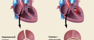
Ultrasound standards for heart valves
As for deciphering the results of a valve examination, it should present a simpler task. It will be enough for you to look at the general conclusion about their condition. There are only two main, most common pathological processes: stenosis and valve insufficiency.
The term “stenosis” refers to a narrowing of the valve opening, in which the overlying chamber of the heart has difficulty pumping blood through it and may undergo hypertrophy, which we discussed in the previous section. Insufficiency is the opposite condition.
If the valve leaflets, which normally prevent the reverse flow of blood, for some reason cease to perform their functions, the blood that has passed from one chamber of the heart to another partially returns, reducing the efficiency of the organ.
Depending on the severity of the disorders, stenosis and insufficiency can be grade 1, 2 or 3. The higher the degree, the more serious the pathology.
Sometimes in the conclusion of a cardiac ultrasound you can find such a definition as “relative insufficiency”. In this condition, the valve itself remains normal, and blood flow disturbances occur due to the fact that pathological changes occur in the adjacent chambers of the heart.
What is better than an ECG or ultrasound of the heart and how they differ
Diseases of the heart and blood vessels are the most common pathologies occurring in people nowadays.
In terms of the frequency of their occurrence, they have left far behind such a terrible disease as oncology. Cardiac problems occur in people of all age groups, including children.
The main thing is not to put off visiting a doctor . Modern diagnostic methods that cardiology has today will allow us to identify disturbances in the functioning of the main organ, determine the correct diagnosis and prescribe appropriate treatment in a timely manner.
The main methods for detecting pathological changes are ECG and ultrasound of the heart.
In some cases, the patient has questions. Which method is better? Do I need to conduct both examinations or can I limit myself to one? What is the difference between the methods?
To answer these questions, you need to consider both diagnostic methods in more detail.
ECG in medicine: what is it?
Electrocardiography (ECG) is a simple and painless method for studying the activity of the heart muscle, its electrical activity over a certain period of time, as well as the physiological state and performance.
How is the examination done?
To take an electrocardiogram, the patient lies on the couch, having previously undressed to the waist. A healthcare worker attaches special electrodes to his wrists, chest and legs.
If it is difficult for the patient to remain in a horizontal position due to severe shortness of breath, the procedure is performed while sitting.
To improve contact between the skin and the electrodes, the attachment points of the latter are degreased with alcohol, after which a gel with electrical conductivity is applied.
All wires from the electrodes are connected to a special device - electrocardiograph , which takes into account and measures the electrical impulses generated by the heart of the subject.
An electrocardiograph is designed in such a way that electrical signals amplified by a galvanometer are displayed as a graphic image on a moving paper tape.
The electrocardiogram recorded on this paper is sent to a specialist for interpretation.
In what cases is an ECG prescribed?
An ECG can be performed both routinely and in cases of emergency. A planned cardiogram is taken:
- Pregnant women.
- Patients with persistent hypertension or hypotension.
- Persons with a history of rheumatism or diabetes.
- After acute poisoning or severe infections.
- Before planned surgical operations.
- When passing the next medical examination, especially for those people whose work is associated with increased stress (drivers, pilots, rescuers, sailors, etc.).
An emergency ECG is done if an acute heart attack is suspected, in cases of a sharp drop or rise in blood pressure, severe arrhythmia, or chest injuries.
What diseases does an ECG detect?
Decoding the cardiogram data allows the doctor to identify pathologies such as:
- Heart rhythm disturbances (bradycardia, tachycardia, extrasystole).
- Myocardial infarction.
- Ischemic disease.
- AV blockade (impaired conduction of electrical impulses).
- Hypertrophy of the myocardium and ventricles.
- EOS offset.
I would also like to note that it is with the help of an ECG that a person can detect disorders of the nervous system and some mental illnesses at an early stage.
Contraindications for performing an ECG
The standard electrocardiography procedure has no contraindications. The only thing is that the process itself may be difficult if the person being examined has a high degree of obesity or severe traumatic damage to the sternum.
The presence of a pacemaker may distort the results of the cardiogram.
If an electrocardiogram is done with a load, then the contraindications to this method will be:
- Malignant hypertension.
- IBS.
- Chronic heart failure.
- Suspicion of aortic aneurysm.
- Acute stage of myocardial infarction.
The transesophageal electrocardiography method cannot be used for patients with diseases of the esophagus (diverticula, tumors, etc.).
Ultrasound of the heart: what is it?
Cardiac ultrasound or echocardiography (Echo-CG) is an instrumental diagnostic method based on obtaining an image of an organ and its structures on a monitor using ultrasound waves passing through tissues and reflected from them.
When is a cardiac ultrasound prescribed?
A referral for examination is issued by a cardiologist or therapist if, during examination and history taking, the patient reveals:
- Noises on auscultation.
- Recurrent episodes of loss of consciousness.
- Systematic increase or decrease in pressure.
- Frequent dizziness, headache.
- Shortness of breath with little physical exertion and at rest.
- Constant general weakness.
- Swelling and coldness of the extremities.
- Cyanosis in the area of the nasolabial triangle.
- Discomfort, pain in the chest or epigastric region.
- Arrhythmias.
- Feeling short of air.
- Previous injuries to the chest.
Echocardiography may also be prescribed for prevention. For example, professional athletes who have a high load on the heart muscle (skydivers, marathon runners, etc.)
Echo-CG is also necessary for pregnant women to identify hidden heart pathologies when natural childbirth may become impossible. In such cases, a caesarean section will be performed.
In no case should you refuse if the doctor gives a referral for an ultrasound of the heart of an infant. This procedure will help determine the presence of congenital defects and other diseases of the organ.
People suffering from thrombophlebitis or varicose veins must undergo echocardiography to eliminate the risk of thromboembolism.
What diseases can be detected by cardiac ultrasound?
Ultrasound can detect pathologies such as:
- Heart failure.
- IHD (various degrees).
- Pre-infarction condition.
- Aneurysm or hematoma of the aorta.
- Inflammatory processes in the heart muscle (endocarditis, myocarditis).
- Myocardial infarction.
- Rhythm disturbance.
- Mitral valve prolapse.
- Hypertrophy of the heart chambers.
- Valve stenosis and insufficiency.
- Cardiomyopathy.
- Thrombosis.
- Rheumatism.
- Pulmonary artery hypertension.
- Heart defects of various etiologies.
In addition, using ultrasound, you can track whether positive dynamics are observed during the period of therapeutic treatment, as well as check the condition of the organ after operations on it (installation of a pacemaker, valve replacement, etc.).
This type of diagnosis has no absolute contraindications. There may be only minor technical difficulties in performing the procedure for women with too large breasts, men with too much hair on the chest, and patients with large scars on the chest after injury.
As for methods such as transesophageal ultrasound or examination with a stress test, the contraindications here are the same as for the electrocardiography procedure.
What do the procedures have in common?
ECG and ultrasound are similar in that both methods are safe, painless, do not harm the body, and can be applied to any, even complex, categories of patients.
Neither procedure has any categorical contraindications.
What is the difference between an ECG and an ultrasound of the heart?
The differences between these types of studies are as follows:
- In the diagnostic process, completely different equipment . When taking an electrocardiogram, it is a cardiograph and electrodes. When performing an ultrasound scan, an ultrasonic sensor is used.
- The ECG result is presented in graphic format on paper. Ultrasound provides the ability to display an organ on a computer screen.
- The time of the procedures differs . Electrocardiography is more often used in cases where the diagnosis needs to be determined urgently (acute infarction, atrial fibrillation, atrial fibrillation). The examination takes 2-4 minutes. Echocardiography can last from 30 to 60 minutes. It is more suitable for routine examination.
- The purpose of the methods is also different . The main purpose of an ECG is to determine the electrostatic activity of cardiac tissue. The ECG perfectly “sees” arrhythmias and disturbances in the conduction of electrical impulses. Ultrasound does not have such capabilities. But ultrasound does an excellent job of identifying anatomical pathologies in the external cardiac sac and internal cavities of the organ.
- Echo-CG easily reveals a variety of heart defects, which cannot be seen on an electrocardiogram.
But, despite all their differences, both methods are successfully used in the diagnosis of cardiovascular diseases, complementing each other.
Which examination is best for whom and when?
- Electrocardiography and ultrasound of the heart occupy the main places in the diagnosis of cardiac diseases.
- None of the methods is superfluous or useless in medicine.
- Each examination has its own indications, which were mentioned above.
- The methods are completely different. They complement each other, but cannot be interchangeable.
- Based on this, it is impossible to give priority to any one method in diagnosing pathologies of the cardiovascular system.
- In any case, the choice of the initial examination always remains with the doctor , to whom the patient turns with symptoms that suggest a malfunction in the functioning of the cardiovascular system.
- Most often, after collecting an anamnesis and examination, the cardiologist first of all sends the patient to take an electrocardiogram.
- If the patient’s complaints do not correspond to the cardiogram indicators, the doctor prescribes an ultrasound procedure to obtain more complete information about the condition of the organ.
- After comparing the results of both research methods, a final diagnosis is made.
- It is impossible to say specifically which type of research is better. Each has diagnostic value and information content.
- ECG and Echo-CG are suitable for almost every person, both for examination for the purpose of prevention and for establishing a diagnosis in the presence of symptoms of the disease.
- For children, starting from infancy, it is preferable to have an ultrasound scan due to its higher information content and content. This also applies to women preparing to become mothers.
- Echo-CG is also recommended for patients with pacemakers.
- But for preventive purposes, electrocardiography is recommended for all people annually, after reaching 40 years of age.
- The availability of both methods of examination and the absence of contraindications allow anyone who wants to maintain the functioning of the heart apparatus at a good level to undergo them at any time.
- But before you start, it would be a good idea to consult with a specialist. It is his responsibility to select the most optimal type of research.
- As practice shows, in most cases both diagnostic methods are used.
Source: https://vchemraznica.ru/chto-luchshe-ekg-ili-uzi-serdca-i-chem-oni-otlichayutsya/
EchoCG and ultrasound of the heart: what is the difference between examinations of the organ
EchoCG is considered one of the most modern methods for examining the heart muscle and its ability to contract. It is this technology that helps to obtain a visual picture of the working organ and blood vessels. The method is based on the influence of ultrasound, which is in no way detected by human hearing.
In addition to standard research procedures, there are other ways to examine the heart, for example, ultrasound. The technology allows you to analyze the activity of the heart in a non-invasive way. Both methods are based on the use of ultrasound, due to which they are characterized by greater information content when compared with other standard procedures.
Most often, an ultrasound is prescribed after an ECG if its results are unclear or questionable.
For cardiac pathologies, echocardiography is often performed
What is the essence of echocardiography?
The ultrasound sensor has a built-in crystal that changes under the influence of electricity and produces high-frequency sound. Passing through the tissue and reflecting from them, the wave returns back in a distorted form. The sensor records this, and then the information is converted into electricity and displayed on the monitor screen.
There are several methods for performing echocardiography:
- Option M is one-dimensional. It allows you to accurately understand the size of the heart chambers and analyze the activity of the ventricles during contraction. The information is displayed in the form of a graph.
- Option B is two-dimensional. Allows you to identify tumors, aneurysms and blood clots. It shows the thickness of the walls and valves, and reveals the degree of contractility of the ventricles.
- Electrocardiography with Doppler. The test is used to look for heart defects and other dangerous disorders.
A Doppler study is performed to examine blood vessels.
What is ultrasound of the heart?
This method is also based on ultrasound, used for diagnostic purposes. During operation, the device produces waves. The doctor uses a sensor to direct them to the desired part of the body.
The sound is reflected from the formations and comes back distorted. The signal is recorded by an ultrasound machine and displayed on the monitor screen for analysis. The procedure is prescribed to people after serious infections, most often bacterial.
Sometimes an ultrasound is needed if a heart tumor is suspected.
Important! Ultrasound and cardiac echo are the same thing. However, the device cannot replace an ECG. These studies are not interchangeable.
It would be more correct to say that the methods complement each other. Studies can characterize different aspects of heart function. An ECG diagnoses the function of the cardiac conduction system and its various disorders. Using the method, you can indirectly judge the size of the cavities of the heart.
An ECG is a test that is performed to study the rhythm of the heart.
Along with this, the method is not informative regarding the structural features of the heart - for example, the valve apparatus, when determining defects, inflammation or blood clots.
In such situations, ultrasound becomes a more preferable study than an ECG.
It helps to directly study the chambers of the heart, valves, layers and possible defects, aneurysms after a heart attack, blood volumes, etc.
For example, a patient’s ultrasound revealed tachycardia (frequent muscle beating), but the doctor cannot yet say anything about the nature of the formation and type. An ECG is required. As a result, it is impossible to say whether an echo or ultrasound of the heart is better. It all depends on the situation.
What are the benefits of the procedures?
Strengths of ECG:
- No pain. The patient does not suffer any discomfort during the examination. Before the procedure, a special gel is smeared on the part of the chest where the sensors are installed, which helps ultrasound pass through the tissue.
- Non-invasiveness. No surgical intervention is required to study the disease.
ECG, like ultrasound, is a safe and informative method
- Safety. The body does not react in any way to sound waves, and therefore health deteriorates. All segments of the population can be examined, regardless of age, illness and other indications.
- Availability. The cost of the procedure is not high, and it can be performed in different medical centers. Prices depend on the status of the institution and the quality of the device.
- Information content. An ECG allows you to obtain the maximum amount of data on the condition of the heart. Standard methods are not comparable to sound testing as the latter provides more information.
An interesting fact is that ultrasound has approximately the same advantages. These are very similar research methods, differing only in the type of information provided.
What are the disadvantages
The methods have the same not only advantages, but also disadvantages:
- Limited resolution. This is explained by the longer ultrasonic wavelength compared to x-ray examination.
- Devices that propagate waves are calibrated by the average speed of transmission through tissue, although in reality the indicator is variable, which can slightly distort the picture.
These techniques are not usually used to examine the lungs.
- Tracing the inverse relationship between survey depth and resolution.
- Problems with the ability to study gas-containing organs and structures (lungs, intestines), which is why ultrasound or ECG are very rarely prescribed for them.
In what cases are ultrasound and echocardiography prescribed?
The doctor may refer the patient for examinations if there are indications for echocardiography:
- Pain in the chest area or heart muscle.
- Noises and rhythm disturbances during heart function.
- Ischemia or acute infarction.
- Symptoms indicating the development of heart failure.
- Shortness of breath, rapid fatigue.
- Lack of air.
- Frequent pale skin.
The doctor will definitely prescribe an ECG for a patient who has undergone muscle surgery or suffered from a chest injury.
What is better to choose: cardiac ultrasound or echocardiography, you can find out if you watch this video:
Patients will receive recommendations if there are indications for ultrasound:
- Frequent headaches that have become chronic.
- Artificial heart valve.
- Hypertension.
- Excessively active sports.
- Atherosclerosis.
How to choose the right method
You cannot choose the examination method on your own if the patient does not have a medical education. It is necessary to see a doctor and undergo a series of procedures (history taking, preliminary tests, etc.) so that the specialist can make a primary diagnosis. Next, the patient will be sent for the necessary examination, with the help of which the doctor will determine the nature of the disorder.
Did the article help? Rate it Loading...
Source: https://infouzi.ru/ultrazvukovoe-issledovanie/serdce/otlichie-ot-jehokg.html