59
Author of the article: Marina Dmitrievna
2017.07.17
27 969
Symptoms
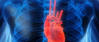
When there is pain in the heart area, most people are wary, because a person’s full life depends on the work of this organ. Many people go to the hospital to see a cardiologist. However, electrocardiography does not always answer all their questions. Can a person’s heart hurt if the cardiogram is good? Why does my heart hurt for more than one month?
In what cases is an electrocardiogram prescribed?
If a person has the symptoms described below, the cardiologist will refer him to an electrocardiogram:
- legs swell;
- fainting conditions;
- there is shortness of breath;
- chest pain, back pain, neck pain.
An ECG is mandatory for pregnant women for examination, for people preparing for surgery, or for a medical examination.
ECG results are also required if you travel to a sanatorium or if you need permission for any sports activities.
For prevention and if a person has no complaints, doctors recommend taking an electrocardiogram once a year. Often this can help diagnose cardiac pathologies that are asymptomatic.
What does an electrocardiogram determine?
- Heart rate - heart rate.
- Rhythms of heart contractions.
- Heart attack.
- Arrhythmias.
- Ventricular hypertrophy.
- Ischemic and cardystrophic changes.
The most disappointing and serious diagnosis on the electrocardiogram is myocardial infarction. In the diagnosis of heart attacks, the ECG plays an important and even the main role. Using a cardiogram, the zone of necrosis, the localization and depth of lesions in the heart area are revealed. Also, when decoding the cardiogram tape, you can recognize and distinguish acute myocardial infarction from an aneurysm and past scars. Therefore, when undergoing a medical examination, it is necessary to do a cardiogram, because it is very important for the doctor to know what the ECG will show.
Most often, a heart attack is associated directly with the heart. But it is not so. A heart attack can occur in any organ. Pulmonary infarction occurs (when lung tissue partially or completely dies if the arteries are blocked).
There is a cerebral infarction (otherwise known as ischemic stroke) - the death of brain tissue, which can be caused by thrombosis or rupture of brain vessels. With a cerebral infarction, functions such as speech, physical movement, and sensation may be completely lost or lost.
When a person has a heart attack, living tissue in their body dies or becomes necrosis. The body loses tissue or a section of an organ, as well as the functions performed by this organ.
Myocardial infarction is the death or ischemic necrosis of areas or areas of the heart muscle itself due to complete or partial loss of blood supply. Heart muscle cells begin to die approximately 20-30 minutes after blood flow stops. If a person has a myocardial infarction, blood circulation is disrupted. One or more blood vessels fail. Most often, heart attacks occur due to blockage of blood vessels by blood clots (atherosclerotic plaques). The area of distribution of the infarction depends on the severity of the dysfunction of the organ, for example, extensive myocardial infarction or microinfarction. Therefore, you should not immediately despair if the ECG shows a heart attack.
This becomes a threat to the functioning of the entire cardiovascular system of the body and threatens life. In the modern period, heart attacks are the main cause of death among the population of developed countries.
can an ecg fail to show heart disease?
An electrocardiogram (ECG) is considered the main diagnostic method for identifying various diseases of the cardiovascular system.
Our heart works in the body under the control of its own pacemaker, which produces electrical impulses and directs them to the conduction system, and they are recorded on the ECG. It turns out that using an electrocardiogram, we can record a peculiar language of our myocardium.
Based on the deviations of the main teeth: P, Q, R, S and T, it is possible to determine which disease is the basis of cardiovascular pathology.
Hypertrophy of the heart chambers occurs as a result of hemodynamic disturbances in the bloodstream, which provoke overload of the ventricles or atria. On the ECG you can see seven main signs of cardiac muscle hypertrophy.
- An increase in the time of internal deviation, since in the hypertrophied myocardium the excitation spreads longer in the area from the endocardium to the epicardium.
- An increase in the amplitude of the R wave, while the excitation vector is larger in magnitude.
- Ischemia of the subendocardial layers of the heart, due to the fact that they lack blood flowing through the coronary arteries.
- Conduction disturbance.
- Deviation of the electrical axis of the heart towards the hypertrophied section, as its mass increases due to the growth of cardiomyocytes.
- Changes in the electrical position of the heart.
- Displacement of the transition zone (V3), manifested by a change in the ratio of the R and S waves in the third chest lead.
The disease is characterized by attacks of anginal pain, lasting from a few seconds to twenty minutes. This disease is one of the formischemic heart diseases. In the classic form of angina pectoris, electrocardiographic signs are manifested by changes in the final part of the ventricular QRS complex.
- Depression of the S-T segment.
- Various changes in the T wave, such as decreased amplitude, biphasicity, isoelectricity, or negativity.
- The focal nature of these changes: they are recorded in one or two leads, since the observed hypoxia is local in nature, developing in the basin of a separate branch of the coronary artery.
During the periods between attacks, the ECG often shows no pathological changes at all. In addition, the above-described deviations are possible in many other heart diseases and pathological conditions. That is why in some cases the diagnosis of angina pectoris can be difficult.
Pathology of the cardiovascular system associated with a violation of the formation of an excitation impulse or its spread throughout the myocardium. In most cases, it manifests itself as an interruption in the rhythm of heart contractions, with periods of acceleration and gradual deceleration. Typically, your heart rate increases as you inhale and decreases as you exhale. The features of the ECG are as follows:
- The frequency of changes in the R - R intervals is more than 0.1 seconds.
- Unlike other rhythm disturbances, there is a gradual change in the duration of the R – R interval, usually due to the T – P segment.
- Small fluctuations of P – Q and Q – T are characteristic.
The most reliable electrocardiographic sign of sinus arrhythmia is considered to be a gradual periodic shortening of the R - R section against the background of an increased rhythm and, conversely, a lengthening of the R - R intervals when the rhythm slows down.
An increase in heart rate is called tachycardia. In this case, the heart rate accelerates to 100-150 beats per minute. A similar disorder can develop due to increased automatism of the sinus node.
The pathology is also inherent in healthy individuals during physical exertion or emotional stress. The cause is often ischemia, dystrophic changes, various infections and toxic effects.
Main ECG signs:
- There is a decrease in the R–R interval as the T–P interval shortens.
- With severe tachycardia, the P–Q segment shortens.
- The degree of increase in heart rate is directly proportional to the decrease in Q – T.
- An upward displacement of the RS – T segment downward from the isoelectric line.
- The amplitude and direction of the teeth correspond to the norm.
A deviation that is manifested by a reduced heart rate (less than 60 per minute).
It occurs with reduced automatism of the sinus node; it can occur even in healthy people, for example in athletes, when exposed to various factors. A common cause is considered to be an increase in the tone of the vagus nerve.
The electrocardiographic picture, in principle, differs little from the norm, only the rhythm is slowed down. The following changes are noted on the ECG:
- The R interval increases due to the T – P shift.
- Q – T increases according to the decrease in rhythm frequency.
- The amplitude and vector of the teeth change slightly.
A cardiac aneurysm is an enlargement of the myocardial cavity due to pathological changes in the muscle layers or abnormalities in the development of the organ at the stage of embryogenesis.
The main signs of a cardiac aneurysm include protrusions in its area due to thinning of the wall, which can rupture. This is what can lead to irreparable consequences, which ECG research helps prevent.
There are two leading signs that allow you to diagnose an aneurysm:
- The QS wave is present in leads where high R is usually recorded.
- A “frozen” ECG curve: instead of Q, a dome-shaped RS-T segment appears, shifted upward from the isoline, and sometimes a negative coronary T-wave appears.
Source: //folkmap.ru/krov/mozhet-li-ekg-ne-pokazat-zabolevaniya-serdtsa/
Causes of heart attack
- Atherosclerosis.
- Rheumatism.
- Congenital heart defect.
- Diabetes.
- Smoking, obesity.
- Arterial hypertension.
- Vasculitis.
- Increased blood viscosity (thrombosis).
- Previous heart attacks.
- Severe coronary artery spasms (for example, when taking cocaine).
- Age-related changes.
An ECG can also identify other diseases, such as tachycardia, arrhythmia, and ischemic disorders.
Arrhythmia
What to do if the ECG shows arrhythmia?
Arrhythmia can be characterized by numerous changes in the contraction of the heartbeat.
Arrhythmia is a condition in which there is a disturbance in the heart rhythm and heart rate. More often, this pathology is marked by an irregular heartbeat; The patient has either a rapid or slow heartbeat. An increase is observed when inhaling, and a decrease is observed when exhaling.
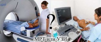
Magnetic resonance imaging
It is performed using an MRI machine, which consists of a large tube surrounded by a magnetic field. The patient lies down on a table that slides inside the device. The images obtained during the scanning process allow the doctor to assess the condition of the vascular bed and check for the presence of blood clots and plaques.
Modern cardiology has a whole range of methods that make it possible to most accurately determine the picture of the disease. Based on the data obtained, the specialist selects individual treatment tactics and decides on the need for surgical intervention. It is important to follow your doctor's recommendations and undergo regular heart examinations to assess the effectiveness of therapy.
Angina pectoris
If the patient experiences attacks of pain under the sternum or to the left of it in the area of the left arm, which can last a few seconds or can last up to 20 minutes, then the ECG will show angina.
The pain usually intensifies with lifting weights, heavy physical activity, or going out into the cold and may disappear with rest. Such pain decreases within 3-5 minutes when taking nitroglycerin. The patient's skin turns pale and the pulse becomes uneven, which causes interruptions in the functioning of the heart.
Angina pectoris is one of the forms of coronary heart disease. It is often quite difficult to diagnose angina pectoris, because such abnormalities can also manifest themselves in other cardiac pathologies. Angina pectoris can further lead to heart attacks and strokes.
CT scan
The tomograph consists of a table with a ring-shaped shell connected to a CT scanner. The patient must lie down so that the area to be examined is inside the ring. Rotating, it takes multi-layer images, looking at which the doctor can check the degree of vasoconstriction. In addition, the three-dimensional image shows the tissues surrounding the heart, which makes it possible to promptly detect the formation of tumors and deformation of the aorta.
A contrast agent is often injected to improve image clarity.
Tachycardia
Many people are very worried when they find out that the ECG showed tachycardia.
Tachycardia is an increase in heart rate at rest. Heart rhythms during tachycardia can reach 100-150 beats per minute. This pathology can also occur in people, regardless of age, when lifting heavy objects or during increased physical activity, as well as during strong psycho-emotional arousal.
Still, tachycardia is considered not a disease, but a symptom. But it is no less dangerous. If the heart begins to beat too quickly, then it cannot have time to fill with blood, which subsequently leads to a decrease in blood output and a lack of oxygen in the body, as well as the heart muscle itself. If tachycardia lasts more than a month, it can lead to further disruption of the heart muscle and an increase in heart size.
Can the heart hurt if the cardiogram is good - answers to frequently asked questions
50
Marina Dmitrievna
2017.07.17
24 136
Symptoms
When there is pain in the heart area, most people are wary, because a person’s full life depends on the work of this organ. Many people go to the hospital to see a cardiologist. However, electrocardiography does not always answer all their questions. Can a person’s heart hurt if the cardiogram is good? Why does my heart hurt for more than one month?
Methods for diagnosing heart failure
There is no medical procedure specifically designed to diagnose heart failure. The doctor usually chooses several methods from existing types of research in each individual case.
- Magnetic resonance imaging. It is considered the safest examination, and also allows you to detect heart failure at an early stage, even in the complete absence of external manifestations.
- Coronary angiography. In this study, a catheter is inserted into the vessels of the neck and upper limbs, which allows the doctor to examine the coronary vessels. The wiring is done using x-rays. The patient requires hospitalization, although the procedure does not cause significant damage.
- Angiography, as a rule, is performed simultaneously with the previous technique, since it also requires the use of radiography. A dye visible in X-rays is injected into the blood, and thus the characteristics of cardiac blood flow are examined.
- An echocardiogram is an ultrasound examination that gives an idea of the presence of cardiac hypertrophy and its functioning.
- Doppler examination is carried out using ultrasound, usually in conjunction with the previous point. Serves to study blood flow and determine the location of insufficiency.
- Nuclear diagnostics. By introducing a substance that emits radiation into the blood, a three-dimensional model of the heart and blood circulation is created. This is a study of very high accuracy; even the slightest glitch can be traced.
- During a stress test, the patient performs special exercises to put stress on the heart while being monitored by echocardiography or nuclear testing.
- An analysis is carried out for the presence of a special peptide in the blood, which increases in heart failure.
- Electrocardiogram (ECG), including its daily monitoring.
Unlike other studies, almost every adult and even a teenager is familiar with ECG. It is one of the mandatory studies and is carried out in many cases, including influenza and respiratory diseases.
Using electrocardiography, it is impossible to diagnose heart failure in the early stages of the disease. However, it can serve as a reason to revise the diagnosis in cases where there is a suspicion of HF, but the cardiogram is normal.
An ECG is performed quickly, does not cause any discomfort to a person, and does not expose him to any traumatic effects or radiation. Therefore, it is carried out even during medical examinations and medical examinations.
How does heart failure manifest?
Depending on which part of the heart is affected, the symptoms of heart failure may vary.
- dyspnea;
- heart rhythm disturbance;
- dizziness, darkening of the eyes;
- veins protrude from the neck;
- the skin turns pale;
- My legs swell in the evenings and start to hurt.
Later, so-called dropsy may occur (fluid accumulates in the abdominal cavity). The patient cannot perform any physical work, after which coughing attacks occur, which many attribute to other reasons, for example, smoking.
However, not all of the above symptoms are unambiguous confirmation of the presence of heart failure in a person. For example, swelling of the legs can be varicose veins; it is also common in older people, as is fatigue. Shortness of breath may be a consequence of pneumonia.
Symptoms can manifest with varying intensity, and there is even the possibility of death. Therefore, if one, or even more so several phenomena occur, you should immediately seek medical help.
First of all, you need to do an electrocardiogram.
Whether heart failure is visible on an ECG remains an open question, but it may help detect other conditions that will contribute to the development of heart failure and require treatment on their own.
Conductivity
In the normal state, the electrical impulse generated in the sinus node moves to the contractile muscle fibers of the myocardium through special muscle cells (conducting system). This stimulates the contraction of the atria and ventricles that pump blood. A short-term physiological delay is observed in the atrioventricular node.
In a state of pathology, the impulse is delayed more than expected, which leads to delayed excitation of the underlying sections, and as a consequence to a disruption of the normal pumping function of the heart.
Impaired conduction (blockade) is shown by a cardiogram of the heart.
Sinoatrial block is a violation of the output of impulses from the sinus node.
It may be congenital or develop against the background of heart valve defects, brain tumors, hypertension, meningitis, encephalitis, leukemia and other diseases.
Pathology can be contributed to by excess potassium in the blood or the use of large quantities of certain medications. Well-defined bradycardia is observed. The patient feels shortness of breath, weakness, dizziness, and sometimes loses consciousness.