Diseases and pathologies of the spinal column require high-quality diagnostics. They have similar symptoms, so when choosing a method, the traumatologist has to decide: X-ray or MRI of the spine will provide more information, which is better in a particular case for the patient?
The methods differ in the principle of impact, the clarity of the images, and have different indications and contraindications.
Radiography and MRI: principles for choosing a method
The spine is most often examined. With early detection of structural damage, it is possible to slow down the progression of the pathological process and maintain mobile functionality.
Both methods provide complete information, but differ in the principle of operation and in the way they achieve results.
Radiography is a clinical, non-invasive type of examination that helps to obtain a projection of the anatomical structures of the body by passing x-rays through them.
The waves emitted by the device are partially absorbed by tissues, and the intensity of penetration depends on the density of the internal organs.
The doctor determines:
- shape;
- localization of the organs being studied;
- peristalsis;
- tone;
- relief of the mucous epithelium.
Magnetic resonance imaging is a method of obtaining layer-by-layer images of organs and tissues necessary for a detailed study.
The method is based on the use of a constant electromagnetic field, which interacts with the hydrogen atoms of the structures under study.
The energy they emit is recorded by a tomograph. The study provides highly accurate visualization of internal organs, the brain, and the spinal cord. The device monitors the flow of cerebrospinal fluid, measures the speed of blood flow, and determines the level of diffusion in tissues.
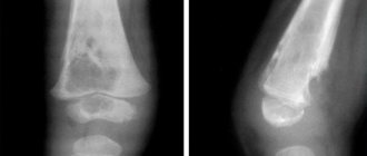
X-ray
MRI
The choice of research option depends not on the wishes of the patient, but on the recommendations of the doctor, who takes into account complaints, the results of the initial examination, and the features of the clinical case.
Principle of X-ray diagnostics
X-rays have long been used in the diagnosis of diseases of the musculoskeletal system. The technique is based on the effect of x-ray radiation, which, penetrating through bones and joints, leaves the outline of the spine on the film. Depending on the density of the fabrics, the pattern becomes clearer and has several shades.
As a rule, the examination is done in 2–3 positions and projections to obtain maximum information. Modern devices take pictures in 1–2 minutes and transfer the results to digital media.
Indications for X-ray diagnostics of the spine
Since x-rays more accurately reflect the condition of bone structures, the use of the method is advisable if:
- the likelihood of intervertebral hernia formation;
- injuries, displacements, fractures of vertebral discs;
- infectious diseases - bone tuberculosis, syphilis;
- formation of tumor formations in bone tissue;
- hematomas, bleeding, pathological accumulation of fluid in the structures of the spine.
The method provides information about spinal curvature (scoliosis) and the development of uncomplicated osteochondrosis.
When to do an MRI or X-ray of the cervical spine?
The cervical spine consists of seven vertebrae and is considered the most mobile. Blood vessels involved in the blood supply to the brain pass through it, so timely diagnosis of any pathological processes in this part of the body is extremely important. When choosing between MRI or X-ray of the cervical spine, the doctor collects anamnesis, evaluates symptoms and identifies possible contraindications.
The cervical spine is examined using MRI in the following situations:
- assessment of the anatomical integrity of the elements of the spinal column, the condition of the cartilage discs and the muscular-articular system;
- pathology of intervertebral discs, inflammatory and infectious process localized in the cervical region;
- suspicion of space-occupying formations of various nature, lesions of the nerve roots;
- preparation for surgery.
Indications for magnetic resonance imaging
Magnetic resonance imaging provides detailed characteristics of changes occurring in soft tissues.
Helps obtain a three-dimensional picture showing the condition of the discs, cartilage, vascular, and nervous tissue of the spinal cord.
During the study, tumor processes and neoplasia are identified, even if they have latent development. MRI diagnostics is justified if it is necessary to identify or exclude:
- pathological disorders in the structure and functioning of the spinal cord and canal;
- inflammatory changes in nerve tissue or soft structures;
- tumor formations;
- infectious lesions of blood vessels or spinal membranes;
- congenital defects of vertebral structures and discs.
Osteochondrosis
Using magnetic resonance imaging, the doctor identifies the development of osteochondrosis, determines the localization, features of the course, monitors the patient’s condition after surgical treatment, and the quality of rehabilitation.
Indications for MRI and X-rays
The applications of MRI are varied. This research option gives better results when examining soft tissues, so the procedure is prescribed for:
- detection of cancer;
- identifying a hernia;
- the presence of developmental anomalies, injuries or pathologies of the spinal cord;
- the course of inflammatory processes;
- identifying foci of infection and the need to assess their intensity;
- the presence of pathological zones in soft tissues.
As for x-rays, specialists resort to its help when:
- joint dislocations;
- broken or cracked bones;
- the presence of diseases of the musculoskeletal system;
- the need to check the body for the presence of foreign bodies.
Get a free consultation Consultation on the service does not oblige you to anything
In some cases, infectious, inflammatory or pathological processes affect not only soft but also hard tissues. In such situations, simultaneous MRI and X-ray are prescribed. The resulting images will allow you to get the most complete picture of the state of a certain area.
Prices for MRI in Moscow may vary. The cost of the procedure will depend on the general pricing policy of the medical center, as well as on the area requiring examination.
The cost of an X-ray will be lower than an MRI. The price of this procedure will depend on the volume of the area being examined, since the main influence on the price indicator will be the format of the film used.
List of restrictions for MRI and X-ray diagnostics of the spine
Since both methods rely on the use of radiation, they are not suitable for everyone.
X-rays are contraindicated in the following categories of patients:
- Children under 15 years of age, if the harm from the procedure is greater than if they refuse it.
- Persons with mental or neurological pathologies who do not control their behavior cannot remain motionless for some time.
- For women during pregnancy, as it poses a danger to the fetus.
The installation accurately diagnoses patients weighing no more than 120-150 kg. In obese people, getting clear images is problematic, since the fat layer prevents the passage of the rays. In this case, it is better to resort to other examination methods.
Even taking into account its relative safety, magnetic resonance imaging has limitations:
- allergy to injected contrast;
- fear of closed spaces - claustrophobia;
- nervous, mental disorders;
- the presence of a metal implant, stent, or pacemaker in the body.
Advantages of MRI over X-ray: list
- Main advantages:
- Wide range of information
- 100% safety
- Efficiency of the finished result
- Lack of preliminary preparation of the patient for the procedure
- Contrast agents may be used
- In addition, it helps to identify difficult-to-diagnose ailments:
- Degenerative-dystrophic pathology (spondylosis, osteochondrosis)
- Scoliotic diseases (with the ability to determine the degree and cause)
- Spondylolisthesis
- Malignant and benign neoplasms
- Infringement (compression) of the spinal nerve nodes and vertebral arteries
- Determines the stage of the disease as accurately as possible in the presence of:
- Malignant tumors in the body
- Multiple sclerosis
- Stroke
- Spinal cord pathologies
- Tendon and muscle injuries
Advantages and disadvantages of both types of diagnostics
A table showing the pros and cons of each of them will help you evaluate the advantages of the methods.
| X-ray | MRI |
| Advantages | |
| — A procedure characterized by simplicity and accessibility — Lack of a preparatory stage — Speedy determination of pathologies of bones and vertebrae — Examination in emergency situations — Availability and reasonable price | — Safety of the examination, including for children and pregnant women — No restrictions on the number or frequency of procedures — Examination of soft tissues, not just bones — Obtaining high-quality images with three-dimensional images of the structures being studied |
| Flaws | |
| — Adverse effects of radiation — Limitations on the number and frequency of procedures — Inability to examine women during pregnancy and breastfeeding — Relatively limited information content | — Impossibility of examining patients with metal implants, stents, pacemakers — Prolonged stay in a state of immobility — High cost of the procedure |
Physical basis of diagnostics
Despite the fact that the final result of an examination using MRI or X-ray can be obtained in an image or in the form of a video recording, the principles of operation of these methods have significant differences. What is the difference and why, when conducting an examination, preference is given to one method or another, you can understand by studying the physical principles underlying X-ray and MRI studies.
How is the MRI procedure of the spine performed?
The principle of x-ray diagnostics is based on the ability of a certain spectrum of electromagnetic rays to pass through the human body. A certain part of the waves emitted by an X-ray unit is absorbed by the tissues of the body, and the degree of their absorption is directly proportional to the density of the structure that falls in the path of the beam.
The final intensity of the beam passing through the body is recorded on photographic paper or using digital technology. Thus, the resulting image reflects the projections of organs, the structure and internal structure of which can be judged by the degree of darkening of the image. Bone tissue, including the spine, having an extremely dense structure, practically does not allow electromagnetic radiation to pass through and appears as a light spot on photographs.
If you imagine a beam of electromagnetic waves emitted by an X-ray machine in the form of a powerful beam of light, then the projection of the bones will be a “shadow”. When using x-rays to diagnose spinal pathologies, you can only see a “shadow”, or, in other words, the outer contour of the bone structure. Using radiographic examination, one can judge exclusively the external anatomical changes occurring in the bone tissue of the spine and paravertebral structures.
X-rays can be used to take pictures of a small portion of the spinal column.
The operating principle of an MRI scanner is based on a temporary change in the polarization of hydrogen atoms, which are found in abundance in the soft tissues of the body, through exposure to a powerful electromagnetic field. The return of hydrogen ions to their original position when the magnets are turned off causes a release of energy, which is recorded and processed by the computer system.
The hydrogen concentration allows us to judge the density of structures and, in accordance with the area under study, allows us to obtain an image of all internal organs. Unlike X-rays, the MRI signal does not project the outline of the bone component, but transmits an image of predominantly soft tissues located inside (the spinal cord, spinal canal) and around the spine.
The contrast between the hydrogen-rich paravertebral tissues and the low hydrogen content of the bones allows a detailed image of all structures of the spinal column to be obtained. MRI can provide an overview of the entire spine.
Important! The main difference between MRI and X-ray is the ability to evaluate not only external changes in the intervertebral discs, but also internal ones.
Purposes of MRI and radiography
Today, radiography remains the standard diagnostic procedure. The study is indispensable before emergency surgery. With its help it is revealed:
- spinal column injuries;
- bone tissue growth;
- curvatures - kyphosis, scoliosis;
- damage, vertebral displacement.
Magnetic resonance imaging is also used in diagnostic testing. The application is suitable for diagnostics:
- spondylosis;
- osteochondrosis;
- tuberculosis or syphilis;
- tumor neoplasms;
- malformations of blood vessels, vertebrae and discs.
Despite the relatively high cost of the procedure, it remains in demand along with radiography.
MRI or X-ray of the cervical spine: general information
When an MRI or X-ray of the cervical spine is needed, you need to understand the main advantages and disadvantages of both techniques. Magnetic resonance imaging uses a powerful magnetic field that specifically affects the hydrogen atoms contained in the body's tissues. During scanning, the tomograph sends a series of radio frequency pulses, which leads to nuclear magnetic resonance and a change in the electromagnetic response of atomic nuclei. The changed parameters are recorded by computer equipment, undergo complex processing and are converted into a three-dimensional image of the area of interest.
X-ray of the cervical spine contains a different principle. The final images are obtained as a result of the attenuation of X-ray waves when passing through media with different densities. On an x-ray, bones appear as white areas, air spaces appear black, and soft tissue structures appear as varying shades of gray.
Which method is more informative?
In an effort to obtain an objective assessment of the accuracy and information content of both types of research, it is taken into account that they are not interchangeable, but are used in different cases and situations. Meanwhile, the differences in MRI are obvious:
- Diagnosis using tomography is more accurate than x-rays.
- It is prescribed to clarify or detail the pathology, if difficulty arises in identifying the etiology of the disease.
- Using magnetic resonance imaging, a specific area is studied while other areas are left untouched.
- Provides detailed images of the spine.
With radiography, it is problematic to obtain detailed images, since the functionality of the installation is limited and inferior to MRI.
The advantage of the procedure is its low cost and examination of patients with contraindications to magnetic resonance imaging.
MRI image
X-ray
Both types of diagnostics are highly accurate and informative, but nevertheless the choice is determined by the nature of the suspected pathology.
MRI is suitable for studying soft tissues, while doctors prescribe X-ray examination for violations of the integrity of bone structures and skeleton.
MRI or X-ray of the cervical spine: contraindications for examination
The doctor includes an MRI or X-ray of the cervical spine in the diagnostic scheme if there are no contraindications to them. Thus, the appointment of MRI is excluded if there are metal prostheses or ferromagnetic products in the patient’s body, with claustrophobia, some neurological syndromes, or in the first trimester of pregnancy. X-rays are prohibited for pregnant women, patients with obesity or disorders that prevent them from being immobile.
The main method of primary diagnosis in vertebrology is radiography, magnetic resonance imaging (MRI) and computed tomography (CT) are used to clarify the diagnosis, as they are the most informative.
Using x-rays, the cause of pain and the nature of spinal damage are determined. This method is indispensable in emergencies and when first seeing a doctor. MRI makes it possible to obtain a spatial, detailed image of an organ. To decide whether it is better to perform an X-ray or an MRI of the spine for diagnostic purposes, you need to know what pathology needs to be excluded.
Diagnostic cost comparison
If there is a difference in the principle of action, in the method of obtaining information, the cost of the procedure differs. The average price of radiography is 1500-2000 rubles, MRI - 3000-6000 rubles. It is formed taking into account the following factors:
- area of the study area;
- the number of images that the doctor will need;
- equipment quality;
- level of training of medical staff;
- status of the medical institution.
A referral from the attending physician is required for an effective examination. Despite the obvious advantages of tomography over x-rays, the use of both techniques is considered justified.
Doctors answer in the affirmative to patients’ interest in what seems better—X-ray or MRI diagnostics, and the compatibility of both diagnostic options.
Both studies provide a comprehensive description of the condition of the spinal column. The information obtained is used to clarify the etiology of the disease and make a diagnosis. As for safety, MRI can be combined with repeated x-ray examinations.
What is more harmful: MRI, CT or X-ray
In the case of MRI, there is no talk of harm at all. When undergoing radiography and CT scanning, the human body receives a certain dose of x-ray radiation.
The x-ray lasts for several minutes, the x-ray is ready in a split second. Computed tomography is accompanied by the reproduction of a series of images; the more slices, the longer the scan and the higher the load - this is the price for the high quality of the final images.
To reduce possible harm from radiation examination, special protective equipment is used (lead aprons, plates, collars).
It is possible to do an X-ray on the same day as an MRI, this is explained by the harmlessness of the second procedure. The situation is different with a combination of radiography and CT.
Since we are talking about studies that involve radiation exposure, diagnostics in a short period of time are not recommended.
Under such circumstances, it is necessary to take a break of at least 1-2 months. Conducting research in a short period of time is justified only if the benefits of diagnostics exceed the possible harm.
How do MRI images differ from X-rays?
On MRI, black and white images provide a clear layer-by-layer image of the spine; they show the condition of the discs, cartilage tissue, spinal cord structures, nerve endings and soft tissues. Images are first taken in a plane dividing the human body into right and left halves.
If a pathology is found, this area is examined in two other planes - frontal (facing the camera) and transverse (as if cutting the body into parts in a horizontal plane). The spinal cord is best visualized with MRI on T1-VI: the structure, white and gray matter, cerebrospinal fluid, and bone marrow of the vertebral bodies are visible.
For a number of diseases, it is necessary to do magnetic resonance imaging with contrast: a paramagnetic contrast agent is injected intravenously, which is distributed throughout the vessels. The difference in blood supply and accumulation of contrast agent distinguishes healthy tissue from tumors and inflammation.
X-ray of the spine (spondylography) is performed in frontal, posterior and lateral projections, to display intervertebral joints - in oblique ones. You can receive pictures in 20-40 minutes. To determine the condition of the intervertebral discs, the examination is done during maximum flexion and extension of the spine, thus identifying the displacement of the overlying vertebrae relative to the underlying ones.
On spondylograms in the lateral projection, the spine is examined along its entire length: the vertebral bodies, their structure and shape, spinous processes, intervertebral discs, joints, and foramina. Using images, fractures and destructive changes in the vertebrae, inflammation and developmental anomalies, pathology of bone tissue are identified and the feasibility of MRI is determined.