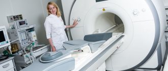
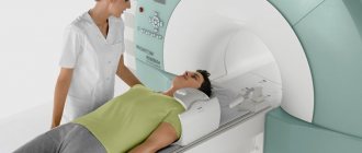
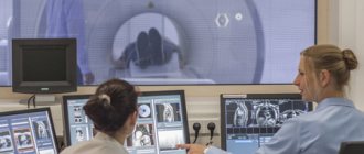
About fluoroscopy of the lumbar region
Radiography is the most accessible and informative examination method. X-rays of the lumbar spine can be performed in almost any medical institution:
- in an outpatient clinic (clinic);
- in a hospital (often hospitals and hospitals have a separate radiology department);
- in the emergency room;
- at a private medical center.
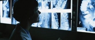
The X-ray result shows structural changes in bone tissue, intervertebral discs and articular areas of the vertebral joints.
Typically, the X-ray result is printed on special X-ray film. It is quite expensive and requires careful handling, but allows the doctor to examine in detail the abnormalities in the structure of the lumbar or sacral region. “Film” X-ray images can be digitized, that is, converted into digital format and stored on electronic media. This will provide relatively greater security than any physically printed photo. Digital radiographs can be printed at any time, and you can also select a specific area of the image.
Modern digital X-ray machines allow you to record X-ray data on electronic media (disk or flash card). To record and subsequently read such an electronic X-ray image, a special program is required, which can cause certain difficulties when presenting the results to other specialists.
The third option for visualizing X-ray results is printing on paper. This method is suitable for obtaining an opinion from a specialist. For example, when the original X-ray film remains in the archives of a medical institution, and the patient needs to consult with another specialist based on examination data of the lumbosacral region. Printing X-rays on paper does not allow us to examine changes in detail, much less identify pathologies in the structure of bone tissue (for example, in osteoporosis). But the picture shows changes in posture, curvature of the spine and the location of the intervertebral hernia.
Why do you need to prepare for the examination?
Not every patient understands why a number of preparatory measures are needed for an X-ray examination of the lumbosacral spine. But in reality everything is simple. For example, if there is feces in the intestines, then the image will be uninformative, since human waste products will also be visible on it . An accurate and correct diagnosis cannot be made.
In some cases, easily excitable people need to take sedatives so that they can lie quietly for a while. Otherwise, the photo will also be useless.
If you want to know in more detail what an x-ray of the spine shows, and also consider what the dangers of x-rays are, you can read an article about this on our portal.
Preparation
Despite the ease of performing x-rays of the lumbosacral region, special preparation is required before the study. As a rule, an X-ray of the spine is carried out in 2 projections (direct and lateral), which makes it possible to determine and identify changes in various planes. Accumulation of gases in the intestines or unresolved stool can cast shadows on skeletal structures, which will significantly complicate the description of photographs. Therefore, it is strongly recommended to prepare for an X-ray of the lumbar or sacral spine:
- 1-2 days before the test, exclude from the diet foods that increase fermentation and contribute to flatulence (gas formation):
- legumes;
- cabbage (especially fresh);
- fresh yeast bread and buns;
- it is recommended to reduce the consumption of sweets, including carbonated drinks;
- milk (does not apply to fermented milk products).
- Avoid eating for 6-8 hours before the test. Therefore, it is preferable to take an x-ray of the lumbosacral region in the morning, skipping breakfast.
- Refrain from taking liquids as with food. You can drink a small amount of water (no more than 100 ml) 1-2 hours before the test.
- Carry out a bowel cleansing procedure by performing cleansing enemas (the evening before the study and in the morning, 1-3 hours before the x-ray).
IMPORTANT! If a patient is diagnosed with chronic flatulence, it is recommended to prepare for the study 2-3 days in advance. You need to start taking carminative medications. This will clear the intestines of gases and allow x-rays to be taken without difficulty.
How to independently prepare for an X-ray of the lumbar spine
If radiography is performed in a hospital setting, then the medical staff (nurse or attending physician) will tell you in detail about all the preparatory procedures. Cleansing enemas are also administered by a nurse. If the examination is scheduled on an outpatient basis, you must independently prepare for x-rays. The principles are the same:
- avoidance of products that cause gas formation;
- refusal from food 7 hours before the study, from water - 2 hours;
- cleansing the intestines with a double enema.
IMPORTANT! For cleansing enemas, boiled water at room temperature is used - from 21°C to 35°C. The volume of liquid depends on the build and age of the person.
X-rays of the lumbosacral region do not take long - from 10 to 30 minutes, depending on the need for re-shooting and the number of functional tests (they will be discussed below). If the examination is prescribed for a child, then parents should talk to the child, explaining that it will not hurt, but you cannot move during the x-ray. Any movement helps to reduce the clarity of the X-ray image and can provoke the formation of shadows on the vertebrae, which can be perceived by the radiologist as a pathology.
Preparation the day before the examination
Preparation for an x-ray begins several days in advance, but it is most important on the day of the procedure. You will need to come to the office on an empty stomach, regardless of the appointed time. Therefore, many advise signing up for the morning hours. In the morning you need to follow the following rules:
- Completely avoid breakfast, coffee, tea or even juice.
- Drink a bag of laxative if there are more than 6 hours before the appointed time or give a cleansing enema.
- Give up bad habits, do not smoke on the day of the event.
- Drink 15 drops of valerian tincture or other sedative to reduce possible stress.
- Take water and food with you so that you can eat immediately after the procedure if it takes place in the afternoon or evening.
- Before undergoing the examination, you must remove any jewelry, especially piercings.
Preparatory procedures are extremely important when examining the lower back or lower back. The described measures will make it possible to more effectively cleanse the intestines of the accumulation of gases and feces, improve visualization, and increase the accuracy of diagnosis.
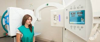
Carrying out
An X-ray image is produced by the difference in emitted and returning electromagnetic waves. Some of the emitted waves are “lost” in the structures under study, forming darkened areas in the final image. The higher the density of the structure, the darker the area of the finished image.
Projections for research
Depending on the expected diagnosis, the doctor prescribes certain positions in which the patient needs to be during the examination. This allows you to take images in different planes. In medical terminology, planes are usually called projections. Accordingly, x-rays of the lumbosacral spine are most often performed in 2 projections.
The main projections for x-rays of the lumbar and sacrum are straight (frontal) and lateral. Direct projection can be performed in two variations:
- anterior (the patient is positioned with his back to the X-ray machine tube, his face is to the fluorescent screen or to the X-ray cassette);
- posterior (the patient is positioned facing the X-ray tube, with his back facing the screen or cassette).
The lateral projection is performed by turning the patient perpendicular to the screen and tube, i.e., the shoulder (right or left) rests against the screen or cassette.
When examining the lumbar and sacral regions, it is important to identify possible displacements of the vertebrae at the time of rotation, therefore two additional oblique projections are often used. Each of them shows the angle of displacement of the vertebrae and is performed in the same way as the lateral ones, but the patient must be facing the fluorescent screen at an angle of 45°.
In addition to direct, lateral and oblique projections, other positions may be prescribed for a more detailed study. They will differ in the angle of inclination of the patient’s body and allow the doctor to determine the degree and stage of the developing pathology.
Indications
There are a lot of indications for X-rays of the lumbar and sacral region. The main ones include:
- Presence of pain of any intensity in the back area.
- Feeling of numbness or crawling (paresthesia) in the lower extremities.
- Diagnosis of spinal injuries.
- Restricted lumbar mobility.
- Rachiocampsis.
- Determination of the localization of intervertebral hernias and areas of pinched nerve roots.
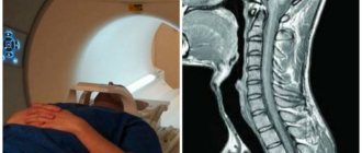
It must be emphasized that radiography is considered uninformative when diagnosing pathological processes in the muscles and ligaments. For these structures, it is preferable to use computed tomography or magnetic resonance imaging.
Contraindications
Modern X-ray machines are considered relatively safe, so there are no absolute contraindications to lumbar X-rays. There is a set of guidelines that experts adhere to when prescribing this study.
X-ray radiation in doses required for X-rays of the lumbosacral region is considered safe, but can have a detrimental effect on the developing fetus. For this reason, x-rays are extremely undesirable for women during pregnancy. It is also not recommended to conduct examinations during menstruation. There are no absolute contraindications for men.
Interesting to know: it is not advisable for people suffering from grade 2-3 obesity to undergo x-rays. There is a high probability of obtaining uninformative images.
Safety and Frequency of Testing
The electromagnetic waves emitted by the X-ray machine are absorbed by the tissues of the human body. This phenomenon causes the occurrence of photochemical reactions in them, leading to changes in the functional activity of cells. That is why, when performing radiography, there are restrictions on the frequency of its implementation.
Radiation exposure when examining different parts of the human body differs in permissible doses. For diagnosing the lumbar region with the film method it is 0.7 mSv (mili-Sievert), with the digital method it is 10 times less. The permissible radiation dose per year is 1 mSv, the maximum is 5 mSv. When these norms are exceeded, so-called long-term effects are observed, characterized by the formation of malignant processes and abnormalities in the development of the offspring.
In conclusion of the above information, I would like to emphasize once again that an x-ray of the spinal column is considered a very valuable technique for diagnosing pathological processes developing in the musculoskeletal system. Qualified specialists will conduct this study competently and correctly interpret its results, and the patient, for his part, must only prepare for it qualitatively.
Functional tests
Functional tests must be understood as functional radiography of the spine. This method of examining the lumbar region includes a set of several projections, selected individually for each patient. Functional tests allow you to obtain the most complete picture of the disease and select the most correct treatment.
A complete functional x-ray examination of the lumbosacral spine consists of three main tests:
- posterior direct projection;
- right and left lateral projections in the position of maximum flexion;
- right and left lateral projections in the position of maximum extension.
The selection of samples and rotation angle should be carried out according to the principle of maximum mutual opposition. This is the only way to determine the true position of the vertebrae at the moment of displacement and calculate the maximum available range of motion.
Functional tests can be carried out in several positions:
It is considered most correct to perform radiography in a standing position. If the patient is unable to remain in an upright position, functional tests are performed in a sitting position. And only if it is absolutely impossible to sit the patient down, the examination is performed in a horizontal position.
IMPORTANT! Functional X-ray examination (FXR) is carried out when examining the lumbosacral region and the sternolumbar junction. This allows you to see changes in all ranges of motion.
In modern medicine, x-rays of the lumbosacral spine are a widely known and accessible method of instrumental diagnostics. Radiography helps to quickly and painlessly diagnose various pathologies. With the help of new medical equipment, the dose of X-ray radiation is reduced to a minimum. In 15 minutes you can get a high-quality image with a transcript, and with it a doctor’s consultation.
X-ray appointment
An X-ray of the lumbosacral spine is performed within 1–2 minutes, and the process itself does not cause pain or discomfort to the patient. Using modern equipment, radiation exposure is reduced to a minimum. The result is printed on a special film, which allows the doctor to see even the smallest details and features. Instrumental research is prescribed in the following cases:
- Suspicion of injury or fracture.
- Rachiocampsis.
- Osteochondrosis.
- Malignant processes in the vertebrae.
- Any pathological processes in bone tissue.
- Numbness of the limbs, chronic lower back pain.
- Diagnosis before surgery.
- Congenital anomalies.
Carrying out the procedure for preventive purposes is not recommended, since the study is quite harmful. In one session, the body absorbs as much rays as from all household appliances in a year. Modern devices cause less harm, and the results obtained can be obtained on a special electronic medium.
Preparation
To obtain a high-quality image, careful but simple preparation for the manipulation is necessary. Accumulations of feces and gas bubbles affect the quality of the study. The specialist giving the referral for the procedure must explain the essence of the manipulation, its features and voice contraindications. Preparation consists of several important steps:
- should begin three days in advance. The person being studied should remove foods that promote gas formation from the menu: fermented milk, vegetables, peas, beans, fresh fruit, sauerkraut and sparkling water. The so-called slag-free diet.
- It is recommended to take only liquid food, broth, tea.
- Before each meal, you should take two tablets of enzyme preparations (Mezim or Pancreatin), and after a meal, drink activated charcoal.
- To feel comfortable and not worry during manipulation, experts recommend using valerian infusion three times a day, 10 drops.
- The last meal should be no later than 18.00 the night before, otherwise the image will be blurred and a repeat x-ray will be required.
- In the evening and on the day of the x-ray examination of the lumbosacral back, it is necessary to do a cleansing enema. If it is ineffective, you can take a laxative (Fortrans) or drink warm, salted water.
- Before the procedure, the patient should not eat or drink water (even still).
First of all, preparation is aimed at cleansing the intestines of feces and gases. Their excessive accumulation complicates the examination and blurs the clinical picture. As a result, the x-ray will need to be repeated. Repeated testing means unnecessary radiation exposure.
You can sign up for the procedure at any clinic. This makes the method accessible to all segments of the population. You can also conduct the study in a private clinic, which is equipped with modern equipment. In a self-help institution, conducting an examination will cost you up to 2,000 rubles.
How to prepare?
Preparation for x-rays of the lumbar spine is not the least important. The result depends on the correct actions. The picture may come out blurry, unclear, and this will not allow the specialist to identify the disease. Patient preparation begins 76 hours before the scheduled event.
The doctor who sends the patient for an X-ray of the lumbar spine talks in detail about all the nuances of preparation.
There are several important steps that you should pay attention to:
- Diet.
- Colon cleansing.
There are other preparatory moments. We will talk about them below.
What diet should I follow?
The diet before an x-ray of the lumbar spine should begin 3-4 days before the expected diagnosis. All foods that contribute to excessive gas production are removed from the menu. These include:
- milk products;
- beans (beans, peas);
- bread (black).
Also eliminate carbonated drinks, garlic and other foods that cause bloating.
Eating is allowed until 6 pm. This refers to the day before the x-ray is taken. Eating food in the morning is also not advisable.
How to cleanse the intestines?
You can cleanse the intestines before x-raying the lumbar region using syringes and enemas.
You will have to do this cleaning more than once. It is advisable to enema twice in the evening (with a half-hour or hour break) and once in the morning.
This procedure will help eliminate all fecal matter from the rectum in the lumbar region, which can become an obstacle to normal viewing of the spine in this area.
Other tips and tricks
Preparation for an x-ray of the lumbar spine also includes the following points:
- A couple of days before the appointed day, take charcoal tablets. Calculate the dosage according to your body weight. Usually there is 1 tablet per 10 kg. Use three times a day.
- If the patient has an unbalanced psyche or is simply worried about the x-ray, then it is allowed to take a sedative. An excellent option would be valerian.
For everything to work out, follow all the above recommendations and come to the x-ray room at the specified time with a referral from the doctor.
Carrying out manipulation
The manipulation is quick and painless, you will receive an image within 15-30 minutes. Unpleasant sensations and discomfort can only be caused by a cool table and fear of finding out the result. The patient must remove outer clothing and all metal accessories and jewelry (belt, piercing, chain), and expose the desired area of the body. When carrying out the manipulation, the subject must take a motionless sitting or lying position, otherwise the picture will be blurry. In order not to receive an extra dose of radiation, areas of the body that will not be visible are covered with a protective apron.
The lumbosacral spine is a very mobile area; it is prudent to study it using functional tests. To do this, the patient is asked to lie on his side and bend as much as possible in the lower back, taking the “embryo” position. To make an accurate diagnosis, photographs are taken in projections: posterior and lateral (in a state of maximum flexion and extension). To help the patient take the correct position and select the desired angle of the X-ray tube, the help of a highly qualified specialist is needed,
Functional tests are an individual indication in each individual case. Their main rule is to bend in the opposite direction. This way we find out the mobility and compression of the vertebrae in the affected area. An important factor is trauma, then the examination is carried out as carefully as possible, in some cases on a stretcher or gurney, without transferring the patient to the X-ray table.
Indications
During the examination, X-rays penetrate deep into the tissue and leave a clear outline of the bones and organs located in the pelvic area. To obtain maximum information, instrumental diagnostics are carried out in three planes. X-rays with a contrast agent are also taken (often when examining the bladder and urinary tract). An additional diagnostic measure is functional tests, which involve maximum curvature of the spine in the opposite direction.
A little about radiography
Radiography is a simple, universal, fast and fairly informative method for studying the tissues of the spine and other organs. This is an affordable and safe way to identify most abnormalities and disorders in this part of the skeleton.
X-ray diagnostics
The method is based on the ability of body tissues to transmit X-rays, and the transmittance of all tissues is different. Due to this, you can get a complete picture of the condition of a certain part of the body, captured on a special film. During the procedure, the person will be between the X-ray machine (X-ray tube) and the same film. The rays passing through the tissue will create an image of a certain nature on it, looking at which the doctor will be able to determine the presence or absence of any abnormalities.
Contraindications
Today there are many ways to examine the lumbosacral spine (magnetic resonance and computed tomography, ultrasound), but radiography is an informative and affordable method. Unfortunately, not everyone can perform this manipulation. X-rays are contraindicated for women during pregnancy and lactation, young children, and overweight people. In case of severe conditions and nervous disorders in the patient, manipulation is also not recommended.
Unfortunately, if it was not possible to refuse an X-ray examination during pregnancy, the abdomen is covered with a special protective screen. Further consultations with a gynecologist are carried out more thoroughly. The procedure is most dangerous in the first trimester, as the formation of all organs and systems in the fetus occurs. According to the World Health Organization, children under 15 years of age should not receive X-rays. If the manipulation has no alternative, then the child is covered with a special protective oilcloth.
In this instrumental study, radiation stops entering the body immediately after the procedure is stopped and the device is turned off. X-ray radiation does not accumulate in the body and does not form radioactive substances. Therefore, no special procedures are provided for their removal from the body.