Enteroscopy - a brief excursion into history
For several decades, the small intestine has remained an area of the gastrointestinal tract that is difficult to reach for endoscopic examination. Accordingly, the diagnosis of small intestinal bleeding, inflammatory, autoimmune and neoplastic lesions of the small intestine has been a challenge for surgeons and gastroenterologists. Therapeutic endoscopic interventions throughout the jejunum and ileum were performed occasionally, and for the most part only during intraoperative intestinoscopy.
For the first time, examination of the small intestine behind the ligament of Treitz using a flexible endoscope was performed by Ogoshi K. et al. [4] in 1973 in patients with small intestinal bleeding. The probe fibroenteroscope was first described by Tada et al. in 1977. The working length of tube enteroscopes used in diagnostics was 250 - 400 cm [6]. Works on enteroscopy using a colonoscope were first published by Parker and Agayoff [5] in 1983, and in Russia presented by V.P. Strekalovsky [3]. But these methods, which are quite time-consuming and labor-intensive for both the doctor and the patient, have not found wide application in clinical practice. The technique of intraoperative enteroscopy, developed in the mid-80s, was for quite a long time the only method for active total examination of the small intestine [1, 2, 7]. But intraoperative intestinoscopy can no longer be classified as a low-traumatic diagnostic method, and its place was reserved at the stage of revision of the abdominal cavity during surgery.
Video capsule endoscopy (VCE) has revolutionized the evolution of diagnostic methods and has significantly improved the diagnosis of small intestinal diseases [8, 9, 16]. The first reports of “wireless” video endoscopy appeared in the mid-90s, and the method began to be widely used in clinical practice since 2001 [13, 14]. In parallel with the introduction of video capsule enteroscopy, the Japanese doctor H. Yamomoto, in collaboration with, created a double-balloon video endoscope in early 2001 and used it in clinical practice. This made it possible not only to observe the condition of the lumen and mucous membrane of the deep parts of the small intestine, but also to perform a biopsy, remove tumors, stop bleeding and perform balloon dilatation of strictures [11].
In 2006, a new single-balloon enteroscope was developed and introduced into clinical practice for the study and treatment of diseases of the small intestine. In 2006-2008 individual clinical centers in Japan, America, Italy and France presented the first positive results of using the new device [13, 14, 15, 16, 17]. According to T. Tsujikawa et al. [11] a single-balloon enteroscope, having all the described positive qualities, has some advantages in comparison with a double-balloon system: it is easier to process and prepare for the study, and provides a shorter duration of the study itself, due to the absence of the need to inflate the second balloon. As you master the technique of single-balloon enteroscopy, it becomes possible to perform it by one doctor, without the involvement of an additional assistant [11, 12]. The first experience of using single-balloon enteroscopy in Russia was published by E.D. Fedorov and E.V. Ivanova in 2008, and in 2009, the results of 98 studies were analyzed, the methodological and technical aspects of the technique when used in clinical practice were considered [18].
New opportunities for access to the distal segments of the jejunum and proximal ileum, which are provided by video capsule and instrumentally assisted (balloon) enteroscopy, forced clinicians to carefully reconsider the indications for these types of endoscopic examination and, first of all, answer the question: “Who needs deep jejunoileoscopy ?
Enteroscopy techniques
The main advantage of enteroscopy is the ability to carry out therapeutic actions during the examination of the intestines. During the examination of the intestinal cavity, if necessary, the doctor can stop bleeding, remove a foreign body, and also excise polyps and even expand obstructed areas.
Enteroscopy is divided into two types:
- Balloon;
- Capsule.
The balloon enteroscopy technique is performed exclusively under general anesthesia. The principle of the study is that during the insertion of the tube, the rectum is straightened using small air balloons. These cylinders are located directly on the tube itself, during the movement of which they are inflated and deflated. For a long time, the enteroscope was equipped with one balloon, but not so long ago, the Japanese doctor Hironori Yamomoto developed new double-balloon enteroscopes. This discovery was a real revolution in endoscopy.
Capsule enteroscopy does not require general anesthesia, which is due to the absence of the need to use an endoscope. This method is more expensive than the first, but also has its significant disadvantages. The essence of the method is that the patient should swallow a special electronic capsule, which is equipped with a video camera and a light source. As the capsule advances, video recording occurs, which is immediately transmitted to the monitor tap. The advantage of the capsule method is that the patient does not experience any discomfort, pain or discomfort. The patient does not even require the administration of local painkillers. It is worth noting some significant disadvantages of capsule enteroscopy:
- the impossibility of performing a biopsy, that is, taking a piece of an organ for laboratory analysis;
- inability to carry out therapeutic manipulations: removal of a foreign object, stopping bleeding;
- obtaining an unclear image during the examination.
The enteroscopy procedure is performed only in specialized institutions that are equipped with the necessary equipment. The patient is admitted to the hospital before the procedure.
Recommendations for performing enteroscopy
According to most experts, balloon enteroscopy is recommended to be performed:
I. For diagnostic purposes
- if you suspect small intestinal bleeding, one of the most important manifestations of which, especially with occult variants of the course, is iron deficiency anemia;
- if malabsorption syndrome is suspected, often manifesting clinically as diarrhea of unknown origin;
- if you suspect a tumor of the small intestine;
- if pathological changes in the small intestine are detected during an X-ray examination;
- to obtain tissue samples of the small intestine for histological examination (forceps biopsy).
II. For medicinal purposes
- to stop small intestinal bleeding using argon plasma coagulation or endoscopic clipping;
- to remove tumors from the small intestine;
- for balloon dilatation for small intestinal strictures;
- for removing foreign bodies from the small intestine
- for retrograde interventions (ERCP) in patients with extensive abdominal surgery (gastric resection, Roux-en-Y surgery, etc.)
III. For surveillance purposes
- in clinical syndromes occurring with multiple lesions of the small intestine by polyps (Peutz-Jeghers syndrome, familial adenomatous polyposis)
- to assess the state of the small intestine over time in patients with previously diagnosed diseases of this organ, in particular to monitor the effectiveness of conservative therapy for Crohn's disease and enteropathies.
Types of enteroscopy
There are several types of enteroscopy. They all provide access to the small intestine in different ways and have other differences:
- Eunoscopy is an examination of an organ during fibrogastroscopy. This method uses tubes that are inserted through the mouth.
- Ileoscopy is an examination of the intestine during fibrocolonoscopy. During the procedure, colonoscopes with a wide channel are used. A controlled thin endoscope moves along it. The devices are inserted through the anus.
- Capsule is a new technique that does not require the use of an endoscope. The patient swallows a small device from which the image is transmitted to the monitor screen.
- The balloon allows you to straighten the intestines as the probe passes. This helps to obtain maximum information, examine even hard-to-reach areas, and obtain a biopsy.
All examinations are performed on an outpatient basis, and the balloon examination is performed under general anesthesia. After this, a two-day hospitalization is required. Anesthesia is necessary because when the intestine expands with gas, severe pain occurs. Intestinoscopy can also be performed using transintestinal guides.
Contraindications to enteroscopy
Contraindications to deep enteroscopy correspond to contraindications to esophagogastroduodenoscopy and colonoscopy.
Limitations that may prevent the full implementation of enteroscopy are:
- Involvement and gross deformation of the small intestine by adhesive process after previously undergone major operations on the abdominal organs.
- Tumor and scar strictures of the small intestine itself.
- Poor preparation of the patient for the study.
Contraindications and possible complications
There are no absolute contraindications to enteroscopy; the use of this method is limited by narrowing of the intestine, constipation, and adhesions. However, the balloon procedure is sometimes used to widen narrowings, so everything is relative.
Be sure to read:
Intestinal erosion: causes, symptoms and treatment methods
Contraindications may include the patient’s general severe condition, in which pain relief is impossible, as well as failure of internal organs.
There are two main complications: bleeding and perforation (breakthrough) of the intestinal wall. This may be the case if a person has developed an ulcer or tumor that did not manifest itself in any way before the study. In this case, they proceed to abdominal surgery, during which the ulcer is sutured or the tumor is removed. Such cases are extremely rare and are considered casuistic.
In the vast majority of cases, enteroscopy allows not only to cure the disease without surgery, but also to detect cancer at an early stage.
In continuation of the topic, be sure to read:
- Endoscopic methods for examining the intestine: description and preparation
- Preparation for gastroscopy and procedure
- Methods for examining the small intestine, their advantages and disadvantages
- How to check the intestines for oncology?
- Checking the intestines for diseases: physical, laboratory and instrumental methods
- How is capsule endoscopy of the intestine performed?
- Preparation and conduction of FGDS (fibrogastroduodenoscopy)
- Intestinal ultrasound or colonoscopy: comparison of techniques and their information content
- MRI of the intestine and colonoscopy: which study is better?
- Details about intestinal colonoscopy: preparation and procedure
Comparison of capsule endoscopy and enteroscopy
Introduction into clinical practice and further study of the capabilities of these methods showed that balloon-assisted and video capsule endoscopy are not absolutely interchangeable, that the disadvantages of one of them are offset by the advantages of the other, and for this reason, VCE and BAE do not compete with each other, but complement each other (Table . 1).
Table 1. Comparative assessment of the diagnostic capabilities of video capsule and balloon-assisted enteroscopy (BAE) techniques.
| Comparable parameters | Capsule enteroscopy | BAE |
| Verification of identified changes | Worse | Great |
| Biopsy | No | Yes |
| Treatment | No | Yes |
| Image quality | Worse | Great |
| Live View | Worse | Great |
| Total examination | Yes | Not guaranteed |
| Need for sedation | No | Yes |
| Tolerability of the study | Great | Worse |
| Duration of intervention | 8-9 hours | 1-2 hours |
| Diagnostic value for TKA | High | Below |
| The need for technical skills to perform the study | No | Yes |
| Need for clinical experience | For image interpretation only | Yes |
Enteroscopy: purpose, procedure and results
Enteroscopy is a procedure that helps your doctor find and treat problems in the digestive system. During an enteroscopy, your doctor inserts a thin, flexible tube with a camera attached into your body. This is called an endoscope. One or two balloons are usually attached to the endoscope. Balloons can be inflated to give the doctor a better view of your esophagus, stomach, and part of the small intestine. Your doctor may use forceps or scissors on the endoscope to remove a tissue sample for testing.
Enteroscopy is also known as:
- double balloon enteroscopy
- double bubble
- capsule enteroscopy
- push-and-pull enteroscopy
There are two types of enteroscopy: upper and lower. In upper enteroscopy, the endoscope is inserted into the mouth. During lower enteroscopy, the endoscope is inserted into the rectum. The type of enteroscopy performed will depend on the type of problem the doctor is trying to diagnose. Your doctor will tell you in advance which type you need.
Enteroscopy allows you to diagnose or evaluate diseases inside the body without making any incisions. It is usually used to identify problems in the small intestine or stomach. Your doctor may recommend an enteroscopy if you have any of the following:
- high white blood cell count
- tumors in the small intestine
- blockage of intestinal passages
- abnormal gastrointestinal bleeding
- intestinal damage due to radiation therapy
- unexplained severe diarrhea
- unexplained malnutrition
- abnormal x-ray results
Your doctor will give you instructions on how to prepare for the procedure. Be sure to follow them carefully. You may need:
- stop taking aspirin or other blood thinning medications
- Avoid solid foods and milk after 10 pm. the night before the procedure
- Drink only clear liquids on the day of the procedure
- avoid all liquids at least four hours before the procedure
Enteroscopy is an outpatient procedure, which means you can go home the same day as the procedure. This usually takes from 45 minutes to two hours.
Depending on the type of enteroscopy being performed, your doctor will either sedate you completely or give you medication to help you relax. These medications will be given through a vein in your arm.
Your doctor will record a video or take pictures during the procedure. They can be viewed in more detail after the procedure is completed. Your doctor may also take tissue samples or remove existing tumors. Removing any tissue or tumor will not cause pain.
Depending on the type of problem, your doctor will perform either an upper or lower enteroscopy. An upper enteroscopy allows your doctor to examine and treat the upper part of the digestive system. Lower enteroscopy allows your doctor to examine and treat the lower part.
Upper enteroscopy
Once your throat is numb, your doctor will insert an endoscope into your mouth and gradually pass it through your esophagus into your stomach and upper digestive tract. During this part of the procedure, you may feel a feeling of pressure or fullness.
You must remain alert throughout the upper enteroscopy. Your doctor may need you to swallow or move to move the tube into place. If growths or other abnormalities are found during an enteroscopy, your doctor may take a tissue sample for further examination.
Lower enteroscopy
After you are sedated, your doctor will insert an endoscope with a balloon at the end into your rectum. When the endoscope reaches the area that the doctor wants to examine or treat, the balloon is inflated. This allows your doctor to see better. If polyps or abnormal growths are found, your doctor may take a tissue sample for testing.
This procedure is also called a colonoscopy.
You may experience mild side effects after the procedure. These include:
- a sore throat
- bloating
- nausea
- minor bleeding
- mild cramps
In rare cases, people may experience complications after an enteroscopy procedure. These include pancreatitis, internal bleeding, and rupture of the wall of the small intestine. Some people may also have an adverse reaction to anesthesia. This is why enteroscopy is generally not recommended for pregnant women, overweight people, or people with heart or lung disease.
Be sure to call your doctor immediately if you experience:
- more than a few tablespoons of blood in the stool
- severe abdominal pain
- hard swollen belly
- fever
- vomit
Abnormal results may indicate that the doctor has found tumors, abnormal tissue, or bleeding in the small intestine. Other possible causes of an abnormal enteroscopy include:
- vitamin B-12 deficiency
- stomach or intestinal virus
- Crohn's disease, inflammatory bowel disease
- lymphoma, lymph node cancer
- Whipple's disease, which is an infection that prevents the small intestine from absorbing nutrients
Your doctor will schedule a follow-up appointment to explain the meaning of your results.
.
Performing an enteroscopy procedure
Balloon-assisted enteroscopy (single-balloon enteroscopy or double-balloon enteroscopy) is performed using a system consisting of an endoscope, a tube with a balloon at the distal end that is placed on top of the endoscope, and a control unit. When using double-balloon enteroscopy, a second balloon is placed on the distal end of the device.
The inner side of the tube has a hydrophilic coating, which facilitates the sliding of the device during the examination. The process of transoral enteroscopy (i.e. examination of the upper parts of the small intestine) consists of 4 main stages:
- Passing the enteroscope through the esophagus, stomach, pylorus into the vertical part of the duodenum.
- Passage of the Treitz ligament.
- Gathering of the small intestine.
- Examination of the small intestine at exit.
Preparation for the procedure
Depends on the research method.
Gastro- and colonoscopy
For fibrogastroscopy, during which jejunoscopy is performed, one prepares according to the general rules for this method. The same applies to colonoscopy followed by ileoscopy.
Be sure to read: Acute and chronic enteritis: causes, symptoms and treatment methods
Read more:
- Preparation for fibrogastroscopy
- Preparing for intestinal colonoscopy
Capsule enteroscopy
Preparation for capsule enteroscopy is simple and takes two days. 2 days before the appointed date, you need to eat according to the rules of a slag-free diet.
| Prohibited | Allowed |
|
|
The amount of food should be minimal - only to satisfy hunger.
On the eve of the examination, breakfast is allowed, but after lunch you are no longer allowed to eat, and you can only drink clear liquids, including weak broth.
From 6 to 8 pm the day before the study, you need to drink the drug Fortrans, dissolved in 2 liters of liquid. At 10 pm you need to take Espumisan.
On the morning of the examination, you cannot eat anything, and drink only with the doctor’s permission (some models of capsules are designed so that you cannot drink water either).
Balloon endoscopy
Preparation for balloon endoscopy begins a day before the study, but 2-3 days before this you need to stop eating foods that take a long time to digest or cause gas formation - nuts, legumes, full-fat milk, meat and fish. During the day before the test, you cannot eat, you can only drink liquids (juices without pulp, weak broth, weak sweet tea). The liquid should be transparent - the printed font is visible through the glass.
On the recommendation of the doctor, either Fortrans is taken the night before, or a cleansing enema is used in the evening and in the morning on the day of the study.
Features of enteroscopy
The technique of transanal colon-ileoscopy at the stage of passage of the rectum and colon is in many ways similar to the technique of colonoscopy, but it also has its own characteristics. The technique of ileoscopy itself, in terms of advancing the device, bringing down, fixing the tube and collecting the ileum, is similar to the technique of performing transoral enteroscopy.
Diagnostic capabilities of single-balloon enteroscopy
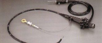
For several decades, the small intestine has remained an area of the gastrointestinal tract difficult to reach for endoscopic examination. Accordingly, the diagnosis of small intestinal bleeding, inflammatory, autoimmune and neoplastic lesions of the small intestine has been a challenge for surgeons and gastroenterologists. Therapeutic endoscopic interventions in the deep parts of the jejunum and ileum were performed occasionally and mostly only during intraoperative intestinoscopy. For the first time, examination of the small intestine behind the ligament of Treitz using a flexible endoscope was performed by K. Ogoshi et al. [10] in 1973 in patients with small intestinal bleeding. The probe fibroenteroscope was first described by M. Tada et al. in 1977. The working length of probe enteroscopes used in diagnostics was 250–400 cm [14]. Works on enteroscopy using a colonoscope were first published by HW Parker and JD Agayoff [12] in 1983, and presented in Russia by V.P. Strekalovsky [3]. But these studies, which are quite lengthy and labor-intensive for both the doctor and the patient, have not found wide application in clinical practice. The technique of intraoperative enteroscopy, developed in the mid-80s of the last century, was the only way for an active total examination of the small intestine for quite a long time [1, 2, 7]. But intraoperative intestinoscopy can no longer be classified as a low-traumatic diagnostic examination, and its place was reserved at the stage of revision of the abdominal cavity during surgery. Video capsule endoscopy (VCE) has revolutionized the evolution of diagnostic methods and has significantly improved the diagnosis of small intestinal diseases [4, 6, 13]. The first reports of “wireless” video endoscopy appeared in the mid-90s, and it began to be widely used in clinical practice since 2001 [8, 9]. In parallel with the introduction of VCE, the Japanese doctor H. Yamomoto, in collaboration with, created a double-balloon video endoscope in early 2001. This made it possible not only to observe the state of the lumen and mucous membrane of the deep parts of the small intestine, but also to perform a biopsy, remove tumors, stop bleeding and perform balloon dilatation of strictures [16]. In 2006, a new single-balloon enteroscope was developed for the study and treatment of diseases of the small intestine. In 2006–2008 individual clinical centers in Japan, America, Italy and France presented the first positive results of using the new device [8, 9, 11, 13, 17]. According to T. Tsujikawa et al. [15], a single-balloon enteroscope, having all the described positive qualities, has some advantages in comparison with a double-balloon system: it is easier to process and prepare for the study, and provides a shorter duration of the study itself due to the absence of the need to inflate the second balloon. As you master the technique of single-balloon enteroscopy, it becomes possible to perform it by one doctor without the involvement of an additional assistant [5, 15]. New opportunities for access to the distal jejunum and proximal ileum, which are provided by video capsule and instrumentally assisted (balloon) enteroscopy, forced clinicians to carefully reconsider the indications for these types of endoscopic examination and, first of all, answer the question: who needs deep jejunoileoscopy. According to most experts, balloon enteroscopy is recommended to be performed : I. For diagnostic purposes · if small intestinal bleeding is suspected, one of the most important manifestations of which, especially in occult variants, is iron deficiency anemia; · in cases of suspected malabsorption syndrome, often manifested clinically as diarrhea of unknown origin; · if you suspect a tumor of the small intestine; · if pathological changes in the small intestine are detected during an X-ray examination; · to obtain tissue samples of the small intestine for histological examination (forceps biopsy). II. For therapeutic purposes · to stop small intestinal bleeding using argon plasma coagulation or endoscopic clipping; · to remove tumors from the small intestine; · for balloon dilatation for strictures of the small intestine; · for removing foreign bodies from the small intestine. III. For the purpose of observation · for clinical syndromes occurring with multiple lesions of the small intestine by polyps (Peutz-Jeghers syndrome, familial adenomatous polyposis) · to assess the state of the small intestine over time in patients with previously diagnosed diseases of this organ, in particular to monitor the effectiveness of conservative therapy for Crohn's disease, enteropathy. Contraindications to deep enteroscopy correspond to contraindications to esophagogastroduodenoscopy and colonoscopy. Limitations that may prevent the full implementation of enteroscopy are: – involvement and gross deformation of the small intestine by the adhesive process after previously undergone major operations on the abdominal organs; – tumor and scar strictures of the small intestine itself; – poor preparation of the patient for the study. Single-balloon enteroscopy is performed using a system consisting of an endoscope, a tube with a balloon at the distal end that is placed on top of the endoscope, and a control unit. The SIF-Q180Y video endoscope (Olympus, Japan) has a working length of 200 cm, an outer diameter of 9.2 mm, is equipped with a standard instrumental channel with a diameter of 2.8 mm and does not have a balloon at its distal end (Fig. 1). The device is compatible with Excera series processors and is capable of operating in wide-screen and narrow-spectrum modes (NBI). The flexible silicone tube (ST-SB1, Olympus) has a length of 140 cm and an outer diameter of 13.2 mm (Fig. 2). The hydrophilic coating on the inside of the tube facilitates the sliding of the device during the examination. At the distal end there is a radiopaque cone-shaped tip, which makes it easy to determine the position of the tube during X-ray control. A single silicone balloon, tightly attached to the distal end of the tube, is inflated and deflated with air, the flow and pressure of which is controlled by a control unit for pumping air into the balloon (MAJ-1725, Olympus). Discharge pressure range from –6.0 to +6.0 mm Hg. Art.; the presence of a control panel allows you to conveniently and quickly control the air supply during the study (Fig. 3).
Technique for performing single-balloon enteroscopy
Transoral enteroscopy. It is performed after adequate preparation of the small intestine using Fortrans and simethicone, preferably in an X-ray endoscopic operating room (especially at the stage of mastering the technique), under general intravenous anesthesia (propofol) while maintaining spontaneous breathing. During the intervention, it is possible, but with active peristalsis of the small intestine, it is necessary to use the parenteral form of buscopan. The transoral endoscopy technique consists of 4 main stages, which involve the following. Stage 1. Passing the enteroscope through the esophagus, stomach, pylorus into the vertical part of the duodenum. Before starting the study, apply gel to the distal end of the endoscope for better sliding of the device. Then pass the enteroscope through the oral cavity into the esophagus and then into the stomach. Lower the tube through the apparatus into the stomach, leaving 10 cm of the distal end of the enteroscope free. After this, pass the enteroscope into the descending duodenum. It is important to emphasize: · the air supply when passing the endoscope through the stomach should be minimal; · it is necessary to prevent the endoscope and tube from “sagging” in the stomach. At the level of the vertical part of the duodenum, the distal end of the endoscope should be bent by 90°, and the endoscope should be pulled up and straightened in the same way as is done when performing endoscopic retrograde cholangiopancreatography. To improve sliding, apply gel to the outer surface of the tube and lower the tube along the endoscope into the duodenum. In this case, it is necessary to strive to keep the endoscope in a straightened state. After the tube is lowered into the duodenum, it is necessary to avoid inflating the balloon in the area of the large duodenal nipple due to the risk of developing pancreatitis. Then you should pass the endoscope into the lower horizontal part of the duodenum and fix it in this position with the curved distal end. Then lower the tube along the endoscope into the lower horizontal section of the duodenum, slightly pulling the endoscope back, inflate the balloon, which will ensure fixation of the tube at this level. Pull up the tube and endoscope and thereby assemble the duodenum. Stage 2. Passing the ligament of Treitz
Pass the endoscope into the jejunum past the ligament of Treitz as far as possible, while slightly pulling the tube. Fix the distal end of the endoscope in the jejunum by bending its distal end. For better fixation of the device, it is important to bend the distal end in a “suitable” bend of the intestine, i.e., where the intestine makes a fairly sharp turn. Then deflate the balloon and lower the tube into the jejunum, slightly pulling the endoscope. Stage 3: Small intestine harvesting
Inflate the balloon and, as a result, fix the jejunum with the distal end of the tube. Pull up the tube and endoscope, thereby straightening the formed loop, and collect the small intestine (Fig. 4 a, b).
Slightly pulling the tube, move the enteroscope further, to the “suitable” bend of the small intestine, which can be hooked onto. Fix the distal end of the endoscope in the “suitable” bowel bend by bending its distal end. Deflate the balloon and lower the tube along the endoscope, slightly pulling the latter (Fig. 4 c, d, e). Repeat similar cycles of advancement and gathering, each time recording the distance that you managed to travel through the small intestine in the distal direction. If the forward advancement of the enteroscope has stopped and is no longer possible, and the loops of the intestine begin to gradually “slip” from the tube immediately after removing air from the balloon, this means that the limit of insertion of the device has been reached, the intestine has been examined as deeply as possible and the next stage of enteroscopy can begin. It is recommended to leave a mark (a metal clip or a submucosal injection of Chinese ink) at this borderline, especially if a transanal ileoscopy is planned. Stage 4. Examination of the small intestine at the exit
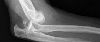
The enteroscope must be removed slowly and gradually, carefully examining the mucous membrane of the small intestine, paying attention to the smallest details of changes in the lumen, structure of the villi, surface, color, and vascular pattern. The collected intestine can easily “jump off” the tube and endoscope, so during exit examination, the enteroscope should be carefully removed and the intestine inspected segment by segment, using the inflated balloon as a retainer. As soon as the distance from the distal edge of the tube to the distal end of the endoscope is approximately 10 cm, deflate the balloon, carefully pull the tube 15–20 cm more proximally, inflate the balloon and continue examining the bowel, removing the endoscope. If necessary, repeat tightening the tube. When the examination of the small intestine is completed and the device is removed into the stomach, it is necessary to deflate the balloon and gradually remove the tube, pulling it out along the endoscope. Then remove the enteroscope. Transanal ileoscopy . The key to successful transanal colonileoscopy is good cleaning of the colon and ileum before the examination. For this purpose, patients receive Fortrans (4 liters over 4 hours in the evening before the examination - when performing colon-ileoscopy in the morning, or 2 liters in the evening and 2 liters in the morning on the day of the examination, if it is performed in the afternoon), as well as espumizan (50 ml of emulsion 30 minutes before starting fortrans + 30 ml 2-3 hours before the test). It is advisable to conduct the study in an endoscopic operating room, under general intravenous anesthesia (propofol) while maintaining spontaneous breathing. During the intervention, fractional intravenous administration of buscopan is used. The technique of transanal colon-ileoscopy at the stage of passage of the rectum and colon is in many ways similar to the technique of colonoscopy, but has its own characteristics. The main one is that a thin enteroscope with a tube attached to it is less suitable for implementing the rotational colonoscopy technique, but this relative disadvantage is compensated by the presence of a “splinting” tube and the unique ability to collect the intestine using a balloon. The technique of ileoscopy itself in terms of advancing the apparatus, bringing down, fixing the tube and collecting the ileum is similar to the transoral enteroscopy technique described above. The main anatomical areas in which the tube balloon is inflated in the colon are: – splenic angle of the colon, – transverse colon, – hepatic angle of the colon. One of the difficult stages of colonileoscopy is the passage of the ileocecal valve and the passage of the enteroscope into the ileum. When the enteroscope reaches the dome of the cecum, you should fix the device, lower the tube, inflate the balloon and pull up the enteroscope along with the tube. Then carefully pass into the terminal ileum through the ileocecal valve. To avoid sudden and unwanted “removal” of the enteroscope from the ileum, it is necessary: · insert the enteroscope gradually, trying to move it as far as possible (more than 15 cm) along the lumen of the intestine, thereby creating a backup for the subsequent reduction of the tube; · bring the tube down smoothly, controlling the location of the distal end of the enteroscope in the ileum. After the tube is reliably lowered into the ileum, the balloon should be inflated and the enteroscope along with the tube should be pulled (straightened). Then you can continue to pass the endoscope into the ileum, sequentially performing cyclic actions to advance it in a retrograde direction by fixing the distal end, bringing down and inflating the balloon and collecting the small intestine until the limit of insertion of the enteroscope or visualization of the mark left during transoral enteroscopy is reached. Removal of the enteroscope, examination of the ileum and, if necessary, colon and rectum are carried out similarly to the technique of the 4th stage of transoral enteroscopy. Own experience of deep jejunoileoscopy To a large extent, the answer to the above question “why” is it necessary to perform deep jejunoileoscopy lies in the cumulative answer to the question “who” needs it. And while the world is accumulating evidence and recommendations verified to level IA, we will try to show what single-balloon enteroscopy brings to the diagnosis and treatment of patients using the example of a separate domestic clinic. Enteroscopy using a single-balloon enteroscope SIF-Q180Y in our clinic began on February 14, 2007. During the period from 02/14/07 to 10/03/08, the study was prescribed in 69 cases, in 62 of them it was carried out in full, and in 7 it was not possible to perform it to the planned extent due to a sharp deformation of the intestinal lumen by the adhesive process (4) and intestinal stenosis in Crohn's disease (3). It was successful and complete in 49 patients - 26 men and 23 women aged from 21 to 89 years, average 53.6±15.1 years. As planned, 62 studies were performed, including 4 repeat studies to monitor the course of previously identified diseases of the small intestine: – 44 oral enteroscopies (including 7 therapeutic); – 18 colon-ileoscopies (including 1 therapeutic). 59 interventions were performed as planned, 3 as an emergency procedure. 16 (32.7%) patients had a history of extensive operations on the abdominal organs, 3 of them had Billroth II, Balfour and Roux reconstruction with the presence of a long adductor loop . For pain relief, endotracheal anesthesia was used in 4 cases, total intravenous anesthesia with preservation of spontaneous breathing was used in 41 cases, and intramuscular premedication (atropine, dolac, relanium, papaverine) was used in 14 cases. For enteroscopy, the total duration of interventions ranged from 20 to 130 minutes (average 74.9±23.3 minutes), for colonileoscopy - from 60 to 110 minutes (average 80.0±14.1 minutes). With oral enteroscopy, it was possible to examine from 100 to 500 cm of the small intestine, an average of 394.0 ± 18.5 cm; with colonileoscopy, in all cases, it was possible to overcome the Bauginian valve and examine from 100 to 250 cm of the ileum (an average of 190.0 ±33.3 cm). The distance traveled by the enteroscope was assessed by cycles of advancement and collection of the small intestine: on average, 9±3 cycles were used for transoral enteroscopy and 6±2 for transanal enteroscopy. The installation of the device was controlled by X-ray endoscopically in 18 patients, in other cases - visually. Indications for performing enteroscopy in the clinic were: – small intestinal bleeding – 14 (28.6%); – formations of the small intestine – 11 (22.4%); – Crohn’s disease (lymphoma?) of the small intestine – 9 (18.4%); – enteropathy – 9 (18.4%); – acute adhesive obstruction – 2 (4.1%); – diseases of the biliary tract in patients with gastric surgery – 4 (8.1%). According to foreign authors, small intestinal bleeding is one of the leading indications for enteroscopy, accounting for from 22 to 75% (on average 42.3%) of the total number of studies undertaken [8, 9, 11, 13, 17]. The most common sources of bleeding are angiodysplasia, erosions and ulcers of the small intestine. According to our data, out of 14 patients with suspected small intestinal bleeding, angioectasia was found in 2 (Fig. 5 a), in 2 cases in combination with phlebectasia, in 4 - erosive hemorrhagic enteritis (Fig. 6). The source of bleeding was not detected in 8 people, 6 of them showed signs of lymphectasia, and 1 had a small intestinal polyp. Diagnostic examination of the small intestine in these patients in most cases was performed some time after the bleeding, which probably determined the rather low level of identification of the source of the latter. If there is a source of bleeding, enteroscopy allows not only to detect and characterize it in detail, but also to perform endoscopic hemostasis by injection, argon plasma coagulation or clipping (Fig. 5b). Tumors of the small intestine (adenocarcinomas, lymphomas, etc.) are detected by enteroscopy in 4.9–30.0% of studies, an average of 16%; polyps are found in 5.0–11.1%, an average of 7.1% [8, 9, 11, 13, 17]. Of the 11 patients we observed with suspected small intestinal polyps, they were found in 6: in 3 they did not require their removal, in 1 a hyperplastic polyp was removed, in 1 patient with familial adenomatous polyposis an epithelial formation of the small intestinal mucosa was detected (Fig. 7) . Its endoscopic removal was routinely performed by resection of the mucous membrane bearing this formation. According to the histological conclusion, a tubular-villous adenoma of the mucous membrane of the small intestine with dysplasia of I–II severity was removed (Fig. 8 a, b). In 1 patient with polyposis of the small intestine due to Peutz-Jeghers syndrome (Fig. 9 a), the largest polyp was removed - up to 5 cm in diameter (Fig. 9 b, c). Thus, in the presence of tumors of the small intestine, enteroscopy allows performing a biopsy to obtain material for a histological report, as well as polypectomy and removing epithelial formations by resection of the mucous membrane. Small intestinal strictures occur in 8.5% of cases (on average 2.0–13.3%), Crohn's disease in 6.2% (3.7–10.0%). [8, 9, 11, 13, 17]. Of the 9 patients with suspected Crohn's disease, the diagnosis was confirmed in 5 cases (Fig. 10 a, b, c) and rejected in 4 cases. In case of inflammatory diseases of the small intestine, using enteroscopy it is possible not only to detect characteristic lesions of the mucous membrane, but also to speak about their localization and extent, to identify inflammatory and cicatricial narrowings and strictures, the presence of which is dangerous when prescribing video capsule endoscopy. According to the observations of a number of authors, balloon dilatation was successfully performed for small intestinal stenosis that developed in Crohn’s disease; no complications were registered during or after the intervention [8, 11, 17]. According to materials from foreign researchers, macroscopic signs of enteropathy diagnosed by single-balloon enteroscopy occur in 5.0–44.4% of cases (on average 19.8%), subsequent histological examination of biopsies of the small intestinal mucosa showed that the most common enteropathy is celiac disease [ 9, 15]. Of the 9 patients with suspected enteropathy, according to the results of enteroscopy and histological examination, 3 had celiac disease (Fig. 11, 12), 1 had eosinophilic enteritis, 1 had exudative enteropathy, 2 had confirmed chronic enteritis, 2 had intestinal changes not detected (including 1 patient with suspected radiation enteritis and a delay in evacuation of the video capsule for more than 8 days). In 2 patients with acute small intestinal obstruction, a violation of passage through the jejunum was confirmed and a probe was installed for its decompression (in one case the obstruction resolved, in the other an operation was performed). In 4 patients after reconstruction according to Billroth II, according to Balfour and according to Roux (2 patients), despite the presence of a long afferent loop, it was possible to reach the biliary tract and in 2 cases carry out their sanitation. There were no side effects or complications during diagnostic and therapeutic enteroscopies, as well as after therapeutic interventions in all the patients we observed.
Conclusion
Experience with single-balloon enteroscopy shows that previously difficult-to-reach areas of the gastrointestinal tract have become accessible for high-quality visual diagnostics and therapeutic endoscopy. The possibilities that have now emerged for a complete endoscopic examination of the small intestine and obtaining material for morphological research will certainly change previously existing ideas about the frequency of occurrence, origin and essence of various pathological changes in this area. Mastering the new method will make it possible to safely and more widely use it in everyday clinical practice, and performing enteroscopy for therapeutic purposes will make it possible to expand the indications for minimally invasive interventions for “surgical” diseases of the jejunum and ileum, and in altered anatomical conditions - on the biliary tract and pancreatic ducts glands.
Popular publications:
- Capsule endoscopy for celiac disease - 01/07/2014 10:42 - Read 7125 times
- Longest duration of endoscopic capsule retention: clinical case and literature review - 10/26/2013 15:12 - Read 6244 times
- Hidden gastrointestinal bleeding. Causes. — 01/28/2015 12:13 — Read 5412 times
- Hidden gastrointestinal bleeding. Diagnostics. — 01/28/2015 12:13 — Read 4641 times
- Capsule endoscopy, history and development. — 06/25/2013 10:54 — Read 3650 times
Key words: enteroscopy, balloon enteroscopy, capsule endoscopy
- < Back
- Forward >