When a patient is diagnosed with disorders or diseases associated with the functioning of the brain, an EEG with sleep deprivation may be prescribed to clarify or confirm the diagnosis. This is one of the most informative methods of examining the brain, which can be used even by children and pregnant women.
So, what kind of procedure is this, how is it performed, are there any contraindications, and how does the doctor decipher the results obtained?
What kind of manipulation is this?
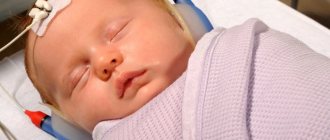
A sleep deprivation EEG is performed to examine the brain in detail without the need for direct access to the cranium . To carry out the procedure, the patient is put on a special cap, to which electrodes are connected at one end.
The second end is connected to a special device - an electroencephalograph, which records impulses coming from neurons.
During the procedure, the patient can sit in a chair or lie down. To determine the reasons why a patient may have disturbances in the functioning of the brain, several types of tests can be performed during EEG sleep:
- Reaction to quickly switched on/off lights and loud sounds. During such an examination, it is possible to identify the causes of brain abnormalities; this can be either a manifestation of hysteria or simulation on the part of the patient.
- Brain functioning when the patient breathes deeply. At this moment, increased release of carbon dioxide begins, which gives rise to signs of an epileptic seizure. Thanks to this examination, it is possible to identify a hidden disease.
- Reaction to flashes of light. During the session, the frequency of light flashes is 10-20 per second. This helps trigger an epileptic attack.
- What brain processes begin to occur when the patient falls asleep. The fact is that in a state of sleep, many processes occur more pronouncedly, so an EEG during sleep provides the most accurate data.
The situation is considered especially favorable when, during an EEG of night sleep, the patient begins to have an attack. The device will record all the changes that appear, and the patient will immediately be provided with all the necessary medical care.
There are 2 types of sleep EEG:
- With deprivation , when the patient is deprived of sleep to better identify pathology. At this moment, the person becomes less susceptible to external stimuli, he may experience a headache, disorientation, and the patient begins to show signs of aggression. Delaying sleep allows you to identify and provoke latent epileptic seizures. It is carried out only under the supervision of a doctor.
- With video monitoring . This method allows you to diagnose epileptic seizures, panic attacks and other disorders that occur during sleep. During the session, all movements of the patient are recorded on video, which are also used in deciphering the device data.
The total duration of the procedure ranges from 30 to 120 minutes.
Electroencephalography with provocative test
To induce an epileptic seizure, the neurologist uses three different methods: hyperventilation (fast breathing), photostimulation and sleep deprivation.
During electroencephalography with hyperventilation, the doctor asks the patient to inhale and exhale as deeply as possible for 3-5 minutes.
During photostimulation, the doctor shows the patient bright flashes of light. During both hyperventilation and photostimulation, the doctor scans the electroencephalography directly.
Many people wonder how an EEG is performed with sleep deprivation? In sleep deprivation electroencephalography, the patient must remain awake throughout the night. The EEG is recorded only 24 hours after the patient initially awakens.
Indications and contraindications for use
EEG sleep monitoring has virtually no contraindications, which allows its use in patients of all ages and categories. It is undesirable to carry out such manipulation in the presence of head injuries, inflammation or fresh postoperative sutures.
Indications for performing a sleep EEG are:
- traumatic brain injuries;
- panic attacks, depression, psychosis, suicidal tendencies, paranoia, atypical actions and behavior of the patient;
- hormonal disorders;
- surgery or brain injury;
- constant headaches, cramps, problems falling asleep;
- VSD;
- if the child has developmental pathologies;
- brain tumors of various nature;
- cerebrovascular accidents, vascular pathologies of the neck and head;
- memory disorders and diencephalic crises.
If at least several of the above problems are present, the patient may be prescribed an overnight EEG to identify hidden diseases or diagnose early detected pathologies.
Is insomnia dangerous?
Sleep deprivation is common. This condition depletes the body and leads to the development of borderline states - neuroses. Depression (persistent decrease in mood) due to insomnia is even more dangerous - they can end in suicide.
If you are sleep deprived, you should immediately consult a doctor. In this case, a neurologist specializing in the treatment of insomnia – a somnologist – will help.
In adults, sleep deprivation is associated with increased anxiety, prolonged stress, insufficient physical activity, or simply the need to perform certain tasks at night.
In children, such disorders indicate a hidden pathology - the consequences of any influences on the central nervous system during intrauterine development, birth injuries, infections suffered in infancy, etc. After three years, such disorders arise due to improper upbringing, lag in school or social conflicts.
How does the procedure work?
You should carefully prepare for a sleep EEG, following all the recommendations of your doctor. The patient continues to take all previously prescribed medications so as not to provoke attacks ahead of time. 12 hours before the manipulation, any foods containing caffeine are excluded from the diet, the last meal is taken a couple of hours before the night EEG.
Before the procedure, you should wash your hair and do not apply any care products to it to avoid weakening the contact of the electrodes and the scalp.
It is also extremely important not to be nervous. For your appointment, you should wear comfortable clothes that do not restrict movement.
A sleep EEG is performed during the day, and in some cases the patient may be advised to remain awake for at least 18 hours the day before the procedure. The patient comes for the procedure, he is seated in a comfortable chair, I put a cap with electrodes on his head, and the places where they come into contact with the skin are lubricated with gel. Then the recording begins.
During the session, irritating factors such as loud sounds or flickering lights may be present periodically. All this activates the electrical activity of the cerebral cortex, its impulses are picked up by electrodes and transmitted to equipment where they are processed and recorded.
After the end of the session, rest is indicated; mental and physical activities can be performed the next day, since the procedure causes severe stress.
Methodology
According to the method of implementation, the electroencephalogram is close to cardiac electrocardiography (ECG). In this case, 12 electrodes are also used, which are symmetrically placed on the head in certain areas. The application and attachment of sensors to the head is carried out in a strict order. The scalp in areas of contact with the electrodes is treated with gel. The installed sensors are fixed on top with a special medical cap.
Using clamps, the sensors are connected to an electroencephalograph - a device that records features of brain activity and reproduces the data on paper tape in the form of a graphic image.
The ventilation test is carried out for children from 3 years of age. To control breathing, the child will be asked to inflate a balloon for 2-4 minutes. This testing is necessary to identify possible neoplasms and diagnose latent epilepsy. Deviations in the development of the speech apparatus and mental reactions will help identify light irritation. An in-depth version of the study is carried out on the principle of daily Holter monitoring in cardiology.
The cap with sensors does not cause pain or discomfort to the child
The baby wears the cap for 24 hours, and a small device located on the belt continuously records changes in the activity of the nervous system as a whole and individual brain structures. After a day, the device and cap are removed and the doctor analyzes the results. Such a study is of fundamental importance for identifying epilepsy in the initial period of its development, when symptoms do not yet appear often and clearly.
How are the results interpreted?
Information obtained during the survey is used to interpret the results. The graphs will show specific neuron activity, which will help to suspect the presence of epilepsy or other pathologies.
To evaluate the results, data from alpha, beta, theta and delta rhythms are used, which may indicate damage to certain parts of the brain . Also, to identify deviations, bioelectrical activity and the presence of strong discharges in certain parts of the brain are taken into account.
All of the above parameters are collected and processed by a specialist, and the patient’s medical history is also taken into account. After this, with a high degree of probability, a diagnosis can be made.
It is important to remember that in the case of epilepsy, an overnight sleep EEG may not always show the presence of pathology. OR the procedure will show normal brain function, although the patient may have previously shown signs of seizures.
Physiological waves in electroencephalography
Depending on the degree of wakefulness, a distinction is made between graphic elements in the electroencephalogram that are not pathological in nature:
- Alpha waves (8 to 12 hertz): awake, relaxed state with eyes closed;
- Beta waves (13 to 30 hertz): concentrated and excited state with eyes open;
- Theta waves (four to seven hertz): tiredness or falling asleep;
- Delta waves (0.5 to 3 hertz): deep sleep.
When a patient opens their eyes or concentrates on a task, they switch from alpha waves to beta waves. This switching is called the Berger effect or “excitation response.”
The newborn baby and older children have slow and also quite irregular EEG waves. Only towards the end of puberty can EEG be recorded because the typical waves become visible.
How safe is the manipulation?
There are many both positive and negative opinions about the feasibility and safety of this procedure.
Proponents make the following arguments:
- With its help, you can detect the hidden course of epilepsy.
- The procedure in some cases helps to cure depression without medication, has a beneficial effect on sleep and improves mental abilities.
- It has a beneficial effect on patients suffering from mental disorders, panic attacks and anxiety.
Opponents make the following arguments:
- The procedure is inhumane. Because it can provoke an epileptic attack. If this happens to a child, then there is a risk that the disease will remain with him forever. If the procedure had not been carried out, the baby could have outgrown the disease and not suffered from it in adulthood.
- In some cases, it can become a catalyst for Parkinson's and Alzheimer's diseases.
Despite all the above arguments, such a study remains today practically the only type of manipulation with which it is possible to identify disorders in the functioning of the brain in the early stages and carry out timely treatment. It is important to carefully prepare for the procedure, not to be nervous and to follow all the doctor’s instructions. Only then will it be possible to obtain the most accurate results.
Prerogative aspects and disadvantages of the EEG method
Neurophysiologists and patients themselves prefer EEG diagnostics for several reasons:
- reliability of the results;
- no contraindications for medical reasons;
- the ability to perform research in a sleeping or even unconscious state of the patient;
- lack of gender and age boundaries for the procedure (EEG is performed on both newborns and elderly people);
- price and territorial accessibility (the examination is low cost and is carried out in almost every district hospital);
- insignificant time costs for conducting a conventional electroencephalogram;
- painlessness (during the procedure the child may be capricious, but not from pain, but from fear);
- harmlessness (electrodes attached to the head record the electrical activity of brain structures, but do not have any effect on the brain);
- the ability to conduct multiple examinations to track the dynamics of prescribed therapy;
- prompt interpretation of results for diagnosis.
In addition, no preliminary preparation is provided for conducting an EEG. The disadvantages of the method include possible distortion of indicators for the following reasons:
- unstable psycho-emotional state of the child at the time of the study;
- mobility (during the procedure it is necessary to keep the head and body static);
- the use of medications that affect the activity of the central nervous system;
- hungry state (a decrease in sugar levels due to hunger affects brain function);
- chronic diseases of the organs of vision.
In most cases, the listed reasons can be eliminated (conduct a study during sleep, stop taking medications, provide the child with a psychological mood). If the doctor has prescribed electroencephalography for your baby, the study cannot be ignored.
Diagnosis is not carried out for all children, but only according to indications
Patient preparation
Computer electroencephalography does not require special preparation of the patient. The procedure is performed both in the hospital and on an outpatient basis. All patients are advised to abstain from alcoholic beverages and caffeine-containing products, including cocoa and chocolate, 24 hours before the study. They stimulate the central nervous system and can lead to false results. Smoking is prohibited for 3-4 hours before the procedure. Nicotine affects the blood vessels in the brain and may cause abnormal results.
If the patient is taking medications that affect the brain (anticonvulsants, antipsychotics, sleeping pills, sedatives, etc.), their use should be discussed with the attending physician. If it is impossible to cancel them, the fact of their use is indicated in the direction of the EEG. The specialist interpreting the results takes into account the patient’s medication intake.
Before the procedure, if it does not involve sleep deprivation, you should get enough sleep. You should wash your hair the day before. The use of varnishes, foams or wax for styling is prohibited. Hair is left loose. Before the EEG, all existing metal objects are removed, including jewelry, hairpins, etc.
Time of deprivation
For preschool children, the recommended deprivation is 4-6 hours. Having raised the child at 2 am, they go for the EEG procedure at 8 am. For younger children, the time increases to 6-8 hours. In such a situation, you will have to wake up the child at 00.00. If the child is over 12 years old, deprivation lasts 18 hours or more.
Sometimes doctors allow partial deprivation to make it easier to prepare the child. Partial deprivation involves extending your waking time by several hours and waking up earlier so that you can nap earlier than usual.
