Projections in X-ray diagnostics
A radiographic projection is an image on a plane obtained when X-rays pass through a given body onto a plane (for example, onto a film in a cassette).
There are many different projections in X-ray diagnostics, but the following standard and additional projections remain the main and frequently used projections. In this case, standard projections are mainly performed in a lying position.
Standard projections:
- Right lateral projection.
- Left lateral projection.
- Ventrodorsal projection.
- Dorso-ventral projection.
Additional projections:
- Oblique projections.
- With a horizontal course of rays - lateral, straight.
Now let's take a closer look.
The right lateral projection (RLP) is performed with the animal lying on its right side, and if the area of study is the thoracic region, abdominal cavity or survey image, then the thoracic limbs should be retracted strictly forward so that they do not block the chest and spine and the sternum should lie parallel to the table, and the ribs should be in superposition.
The left lateral projection (LLP) is performed similarly to the right lateral one, but with this projection the animal lies on its left side.
The position of the animal when performing the lateral projection.
Ventro-dorsal projection (VDP) is the position of the animal lying on its back, while the sternum and spine should be in superposition throughout, and the pelvic limbs should be retracted evenly back.
The position of the animal when performing the ventro-dorsal projection.
The dorsoventral projection (DVP) is similar to the ventrodorsal one, but the animal lies on its stomach.
The position of the animal when performing a dorsoventral projection.
Oblique projections are projections when the animal lies on its side at an angle of 45º to the table surface (see figure). Such projections can be performed both on the right side and on the left. And other variations are also possible depending on the area being studied.
The position of the animal when performing oblique projections.
With horizontal ray path - with this projection, X-rays pass horizontally (from the side). These projections can be performed both in a lying and standing position, and can be lateral and straight.
The position of the animal when performing direct projections with horizontal rays.
The position of the animal when performing lateral projections with horizontal rays.
The position of the animal during lateral projections with horizontal rays.
Projections in X-ray diagnostics are selected based on the area of study and preliminary diagnosis.
It should be borne in mind that if the area of interest is the chest, then only the chest is imaged. From a survey image, it is impossible to evaluate any indicators for examining the chest due to the fact that, firstly, the image is distorted due to the inclination of the X-rays passing along the periphery (the further the object is from the center to the edge, the greater the distortion). Secondly, we will not see small details that should be taken into account when describing the chest and making a correct diagnosis. The same situation occurs when examining the abdominal cavity, head, etc. Therefore, survey photographs should be used very rarely and only in emergency cases.
Therefore, before the owner and the animal go for X-ray diagnostics, it is necessary that the doctor, at a minimum, conduct a full clinical examination, make a preliminary diagnosis and, only on this basis, determine the area of study and the necessary projections.
To identify pulmonary pathology, the following are necessarily used: right lateral and ventro-dorsal projections.
To identify metastases: right lateral, left lateral and ventro-dorsal projections.
For cardiology: right lateral and dorsoventral projections.
To examine the trachea (including to identify collapse): lateral during inhalation and exhalation, straight, oblique cranio-caudal tangentially.
To examine the abdominal cavity: right lateral and ventro-dorsal.
However, do not think that for the same pathology there will be a standard number of images, this is not the case. For each specific case there will be a different number of projections due to the pathophysiology of the ongoing process and the anatomical structure of the animal. In some cases, in order to identify and correctly make an x-ray diagnosis, 2 projections are enough, but in some cases, 4 projections are completely insufficient. Therefore, the main standard projections are initially performed, and then additional projections are made as necessary.
It should also be taken into account that in order for the image to be obtained qualitatively, a well-positioned and fixed animal is important; for this it is necessary that at least 2 people come with the animal for X-ray diagnostics.
I would also like to note that when you come for X-ray diagnostics, you must have a referral for X-ray diagnostics with you, which you can read and print on our website or in the VKontakte group.
You can take an x-ray in Tver at our veterinary clinic “Doctor Ay and Oy” at the address: Tver, T. Ilyina St. 1 B. If you have any questions, please call; (4822)737-077
Source of the VK article “Doctor Ay and Oy” - 2015
radiologist Alexandrova Maria Sergeevna
TRG - lateral and direct image of the skull
TRG, teleroentgenogram, is an x-ray of the head. It is also called cephalometric, that is, measuring the head.
TRG is most in demand in orthodontics: for diagnosing occlusion and planning treatment: methods, stages, timing. To tell your doctor how long you will have to wear braces, bring him a TRG.
What will the TRG show in lateral projection?
The lateral projection of a teleroentgenogram is a profile shot of the skull. Based on this, the orthodontist will determine:
- Direction of growth of maxillofacial structures;
- Jaw angle;
- Size of jaws and teeth;
- Type of bite;
- Hidden injuries of the dental system;
- Soft tissue features (to improve profile aesthetics)
Front projection - more information
For this type of TRG, photographs of the skull are taken from the frontal view, from the side of the face and from the back of the head. Based on the results of TRG in direct projection, the orthodontist will determine:
- Features of the structure of the dentition (narrowing, widening);
- The plane of teeth closure.
- Degree of facial asymmetry.
Calculation and analysis of TRG
An integral part of orthodontic treatment planning is teleradiogram analysis. By directing you to the image, the orthodontist will indicate in the direction whether interpretation is required.
- Required - the calculation and analysis will be carried out for you by the orthodontist CosmoStom.
- Not required - your doctor will calculate and analyze TRH yourself.
Previously, calculations were done manually. The picture had to be printed, and the orthodontist would find key points on it. Next, the doctor, using tracing paper, a ruler and a protractor, calculated angles, distances, and proportions. The problem of measurement error inevitably arose, because even the thickness of a pencil introduced distortions into the calculation.
The digital revolution in dentistry has made it possible to automate these steps. TRG results are loaded into the program, where more than 80 points are set according to anatomical landmarks. The points are connected into diagrams, and then the doctor chooses which indicators he needs. The accuracy and reliability of such measurements have increased manifold. Now the orthodontist analyzes each case in more detail, and the effectiveness of treatment increases.
CosmoStom uses Romexis programs to calculate and analyze TRG, as well as its own unique software module “Cosmos”.
5 reasons to do TRG at CosmoStom
- Precision equipment: Planmeca ProMAX 3D (Finland) with high sensor resolution (5 pairs of lines per mm).
- Low radiation exposure: from 1.8 to 3.1 μSv. A person receives a similar dose after flying on an airplane or spending a weekend watching TV.
- We do the decoding, use our own TRG analysis module
- On weekdays and weekends we are waiting for you in the X-ray room.
- Convenient location: you can quickly get to CosmoStom on Volochaevskaya 19/1 from anywhere in Omsk.
Harmony of facial features
and a healthy, correct bite are calculated mathematically.
How does a teleradiogram work?
You can do TRG of teeth in 15 minutes. No pre-registration is required at CosmoStom. You just come to the clinic on Volochaevskaya 19/1 (stop “Rabinovicha Street”) in the X-ray room operating mode:
- Mon-Sat from 9:00 to 19:00
In the X-ray room:
- The patient usually stands. The head is fixed motionless so that the image is clear. Also, the same position of the jaw will allow you to take a repeat image in the future and measure the result of the treatment.
- The X-ray technician places the cephalostat in the desired position, asks you not to move, and starts the scan.
- The patient is waiting for the results in the lobby. In a few minutes he will be given a printed photograph or a disc with the recording.
- If calculation and analysis of TRG is required, the results will be ready within a week.
Price TRG
The cost of a teleradiogram depends on 2 factors:
- Only side projection or both?
- Is decoding necessary?
The answers to both questions should point in your direction. Be sure to take it with you to the clinic.
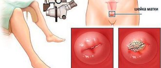
X-ray procedure of the sinuses
Nasomental projection. The subject is in an upright position. The tip of his nose and chin are pressed tightly against the cassette, perpendicular to which the central beam generated by the X-ray machine is directed. The light stream passes in the sagittal projection, at the level of the outer edges of the eye sockets. The resulting image shows the pyramids and the base of the skull, along with the paranasal sinuses.
Chin projection. The patient must sit. The chin touches the cassette. When examining for sinusitis, this position is the most informative, as it displays the sinuses as openly as possible, without shading.
Nasofrontal projection. Instructs the patient to be in a standing or sitting position. The tip of his nose and forehead should touch the cassette. From this position it is possible to obtain a reliable image reflecting the state of the frontal sinus and ethmoidal labyrinth.
Lateral projection. The patient's head is positioned so that the sagittal plane is parallel to the cassette. From this position, the radiologist is able to take a picture showing the condition of the walls of the frontal sinuses. This study has great diagnostic value in differentiating a pathological process localized in the frontal sinus from inflammation of the posterior cells of the ethmoidal labyrinth.
Types of radiography of the sinuses
X-rays of the sinuses are performed in several projections:
- Nasomental: used to visualize the anterior sinuses, including the maxillary sinuses.
- Mental: helps to avoid the shadow of the pyramids of the temporal bones on the maxillary sinuses.
- Nasofrontal: required when it is necessary to obtain a detailed picture of the condition of the frontal sinuses, orbits and ethmoidal labyrinth.
- Lateral: considered indispensable in assessing the shape and size of the paranasal sinuses and other elements of the facial skeleton (often prescribed before trephine puncture).
The choice of installation method is determined by the indications for the procedure. Correct positioning of the patient during the examination helps obtain a symmetrical image. Any inaccuracies and deviations may lead to incorrect interpretation of the results.
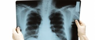
What does the examination show?
On an image of the lungs, a specialist may find:
- fluid in the pleural cavity;
- diaphragm defects;
- cavity in the lung;
- malignant oncological formation;
- focal eclipses;
- large round shadow.
A cavity in the lung, for example, is necrosis of lung tissue. Small focal eclipses indicate pneumonia or tuberculosis. Major eclipses – cancer. A large round shadow on the lung may be progressive tuberculosis or a tumor.
In addition, by analyzing the images, the radiologist can exclude suspicion of pleurisy or malignancy.
Decoding the results
Studying the images can lead a specialist to different conclusions. Eclipses or white spots may indicate tuberculosis, pleurisy and occupational diseases. Black spots on the lungs indicate chronic inflammation. In addition, such a nuance may indicate malignant tumors in the examined organ.
With pneumonia, an x-ray shows global spotty formations over the entire surface. The more distinct the spots, the more advanced the process.
The doctor diagnoses bronchitis if the photo shows root deformation, changes in the pattern and lamellar lesions. In addition, after irradiation, areas of fluid accumulation are read on the monitor.
If the trachea is pulled forward or displaced, and a thin darkened strip is visible on the picture above the edge of the lower costal arch, then this is evidence of exudative pleurisy. Pulmonary edema will be indicated by unevenly distributed shadows, similar to flakes.
Having seen the “butterfly wings” formed by the expansion of the roots in the image, the specialist diagnoses venous congestion of the pulmonary circle. If there are no light areas under the dome of the lungs, and an accumulation of gases is visible in the abdominal cavity, then this indicates peritonitis.
When a darkening of the posterior mediastinum is visible on a lateral X-ray, this indicates a collapse of the lung lobe or atelectasis. Heart disease can be recognized by the enlargement of the rounded border of the heart shadow. Shadowing on the right side means problems with the left ventricle and vice versa.
Having seen on the monitor in frontal and lateral projections at the apex of the right lung a mid-focal infiltrative shadow up to 0.6 cm in size and a path extending from it to the right root, the doctor concludes that this is pneumonia.
In addition, the right lateral projection image shows small, focal shadows in S1 and S2. What convinces the specialist about the presence of infiltrative tuberculosis in the patient’s upper lobe of the right lung. At the same time, no changes in the contours of the diaphragm and sinuses were detected in the subject. The cardiac shadow does not depart from the norm.
A note in the patient’s referral that he complains of a persistent cough and sputum with blood, and wheezing in the right lung identified by the therapist confirms the radiologist’s diagnosis.