What is an abdominal ultrasound?
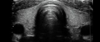
Abdominal ultrasound (transabdominal) is one of the types of ultrasound diagnostics that involves the use of an external sensor (transducer) to study the abdominal and pelvic organs. The device generates high-frequency waves that pass through the anterior wall of the peritoneum and, reflected from tissues of various densities, return back. The sensor analyzes the nature of their reflection and displays the result on the monitor. In this way, the organs being examined are visualized, based on which the diagnostician can make a diagnosis.
Advantages of the method
The advantages of this type of examination:
- during the procedure, it is possible to examine several organs at once and assess their general condition;
- absolutely painless;
- can be carried out for people of any age: both newborns and children, and adults;
- has no contraindications for most patients.
Indications for ultrasound
Indications for abdominal ultrasound include:
- increased gas formation;
- bloating;
- suspicion of a tumor of a malignant or benign type;
- symptoms of appendicitis;
- abdominal pain;
- diabetes;
- menstrual irregularities;
- a feeling of heaviness and aching pain in the right side;
- hepatitis symptoms;
- inflammation of the prostate;
- urolithiasis disease;
- the presence of stones in the gallbladder;
- hormonal imbalances associated with the functioning of the ovaries;
- disturbances in fetal development during pregnancy;
- preparation for surgery;
- for preventive purposes or as part of medical examination.
Contraindications
TA ultrasound (transabdominal ultrasound) has no specific contraindications for most people. In rare cases, some patients have an allergic reaction to the gel used during the examination. Experience shows that the likelihood of such a symptom occurring is extremely low, so there is no need to be afraid of the procedure. In addition, if the outcome is unfavorable, the diagnostician will be able to provide first aid and stop the manifestation of allergies.
Women during menstruation are advised to reschedule an abdominal ultrasound to a later date, as during this period the results of the study may be distorted.
Contraindications
Abdominal ultrasound diagnostics has no special contraindications. There are some restrictions associated with menstruation in women that should be discussed with your doctor. Some pathologies can be reliably confirmed only after the end of the cycle, and to identify others, the study is carried out only in the middle.
Doctors still cannot definitively answer whether ultrasound diagnostics harm the body. Most of them agreed that ultrasound is a safe method. Even pregnant women are recommended to carry out such a study at least three times during the entire gestation period. However, you should not do it too often.
Read also: how ultrasound affects the body.
Organs examined and ultrasound capabilities
During the procedure, doctors look at the condition:
- stomach;
- kidney;
- adrenal glands;
- ureters;
- prostate gland;
- liver;
- gallbladder;
- duodenum and other parts of the intestine;
- female pelvic organs;
- spleen;
- pancreas;
- lymph nodes, etc.
During the study, the following organ indicators are analyzed:
- size;
- homogeneity of structure;
- location;
- presence of pathological inclusions;
- anatomy.
Ultrasound of the abdominal organs
Abdominal ultrasound of the abdominal cavity includes examination of the following organs:
- Liver. Excessive density of this organ indicates the presence of fatty hepatosis. If the liver and splenic vein are enlarged, then cirrhosis can be assumed. Violation of the normal echostructure of an organ, as a rule, means the presence of malignant neoplasms.
- Gallbladder. In a healthy state, it should have the shape of a pear, the width of which does not exceed 5 cm, and the length - no more than 10. The presence of dark spots in the structure of the organ indicates a large number of stones. If the thickness of the walls of the bladder exceeds 4 mm, this indicates an inflammatory process.
- Stomach. A change in the color and structure of the walls of the organ means the presence of ulcerative or erosive formations. Lumps in the stomach and its deformation may be the result of cysts or polyps.
- Spleen. A study of this organ will help to more accurately diagnose diseases of the body’s hematopoietic systems, and will also indicate liver pathologies. The normal dimensions of the spleen should be no more than 12x8x5 cm.
- Pancreas. This organ is examined, as a rule, with a comprehensive ultrasound of the abdominal cavity. Violation of the echostructure of the gland indicates the deposition of calcium salts in its tissues. According to standards, the length of the organ cannot exceed 22 cm, thickness - no more than 3 cm. Changes in the size of the pancreas are a consequence of pancreatitis.
The process of performing ultrasound of the abdominal organs
Ultrasound of the pelvic organs
Features of TA ultrasound of the pelvic organs:
- Rectum. The process of examining the pelvic organ abdominally occurs in two ways: standardly or using irrigoscopy. In the second case, a special liquid is poured into the intestines - contrast. It simplifies diagnosis, allowing a more detailed assessment of the condition of the walls of the rectum. Normally, their thickness should be no more than 9 mm, without seals.
- Bladder. The presence of echogenic inclusions in the walls of the organ indicates acute cystitis. If the contours of the bladder are uneven and thickened, then the infectious disease has already acquired a chronic form, which happens in the absence of proper treatment. Sometimes, using an abdominal sensor, you can see moving formations in the structure of the organ - this means the presence of stones or other foreign objects.
- Prostate. Normally, the prostate measures no more than 4.5 x 4.5 x 2.5 cm. If two of the three indicators exceed the standard, this indicates the presence of prostate diseases, for example, adenoma or hyperplasia. It is also important to determine the structure of the tissues of the organ, since any protracted inflammatory processes lead to fibrosis. This pathology is characterized by the formation of connective tissue instead of healthy working tissue and leads to disruption of genitourinary function.
Kidney ultrasound
During an abdominal examination of the kidneys, the following diseases and pathologies can be identified:
- kidney stones;
- chronic or acute pyelonephritis;
- urethritis;
- cystitis;
- pyeelectasis;
- congenital pathologies of organ development;
- cysts and tumors.
Gynecological ultrasound
The gynecologist prescribes an abdominal ultrasound based on the woman’s menstrual cycle. In the first phase, the uterine mucosa is usually examined, in the second phase, the presence of ovarian pathology and inflammation of the fallopian tubes is checked. After artificial termination of pregnancy, it is recommended to undergo this study in order to monitor the possible negative consequences of abortion.
During pregnancy, TA ultrasound is performed in the second and third semester, with its help you can:
- establish the exact duration of pregnancy;
- determine the gender of the unborn child;
- assess the condition of the cervix;
- measure the proportions of the unborn baby (fetometry);
- measure the amount of amniotic fluid;
- examine the child’s brain for possible pathologies;
- analyze the position of the fetus in the womb.
Timing for analysis of female genital organs
— In case of urgent diagnosis, the girl must name the date of her last menstruation.
— If there is a suspicion of an inflammatory process in the uterine appendages, then this analysis can be performed on any day of the cycle.
— After an abortion, an abdominal ultrasound is performed at the end of the next menstruation. If a woman has pain or bleeding, then a scan is performed any day.
— If there is a suspicion of fibroids, then the study is done in the first phase of the cycle.
How to prepare?
Basic recommendations for preparing for the procedure:
- twelve hours before the TA ultrasound, you cannot eat anything, you can only drink water in small quantities;
- immediately before diagnosing the pelvic organs, you need to drink 2 glasses of liquid to fill the bladder - this will improve the quality of the procedure;
- 2-3 days must pass after an X-ray with contrast, otherwise the results of the study may be distorted;
- Before examining the gallbladder, you should not smoke for 2 hours, as this provokes spasms of the organ.
For children, the recommendations for preparation are similar, only the fasting time before the procedure is reduced: 3 hours for infants and 6 hours for an older child. During the examination, you should not allow the small patient to move on the couch, so you need to take your baby’s favorite toy with you to the ultrasound to distract him. In addition, you should not actively play with your child before the procedure, as this can provoke excitement of the nervous system and lead to restlessness.
This video provides a reminder for a patient preparing for an abdominal ultrasound. The video was prepared by the “Medical in Dnieper and Mariupol” channel.
What medications can be used before the procedure?
In preparation for an abdominal ultrasound examination, you should, if possible, completely avoid the use of medications. The only exceptions will be vital medications and those that need to be taken to cleanse the digestive tract.
Nutritional Features
Before performing a TA ultrasound, you should eat right for 2 days and follow a special diet that allows you to minimize the amount of gas in the intestines. Excess air in this organ will impair the quality of the abdominal examination.
When preparing for it, you need to exclude the following products:
- fried and spicy foods;
- milk and cottage cheese;
- baked goods;
- sweet berries;
- legumes;
- vegetables, especially cabbage;
- carbonated drinks;
- coffee;
- alcohol.
You should eat food in small portions 5-6 times a day, without overeating. It is also necessary to drink at least 2 liters of clean water per day. These measures will relieve the digestive system and increase the reliability of ultrasound results.
Cleansing
On the eve of the procedure, doctors recommend carrying out cleansing preparatory measures, for example, cleaning the intestinal tract using an Esmarch mug or a microenema with a rectal solution. You can prepare for the examination in an alternative way, using laxatives, for example, Bisacodyl. Sorbents such as Smecta and activated carbon are used to absorb toxins and remove excess gas bubbles.
Bisacodyl 28 rubles
Smecta 129 rubles
How is the procedure done?
Procedure for conducting the examination:
- The patient undresses to the waist and lies down on the couch.
- The doctor applies a small amount of gel to the area of the body being examined.
- He places the sensor against the skin and moves it over it, pressing lightly in some places to get a better look at the organ.
- During the procedure, the specialist examines the image on the monitor transmitted by the transducer.
- Having chosen the best angle for displaying the insides, the doctor records the picture, which allows him to take measurements of the organs and assess their condition.
- Periodically, the doctor asks the patient to hold his breath or change his position.
- At the end of the study, excess gel is removed using napkins or towels.
- The diagnostician generates and prints out the results of the ultrasound examination (sonogram), certifies them with a seal and signature, and gives them to the patient.
Immediately after an abdominal ultrasound, the specialist must prepare a preliminary diagnosis. Only the attending physician can give a final conclusion, based on the medical history.
What device is used to conduct the study?
The diagnosis described in this article is done using a special device called an abdominal ultrasound probe. This device, which looks like a small stick with a cap, can be of different sizes. Depending on who the study will be conducted on (children, newborns, adults) a specific sensor is selected. Such a device is called convex. The sensor may also remind many people of a microphone that is connected to the scanner using a cable. During an abdominal ultrasound, the device does not need to be protected with anything (unlike transvaginal diagnostics, when a condom is placed on the device).
How much does an abdominal ultrasound cost?
Cost of abdominal ultrasound in different cities of the country:
| Name | Price, rub |
| Comprehensive ultrasound (liver, gallbladder, pancreas, spleen) | 1800-2200 |
| Ultrasound of the prostate gland | 1100-1500 |
| Ultrasound of pregnant women II, III trimester | 1450-1800 |
| Gynecological ultrasound | 700-900 |
| Ultrasound of the bladder | 600-700 |
| Ultrasound of the genitourinary system: kidneys + adrenal glands | 700-800 |
Prices are valid for regions:
- Moscow;
- Chelyabinsk;
- Krasnodar;
- Omsk;
- Samara.
Video
This video explains the capabilities of a comprehensive abdominal ultrasound examination and the correct technique for performing it. Filmed by the Moscow Doctor Clinic channel.
Do you have any questions? Specialists and readers of the HROMOSOMA website will help you ask a question
Was this article helpful?
Thank you for your opinion!
The article was useful. Please share the information with your friends.
Yes (100.00%)
No
X
Please write what is wrong and leave recommendations on the article
Cancel reply
Rate the benefit of the article: Rate the author ( 1 vote(s), average: 5.00 out of 5)
Discuss the article:


