Who is being tested?
Most often, an ultrasound of the bladder is performed in women with clear signs of genitourinary dysfunction. These include:
- frequent urge to go to the toilet;
- difficulty urinating;
- presence of blood in the urine;
- presence of stones;
- vesicoureteral reflux;
- pyelonephritis;
- cystitis;
- pain above the pubis.
Ultrasound of the bladder is used as a complement to a gynecological examination, to assess the patient’s condition after undergoing surgery on the genitourinary system, to monitor the functioning of the kidneys.
How to prepare for a bladder ultrasound
The procedure for examining the bladder in women is carried out on a full bladder, therefore it is necessary to prepare for the ultrasound in a certain way.
- Some time before the appointed time, you need to drink a noticeable liter of water, compote or tea. It is important to remember that water should not contain gases. Do not replace liquid with milk. To keep your bladder full, you cannot urinate. If it is impossible to endure the urge to urinate, you can empty the bladder, but then you need to drink a few glasses of water again and by the appointed time the bladder will be filled to the desired level;
- You don’t have to drink the water, but wait for the bladder to fill on its own. To do this, you need to not empty it for three to four hours. Often the procedure is scheduled for the morning. At the same time, you can prepare for an ultrasound if you don’t urinate in the morning. If this is too difficult, you can go to the toilet a few hours before you finally wake up, but after getting up you shouldn’t do this anymore.
It is important to take into account the fact that a gas-filled intestine can prevent a correct procedure for diagnosing the bladder. For those people who suffer from bloating or constipation, it is recommended that a few days before the scheduled procedure follow a diet in which fresh fruits and vegetables, legumes, gas-containing drinks and alcoholic beverages are excluded from the diet.
If even before the start of the ultrasound it becomes known that the procedure will be carried out through the rectum, a few hours before visiting the office you should do a cleansing enema or use special suppositories.
Preparation
Many women have a question about how to prepare for an ultrasound examination in order to get the most accurate results. Preparation for an ultrasound of the bladder is based on good filling of the organ. This is a basic rule that applies to patients of any age. The main points are:
- two hours before the procedure you should drink at least 2 liters of liquid. This could be clean water, weak tea, dried fruit compote;
- you must refrain from going to the toilet 2 hours before the test;
- people who do not suffer from cardiovascular pathologies can take diuretics.
The basis of preparation is to fill the bladder with water.
The above measures will help the doctor conduct a quality study. If the diagnosis is performed transvaginally or transrectally, then filling of the bladder is also required. In addition, to carry out diagnostics, the last type of treatment is to do an enema cleansing. You can get unreliable results if there is increased gas formation in the intestines. Therefore, 3 days before the test, it is necessary to avoid eating a diet that increases the number of gases.
To do this, you need to adhere to a special diet that excludes the use of:
- legumes;
- tomato;
- cabbage;
- carbonated drinks;
- alcohol;
- dairy products.
If a woman has difficulty refraining from urinating, which often happens during pregnancy, then she can visit the toilet. Next, you should drink 1 liter of water so that your bladder is well filled during the study. Women are examined on any day of the menstrual cycle.
Examination technique
Ultrasound of the genitourinary system in women, men and children has some peculiarities. If you only need to examine the kidneys and ureters, an external ultrasound is performed. To determine the condition of the internal reproductive system, the examination is carried out through the vagina or rectum.
In children
In childhood, only an ultrasound of the urinary system is performed; examination of the prostate and uterus is required as a last resort. Children undergo an external examination. The kidneys are examined from the lower back and through the abdominal wall, the bladder - through the abdomen.
The skin is treated with sound-conducting gel. The examination is carried out with a standard sensor, first examining a full bladder. Then the child is asked to have a bowel movement and is re-examined.
In the video, a child has urolithiasis:
Among women
How an ultrasound of the female genitourinary system is performed depends on what needs to be examined. There are two methods of examination.
- Transabdominal method - examination through the abdominal wall. Used if you want to examine only the kidneys and ureters with a bladder. Also used to examine girls who are not sexually active.
- The transvaginal method is used to study the entire genitourinary system, when the uterus and ovaries are examined. The sensor is inserted into the vagina.
Both methods are highly accurate. Transvaginal examination is accompanied by mild discomfort.
In the following video, an ultrasound specialist talks about these diagnostic methods:
In men
For ultrasound examination of the male genitourinary system, two methods are also used. The transabdominal method is used more often. This is how an ultrasound of the ureters, kidneys, and bladder is done.
To examine the prostate, the transrectal method is used. The sensor is passed through the rectum. The procedure is accompanied by minor discomfort. In preparation for the study, it is recommended to do an enema.
Additionally, read the article about simultaneous examination of the prostate and bladder.
Below is a video about how an ultrasound scan is performed in men:
How the research is carried out
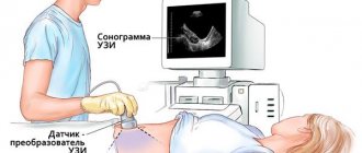
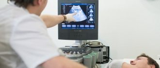
Most often, diagnosis is carried out transabdominally, through the abdominal wall. During the procedure, the patient lies on the couch on her back. The doctor uses an ultrasound sensor with a special gel applied to it to perform the procedure. If during the diagnosis it is suggested that there are stones, sand, or a tumor process, then the patient may be asked to conduct the examination while standing.
To assess the condition of the bladder mucosa, it is recommended to conduct an examination on the side.
Typically the diagnosis lasts no more than 15 minutes. It may be necessary to assess how completely the organ is being devastated. To do this, the woman may be asked to visit the toilet and then examine the bladder again. If the patient is obese or there is an assumption of the presence of a tumor, then ultrasound can be performed using the following methods:
- Transvaginally. The sensor is inserted into the vaginal opening. The study can only be carried out on sexually active women.
- Transrectal. Using a special sensor inserted into the rectal opening. Diagnosis can be carried out both for women who are sexually active and for virgins.
Transvaginal diagnosis allows for simultaneous gynecological examination
Ultrasound of the genitourinary system
The diagnostic procedure is aimed at the early detection of diseases of the excretory and reproductive systems in male or female patients. It is most often used to identify developmental anomalies of various organs, kidney diseases, problems in the reproductive area, cancer, and urolithiasis.
Doppler sonography is also performed at the same time . It allows you to study the condition of blood vessels or the usefulness of the circulatory system of various tissues. This type of examination of the patient helps to determine the presence of an infectious or inflammatory process, as well as to find the exact location of the tumor.
General description of the procedure
Ultrasound of the genitourinary system is based on the individual susceptibility and conductivity of various body tissues in relation to the echo signal supplied to them. As a result of the contact that occurs, a corresponding image appears on the monitor of the ultrasonic device. Using it, the specialist evaluates the volume, outline and condition of the organ being examined, and also observes the possible presence of pathological changes in it.
Ultrasound equipment
Diagnostic equipment is a computer installation connected to special electronics. The kit also includes a monitor and an ultrasound sensor for contact with the organ being examined.
The echo signal it transmits is sent to a scanning device, which analyzes it and displays the processed image on the display of the diagnostic device. Its essence lies in the fact that reflection from a number of tissues has different wave characteristics.
Subsequently, it is studied by a sonologist, who draws up an expert opinion on the patient’s condition.
Contraindications
Contraindications to an ultrasound examination of the bladder include in the abdominal form: urinary incontinence, since diagnosis is carried out exclusively on a full bladder, the presence of excess weight (since with an excess amount of subcutaneous fat, information content decreases), skin lesions in the area being examined, the presence of scars on bladder.
Transrectal examination is not done for intestinal inflammation, anal fissures, intestinal obstruction, or latex allergy. The transvaginal method is not indicated for allergic manifestations to latex, the presence of virgin pleura, pregnancy in the 2nd and 3rd trimesters, and infectious diseases of the genital organs.
results
During the study, the doctor, assessing the parameters of the bladder, can evaluate the following parameters:
Ultrasound OMT in women
- what shape is the bladder, deformation may indicate the presence of neoplasms;
- size. A reduced organ indicates fibrosis, frequent cystitis, an enlarged organ indicates hyperplasia, narrowing of the urethra, the presence of stones;
- contours;
- what contents does the organ have? This may be clots of pus, blood, hematomas, urine;
- the presence of neoplasms and their size, shape, mobility;
- integrity of the organ or the presence of damage.
If a woman has cystitis, then an ultrasound may show uneven contours and enlarged walls. Ultrasound examination makes it possible to identify various neoplasms, which include polyps, cysts, and tumor processes. As a result of the study, it is possible to diagnose the presence of patency of the ureteral canals, foreign neoplasms, sediment, inflammation, increased tone, atony, prolapse of the bladder, diverticulosis and pathology in the genital organs.
The attending physician deciphers the study picture
Indications
Indications for diagnosis are:
Transrectal ultrasound of the prostate gland + video
- night and daytime urinary incontinence;
- pain during and after urination;
- bad OAM;
- increased blood pressure;
- endocrine disorders;
- presence of space-occupying formations;
- abdominal trauma;
- abnormal development of the genitourinary system;
- infertility;
- impaired erection;
- lower back pain;
- the presence of chronic and acute inflammatory process.
This study allows you to diagnose the following organs:
- kidneys;
- bladder;
- urinary tract;
- uterus;
- ovaries;
- appendages;
- prostate gland;
- prostate;
- penis;
- scrotum
Norm
After receiving the study picture, the doctor evaluates the results with normal indicators. A healthy woman is diagnosed with the following parameters:
- the bladder should be pear-shaped when the organ is full, and saucer-shaped after urination;
- on the device screen, the normal structure appears as dark spots;
- urine volume varies between 250–550 ml;
- walls with a thickness of 2 to 4 mm;
- filling speed is about 50 ml per hour;
- residual urine should have a volume of no more than 40 ml.