- home
- Blood analysis
- General blood analysis
Most laboratory tests are known to humans since childhood, from routine vaccinations and medical examinations. Everyone knows why a sugar test, liver or kidney tests are performed. The wording “LE-cell” makes many people wonder what kind of indicator this is? This designation hides a special type of white blood cell, a cell that has been partially destroyed and cannot perform its functions. To detect LE cells, the test is performed using blood from a vein.
What is it and what does diagnostics show?
LE cells are also called lupus erythematosus cells (from the Latin Lupus Erythematosus cells) or “lupus cells.” Their detection in the blood is considered reliable evidence of the development of a dangerous autoimmune disease: systemic lupus erythematosus (SLE).
In a sick body, pathogenic mechanisms lead to the appearance of antibodies that behave in an abnormal way and attack the “correct” tissues and cells. They are called autoantibodies.
One of these specific immunoglobulins provokes pathological changes in individual leukocytes. They contain the sculpted nuclear substance of other cells, consisting of depolymerized DNA, RNA and proteins. This substance is called the LE factor, and the changed leukocytes themselves are LE cells , which act as markers of TFR.
The own segmented nucleus of neutrophil leukocytes is pushed aside. Its place is taken by a homogeneous inclusion of absorbed nuclear material. In addition to neutrophils, monocytes, another type of leukocyte with one polymorphic and eccentrically located nucleus, can also become LE cells.
Important! In addition to real LE cells, there are “Tart cells” similar to them. Usually they do not have diagnostic value when examined for SCR, but a number of specialists consider this type of neutrophils as a stage preceding the appearance of lupus erythematosus markers in the blood.
The main difference between Tart cells and LE cells is that the inclusion they absorb is not homogeneous, but has signs of structure.
general description
Systemic lupus erythematosus (SLE) is the most common chronic systemic autoimmune disease belonging to the group of large collagenoses, which is characterized by diffuse damage to connective tissue and blood vessels. Early diagnosis of this pathology is a serious problem, since SLE can begin under the “mask” of other diseases. Since SLE is an autoimmune disease, the mechanism of its clinical manifestations, according to modern concepts, is explained from the following positions:
- circulating immune complexes (CIC), which include antinuclear antibodies, deposited in the microcirculatory link, lead to the development of vasculopathies and tissue damage;
- autoantibodies to blood cells lead to leuko-, lymphothrombopenia and anemia;
- antiphospholipid antibodies lead to the development of antiphospholipid syndrome (APS).
Modern methods of immunological laboratory diagnostics make it possible to identify all components of the pathogenesis of SLE and thereby verify the diagnosis of the disease with extraordinary, almost 100% accuracy. However, the presence of any changes in the analyzes allows them to be interpreted only taking into account the individual clinical picture.
It should be noted that the previous method of diagnosing SLE by the presence of LE cells in the blood did not stand the test of time, showing extremely low sensitivity and specificity, and therefore it was abandoned. LE cells are not even included in the SLE criteria system.
The main markers of SLE are:
Indications for prescribing a blood test for markers of systemic lupus erythematosus
Blood is drawn from the ulnar vein on an empty stomach in the morning.
8 hours before the start of the study, it is recommended to exclude fatty and fried foods, as well as alcohol, from the diet. You are allowed to drink only plain water.
Lupus anticoagulant
Lupus anticoagulants (LA) is one of the important screening and confirmatory tests for diagnosing APS. VAs are formed in the body as a result of the development of autoimmune processes after infectious influences and suppress the reaction of conversion of prothrombin into thrombin in the blood. When these antibodies are detected in the blood by prolongation of coagulation tests, they are defined as “lupus anticoagulant.”
Decoding the analysis result
Antinuclear factor
Antinuclear factor on the HEp-2 cell line (ANF HEp-2, titers; ANA IF, titers). A positive result of ANF is observed in more than 90% of patients with SLE and cutaneous forms of this disease, scleroderma, mixed connective tissue disease, Sjögren's syndrome. The result of determining ANF is the titer, which is the value of the final dilution of the serum at which significant fluorescence of the nucleus remains. The higher the denominator of the fraction, the greater the dilution of the serum, the more antibodies in the patient’s serum. The sensitivity of this test for SLE is 95%.
Decoding the analysis result
High ANF titers (1/640 and above):
- high probability of systemic rheumatic disease;
- high probability of autoimmune liver disease;
- an increase in ANF titers over time indicates an exacerbation of a systemic disease;
- in SLE, the titer correlates with the severity of the disease and decreases with effective therapy.
Low ANF titers (up to 1/160):
Antibodies to nucleosomes
Nucleosome antibodies (NCAs) are one of the first antibodies to form in the body during the development of SLE. NCA titers correlate with disease activity. The specificity of determining these autoantibodies for diagnosing SLE is more than 95%.
Decoding the analysis result
High levels of antibodies to nucleosomes (positive):
Low levels of antibodies to nucleosomes (negative):
- low probability of SLE;
- low risk of kidney damage in SLE.
IgG antibodies to double-stranded DNA
The presence of IgG antibodies to double-stranded DNA (anti-dsDNA IgG; anti-dsDNA IgG) is highly specific for SLE, and to a lesser extent for other diffuse connective tissue diseases or drug-induced SLE. The sensitivity of the test for SLE is 85%. Quantitative determination of anti-dsDNA IgG antibodies is most suitable for monitoring the condition, prognosis and control of therapy in patients with SLE, as it correlates with its activity and the severity of glomerulonephritis.
Indications for the study
The reason for getting tested for LE cells is alarming symptoms indicating lupus. They are so heterogeneous and manifest themselves with such different intensities that it is impossible to predict which medical specialist will suspect TBS and refer for differential diagnosis. A common feature of the symptoms is that the disease affects connective tissues and microcirculation of biological fluids.
On a note. Quite often, a study to identify LE cells is initiated by a rheumatologist, since lupus erythematosus typically provokes inflammatory processes in the joints.
Symptoms of SCR:
- General prolonged weakness, a sharp decrease in normal body weight, an irritable-depressive state, a long-term (more than a week) slight increase in temperature.
- A classic sign observed not in all patients is local redness of the skin due to maculopapular rashes with clear outlines. Most often they appear on the cheeks or bridge of the nose. The shape of the spots resembles a butterfly, but the name of the disease was given by the medieval comparison of the redness with the mark of a wolf bite.
- Hemorrhagic rashes of various colors and sizes on the abdomen and the inside of the limbs, on the mucous membranes. The appearance of red spots on the arms, neck and torso - dense scaly formations that can develop into local pathological atrophy of the skin. Increased skin sensitivity to sunlight (photodermatosis). The occurrence of trophic ulcers, hair loss, increased brittleness of nails.
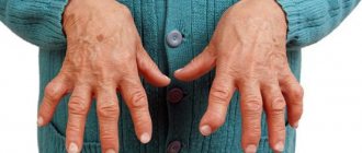
- Painful, “aching” sensations in the joints (arthralgia). Inflammatory processes in synovial membranes and degradation of cartilage tissue. Development of polyarthritis. Diffuse myalgia (muscle pain). Small joints are predominantly affected.
Reference. Although arthropathy in SBS does not involve deep damage to the subchondral bone tissue, damage to the joints leads to irreversible deformation in 20% of cases.
What is immunoglobulin E (IgE)
Our body has a special substance that is located in the mucous layer of many human organs and tissues. In the body, as a rule, it is in a bound state and is not detected in the blood in a free form.
In humans, this substance is primarily responsible for the development of allergic reactions. If we consider immunoglobulin as a whole, we can distinguish four types, each of which is capable of protecting our body:
- IgA – antibodies that ensure the formation of local immunity. It is activated if a person falls ill with an acute respiratory infection, or skin rashes occur on the body.
- IgE – destroys various viruses and fungi. Immunoglobulin E has antiparasitic and anti-infective properties. In addition, it eliminates various toxins that enter the body with food and drink.
- IgM are “alarm” antibodies. Once the disease affects a person, their number increases significantly. They are the first to encounter the infection and resist it.
- IgE and IgD are antibodies of a specific nature. They appear in the blood after allergies or worms.
By performing a blood test for immunoglobulin E, allergic diseases can be identified and the correct treatment can be prescribed.
Norm and interpretation of results
The analysis is performed both for preliminary diagnosis and during treatment: to determine the results and condition of the patient. The detected 10-20 LE cells per 1000 neutrophils are no longer considered simply as a positive result, but as evidence of the active manifestation of SCR. The picture is complemented by the presence of free nuclear substance in the plasma and the observation of “rosettes” (accumulations of neutrophils around LE cells).
An increase or decrease in the previously recorded concentration of LE cells in a patient can shed light on the course of the disease and indicate the degree of success of treatment measures.
Important! The norm for a healthy person is the complete absence of lupus cells in the blood taken for analysis.
Preparation for analysis and collection of material
Hemorrhoids kill the patient in 79% of cases
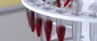
For the LE cell test, blood is drawn in the morning, before meals. Preparation for the procedure is standard: the day before, you should stop drinking alcohol and doing heavy physical work, you should not smoke for the last half hour, and spend the last half hour in a calm environment. Blood is taken from the ulnar vein using the puncture method. To prevent it from coagulating and changing its properties, an anticoagulant (ethylenediamineacetic acid) is added to the test tube. The container is filled halfway with blood, then carefully mixed and filled to the mark. Transportation to the laboratory is carried out in special boxes that protect against temperature changes and shocks - factors that can damage LE cells and other formed elements.
There are several methods for determining LE cells in the blood; currently, chemiluminescence immunoassay is the most common; some laboratories use the Zinkhoma-Conley method modified by E. N. Novoselova. ICHLA is based on the reaction between the cells under study and a specific substance that forms “antigen-antibody” complexes with these cells. The latter bind to the enzyme, then a reagent is added to the mixture, which colors the labeled complexes. The optical density of the sample determines the presence and concentration of LE cells. The Zinkhoma-Conley method, as modified by E. N. Novoselova, involves a mechanical effect on the blood, which facilitates the appearance of the LE phenomenon. The blood is mixed with sodium oxalate and left to stand at room temperature for an hour. Then glass beads are introduced into the test tube, it is tightly closed and shaken vigorously. After an hour of settling, the biomaterial is divided into layers, the plasma is placed in a centrifuge, smears are made from the resulting residue, they are stained and examined under a microscope. It takes up to 3 business days for LE cell test results to be ready.
LE cells (EDTA blood)
What are LE cells?
LE cells
– defective neutrophils with nuclear inclusions of leukocytes lysed by them. These cells are named after the first letters of the Latin name for systemic lupus erythematosus (Lupus erythematosus), since they were first identified in patients with this disease. The highest percentage of LE cells is detected in SLE, but they also appear in patients with other systemic diseases:
- rheumatoid arthritis,
- dermatomyositis,
- systemic scleroderma.
The formation of LE cells is a consequence of a violation of the immune system, evidence of its autoaggression, and is caused by the presence of the LE factor in the blood.
LE factor is a complex of specific circulating immune complexes (CIC) with antinuclear activity, which destroy not only the nuclear material of protective cells, but also settle on articular surfaces, vascular walls, are retained in the kidneys (lupus nephritis) and have a damaging effect on these tissues .
Analysis methodology
Blood for LE cell analysis
taken from a vein on an empty stomach in a volume of 10 ml.
To prevent the blood from clotting longer and changing its properties, the anticoagulant ethylenediamineacetic acid
(EDTA) is added. This allows you to extend the shelf life of blood and obtain more accurate data.
Video, Live healthy: ESR, what your blood tells you
Why is there leukocytosis in the analysis, an increase in the level of leukocytes?
- Exceeding the normal level of leukocytes occurs with temperature changes (hypothermia/overheating), increased stress conditions or physical activity.
- An increase in the number of leukocytes provides grounds for examination for the presence of an inflammatory process in the body, blood diseases, epilepsy, toxin poisoning, cancer, and allergies.
Why is there leukopenia in the analysis, a decrease in the level of leukocytes?
If the level of leukocytes is low, with corresponding symptoms, prescribe an examination for lupus erythematosus, metastases in bone marrow tissue, nervous disorders at the functional level, cirrhosis of the liver, plus inflammatory and infectious diseases.
Blood test interpretation is carried out only by a qualified specialist. Based on the data obtained, a diagnosis is made and a course of treatment is developed.
Share on social networks
During remission, as well as during an exacerbation of the process, but treatment has already begun, in particular hormonal treatment, LE cells may not be detected. If there are no cells, it is recommended to conduct several repeat studies. While the presence of LE cells in suspected SLE confirms the diagnosis, their absence does not allow it to be rejected.
Kernel segmentation. The cell size is mostly increased. Vacuolization occurs more often in the cytoplasm, sometimes in the nucleus. The presence of its nucleus indicates deeper changes in the cell and the severity of the pathological process.
Systemic lupus erythematosus (SLE) (continued...)
Minor diagnostic criteria
- Fever over 37.5 ºС for several days;
- unmotivated weight loss (5 kg or more in a short time) and impaired trophism;
- capillaritis on the fingers;
- nonspecific skin syndrome (erythema multiforme, urticaria);
- polyserositis - pleurisy, pericarditis;
- lymphadenopathy;
- hepatosplenomegaly;
- myocarditis;
- damage to the central nervous system;
- polyneuritis;
- polymyositis, polymyalgia;
- polyarthralgia;
- Raynaud's syndrome;
- increase in ESR (over 20 mm/h);
- leukopenia (less than 4 × 10⁹/l);
- anemia (hemoglobin less than 100 g/l);
- thrombocytopenia (less than 100 × 109/l;
- hypergammaglobulinemia (more than 22%);
- ANF in low titer;
- free LE bodies;
- persistently positive Wasserman reaction;
- altered thromboelastogram.
The diagnosis of systemic lupus erythematosus is reliable when three major signs are combined, one of which is mandatory - the presence of a “butterfly”, large numbers of LE cells or high titer ANF, hematoxylin bodies. When only minor signs are present or when minor signs are combined with lupus arthritis, the diagnosis of systemic lupus erythematosus is considered probable.
The diagnostic criteria for APA should also be taken into account. The presence of 4 out of 11 signs makes the diagnosis of systemic lupus erythematosus reliable.
Laboratory data
- General blood test: almost all patients have a significant increase in ESR, more than half have leukopenia with a shift in the blood count to promyelocytes, myelocytes and young in combination with lymphopenia, quite often hypochromic anemia, in rare cases hemolytic anemia develops with a positive Coombs test , thrombocytopenia may occur.
The detection of a large number of LE cells is pathognomonic. LE cells are mature neutrophils, the cytoplasm of which is almost entirely filled with the phagocytosed nucleus of a dead leukocyte, while its own nucleus is pushed to the periphery (the detection of at least 5 LE cells per 1000 leukocytes is diagnostically significant).
LE cells are formed in the presence of antibodies that react with the DNA-histone complex and complement.
- Single lupus cells are also found in other diseases. Freely lying destroyed leukocyte nuclei (hematoxylin, lupus bodies), sometimes surrounded by leukocytes, can also be detected - the rosette phenomenon!
Diagnostic criteria for systemic lupus erythematosus, ACR (1982), updated by ACR (1997)
| Criterion | Meaning |
| Rash on cheekbones and cheeks | Fixed erythema, flat or raised above the skin, not affecting the nasolabial folds |
| Discoid rashes | Erythematous raised patches with adjacent scales and follicular plugs, developing atrophic scars over time |
| Photosensitivity | Skin rashes resulting from an unusual reaction to sunlight - a medical history is required or photosensitivity should be monitored by a physician |
Source: https://www.eurolab.ua/encyclopedia/295/1340/?page=3
LE cells :: description, norms, causes, clinics
Name: LE cells.
LE cells
LE cells.
A type of white blood cell that has been destroyed by a specific immunoglobulin (LE factor). They are markers of autoimmune diseases. This indicator has independent diagnostic value. Detection of LE cells is essential for diagnosing systemic lupus erythematosus and monitoring the effectiveness of treatment for this disease.
Of secondary importance is the analysis of rheumatoid arthritis, dermatomyositis, and systemic scleroderma. Blood is collected for testing through veins. The most common method for detecting LE cells is immunochemiluminescence (a type of ELISA). As a rule, the test result is negative.
Test results are ready within 2 to 3 business days.
LE cells are neutrophilic leukocytes that contain the destroyed nuclei of other leukocytes that died during their interaction with the LE factor. In clinical practice, their detection in the blood is called systemic lupus erythematosus.
This disease is autoimmune, meaning the pathogenic mechanism is the production of proteins specific to autoantibodies that attack the tissues and cells of your body. In SLE, connective tissue and blood vessels are damaged. The chromatin of destroyed nuclei is phagocytosed by neutrophils - this is how LE cells appear.
They discard their own functional core, the central part is occupied by the material of a dead neutrophil. LE cells are formed with the participation of the LE factor. This immunoglobulin is produced in the body with systemic lupus erythematosus and is found in various body fluids, including blood, bone marrow puncture, exudate and urine.
Occasionally, small numbers of LE cells are detected in other autoimmune diseases, such as systemic sclerosis or dermatomyositis. The analyzed biomaterial is whole vein blood.
The level of LE cells is determined by the chemiluminescent method or the Zinkhoma-Conley method. The test is very specific and therefore has a limited scope. Most often it is used in general therapeutic practice and in rheumatology.
LE cells
Analysis of LE cells in the blood is one of the specific tests for detecting systemic lupus erythematosus. The study is designed to diagnose this disease and monitor the effectiveness of therapy. The results obtained make it possible to determine SLE and determine the stage of the pathological process and its activity.
With a good response to treatment, the level of LE cells in the blood often drops to zero. For the diagnosis of other systemic diseases, a blood test for LE cells is not suitable, since an increase in indicators is observed in a small part of patients and is unstable.
It is worth remembering that a negative result of blood tests for LE cells does not exclude the presence of systemic lupus erythematosus. Most often, the indicators increase at the onset of the disease and during its exacerbation, and during periods of remission they correspond to the norm. In the acute period of SLE, LE cells are found in 40-95% of patients.
The analysis is complemented by the identification of lupus cells and clusters of neutrophils around them, and the study of free nuclear substance (hematoxylin bodies, Hargreaves bodies). Currently, the LE cell test is used less and less in clinical practice; it is being replaced by studies of antinuclear antibodies in the blood and genetic methods.
They are more sensitive and accurate. The advantage of the LE cell test is its distribution and the cost-effectiveness of the assay procedure.
For the LE cell test, blood is drawn in the morning, before meals. Preparation for the procedure is standard: the day before, you should stop drinking alcohol and doing heavy physical work, you should not smoke for the last half hour, and spend the last half hour in a calm environment. Blood is taken from the ulnar vein using the puncture method.
To prevent it from coagulating and changing its properties, an anticoagulant (ethylenediamineacetic acid) is added to the test tube. The container is filled halfway with blood, then carefully mixed and filled to the mark.
Transportation to the laboratory is carried out in special boxes that protect against temperature changes and shocks - factors that can damage LE cells and other formed elements.
There are several methods for determining LE cells in the blood; currently, chemiluminescence immunoassay is the most common; some laboratories use the Zinkhoma-Conley method modified by E. N. Novoselova.
ICHLA is based on the reaction between the cells under study and a specific substance that forms “antigen-antibody” complexes with these cells. The latter bind to the enzyme, then a reagent is added to the mixture, which colors the labeled complexes. The optical density of the sample determines the presence and concentration of LE cells. The Zinkhom-Conley method modified by E.N.
Novoselova is a mechanical effect on the blood, which facilitates the appearance of the LE phenomenon. The blood is mixed with sodium oxalate and left to stand at room temperature for an hour. Then glass beads are introduced into the test tube, it is tightly closed and shaken vigorously. After an hour of settling, the biomaterial is divided into layers, the plasma is placed in a centrifuge, smears are made from the resulting residue, they are stained and examined under a microscope. It takes up to 3 business days for LE cell test results to be ready.
Usually the analysis data is negative. If smear microscopy reveals two or more LE cells per 100 white blood cells, the result is considered positive. The higher the score, the worse the prognosis of the disease. The level of LE cells in the blood does not depend on age, gender and physiological factors (physical activity, stress, diet, etc.).
The main reason for the increase in the number of LE cells in the blood is systemic lupus erythematosus. The highest concentrations are determined at the onset of the disease and during periods of exacerbation (in 60–80% of patients). In rare cases, other autoimmune conditions cause elevated levels of LE cells in the blood.
Positive test results are seen in rheumatoid arthritis, scleroderma and acute rheumatic fever.
In less than 10% of cases, LE cells are found in patients with periarteritis nodosa, benign thrombocytopenia, hemolytic and megaloblastic anemia, erythroderma, hepatitis and plasmacytoma.
The absence of LE cells in the blood is normal, so a decrease in their number is diagnostically significant only if the levels are initially elevated. In these cases, the reason for the decrease in the level of LE cells in the blood is the success of treatment.
| Clinic | Price | Telephone |
| MediScan in Domodedovo Domodedovo, Shkolny Prospect, 1m. | +7(929) 910..show+7(929) 910-90-10+7(926) 910-90-10+7(496) 794-11-55 | |
| Vitbiomed+ in Beregovoy Proezd, Moscow, Beregovoy Proezd, 7m. Fili | +7(499) 519..show+7(499) 519-36-63+7(495) 152-44-49 | |
| Medical Center for Immunocorrection named after. Khodanova RN Moscow, st. Davydkovskaya, 6m. Slavyansky Boulevard | +7(495) 152..show+7(495) 152-58-36+7(499) 445-40-83+7(495) 740-61-75 | |
| Polyclinic CG 'Lapino' in Odintsovo Odintsovo, st. Marshala Nedelina, 15m. | +7(499) 519..show+7(499) 519-36-94+7(499) 116-77-61+7(800) 700-70-01 | |
| City Hospital No. 33 in Kolpino Kolpino, st. Pavlovskaya, 16Am. | +7(812) 461..show+7(812) 461-34-51+7(812) 409-84-39+7(812) 242-38-02 | |
| KB No. 119 Moscow, Khimki, Novogorsk microdistrict. Pyatnitskoe highway | +7(499) 519..show+7(499) 519-39-17+7(495) 575-62-68+7(495) 575-61-95+7(495) 575-60-63 | |
| Polyclinic No. 2 of the Federal State Budgetary Institution Federal Clinical Center VMT FMBA of Russia Moscow, st. Novozavodskaya, 14Am. Fili | +7(495) 749..show+7(495) 749-95-37 | |
| Vitbiomed+ on Academician Pilyugin, Moscow, st. Academician Pilyugina, 14, bldg. 2m. New Cheryomushki | +7(495) 152..show+7(495) 152-58-54+7(499) 134-27-48 | |
| Vitbiomed+ on Donskoy, Moscow, st. Donskaya, 11, building 2m. Oktyabrskaya | +7(495) 152..show+7(495) 152-58-41+7(495) 104-40-03 | |
| Vitbiomed+ on Novorogozhskaya, Moscow, st. Novorogozhskaya, 8m. Roman | +7(499) 519..show+7(499) 519-37-25+7(495) 132-33-92 | |
| Vitbiomed+ on Novoyasenevsky Prospekt, Moscow, Novoyasenevsky Prospekt, 25m. Novoyasenevskaya | +7(499) 116..show+7(499) 116-79-74+7(495) 426-14-93 | |
| EndoMedLab on Novodmitrovskaya, Moscow, st. Novodmitrovskaya, 5A, building 2m. Dmitrovskaya | +7(499) 116..show+7(499) 116-79-74+7(495) 773-44-97+7(499) 270-33-71+7(495) 627-61-03 | |
| Vitbiomed+ on Bolshaya Marfinskaya, Moscow, st. Bolshaya Marfinskaya, 2m. Fonvizinskaya | +7(499) 116..show+7(499) 116-79-74+7(495) 104-40-03 | |
| Vitbiomed+ on Narimanovskaya, Moscow, st. Narimanovskaya, 8, bldg. 1m. Rokossovsky Boulevard | +7(499) 116..show+7(499) 116-79-74+7(495) 603-18-05 | |
| Vitbiomed+ in Bronnitsky Lane, Moscow, Bronnitsky Lane, 2m. Nizhny Novgorod | +7(495) 152..show+7(495) 152-58-90+7(495) 104-40-03 | |
| Vitbiomed+ on Pronskaya, Moscow, st. Pronskaya, 6, bldg. 2m. Lermontovsky Prospekt | +7(499) 519..show+7(499) 519-39-17+7(495) 104-40-03 | |
| OCM clinic on Bolshevikov Avenue, St. Petersburg, Bolshevikov Avenue, 22, bldg. 5m. Dybenko Street | +7(812) 586..show+7(812) 586-76-06 | |
| OCM Clinic on Professor Popov, St. Petersburg, st. Professora Popova, 27m. Petrogradskaya | +7(812) 327..show+7(812) 327-60-00 | |
| Medical center in 2nd Botkinsky Proezd Moscow, 2nd Botkinsky Proezd, 5, bldg. 5m. Treadmill | +7(499) 519..show+7(499) 519-37-39+7(495) 945-79-82+7(495) 653-14-57 | |
| Clinic of Doctor Klimenko on 3rd Tverskaya-Yamskaya, Moscow, st. 3rd Tverskaya-Yamskaya, 10m. Mayakovskaya | +7(495) 255..show+7(495) 255-21-88+7(926) 204-11-75+7(963) 676-06-88 | |
| Medical center in 4th Dobryninsky lane, Moscow, 4th Dobryninsky lane, 4m. Dobryninskaya | +7(495) 933..show+7(495) 933-86-48+7(495) 933-86-49+7(499) 237-38-52+7(499) 237-40-04 | |
| Research Institute of Phthisiopulmonology of St. Petersburg on Ligovsky Prospekt, St. Petersburg, Ligovsky Prospekt, 2-4m. Chernyshevskaya | +7(812) 775..show+7(812) 775-75-55 | |
| Research Institute of Phthisiopulmonology of St. Petersburg on Polytechnic St. Petersburg, st. Politekhnicheskaya, 32m. Polytechnic | +7(812) 775..show+7(812) 775-75-55 | |
| Clinic of St. Petersburg State Medical University, St. Petersburg, st. Litovskaya, 2m. Vyborgskaya | +7(812) 542..show+7(812) 542-93-57+7(812) 248-18-40+7(812) 295-46-23+7(812) 295-40-31 | |
| Clinic of functional disorders on Gabrichevsky, Moscow, st. Gabrichevskogo, 5, bldg. 3m. Shchukinskaya | +7(495) 152..show+7(495) 152-58-35+7(495) 363-07-54+7(499) 654-08-00 | |
| Muscovy in Stupino Stupino, st. Pervomaiskaya, ow. 59m. | +7(496) 647..show+7(496) 647-07-05+7(496) 647-07-10 | |
| GBUZ LenOblTsentr on Rizhsky Prospekt, St. Petersburg, Rizhsky Prospekt, 43m. Narvskaya | +7(812) 251..show+7(812) 251-15-26+7(911) 749-08-57 | |
| Research Institute of Pulmonology of the Federal Medical and Biological Agency of Russia on Orekhovy Boulevard, Moscow, Orekhovy Boulevard, 28m. Zyablikovo | +7(495) 395..show+7(495) 395-05-11+7(495) 395-63-93+7(495) 395-06-78+7(495) 395-63-93 | |
| MC Healthy Woman on Ivankovskoye Shosse, Moscow, Ivankovskoye Shosse, 3m. Shchukinskaya | +7(495) 488..show+7(495) 488-32-56+7(499) 193-96-56+7(499) 520-83-16+7(499) 193-52-01+ 7(495) 942-40-43 | |
| City Clinical Hospital No. 29 named after. N.E. Bauman Moscow, Hospitalnaya sq., 2m. Baumanskaya | +7(499) 263..show+7(499) 263-23-47+7(499) 263-03-84+7(495) 360-40-93+7(985) 769-63-94 | |
| Antel on Marshal Zhukov Avenue St. Petersburg, Marshal Zhukov Avenue, 28, bldg. 2m. Avtovo | +7(812) 384..show+7(812) 384-68-88 | |
| Military Medical Academy named after. S.M.Kirova St. Petersburg, st. Academician Lebedeva, 6m. Lenin Square | +7(812) 292..show+7(812) 292-34-35+7(812) 292-32-86 | |
| Alexandra Med on Kastanaevskaya, Moscow, st. Kastanaevskaya, 9, bldg. 1m. Bagrationovskaya | +7(499) 519..show+7(499) 519-37-25+7(495) 796-39-06 | |
| Apollonia in Krasnoarmeysk Krasnoarmeysk, st. Komsomolskaya, 4, bldg. 1m. | +7(495) 135..show+7(495) 135-22-03+7(496) 523-58-40+7(916) 811-02-04+7(926) 811-59-70 | |
| Accurate diagnosis in Bolshoi Demidovsky Lane, Moscow, Bolshoi Demidovsky Lane, 17/1m. Baumanskaya | +7(495) 152..show+7(495) 152-58-41+7(499) 397-01-57+7(925) 049-92-11 | |
| Clinic of topical medicine on 3rd Frunzenskaya, Moscow, st. 3rd Frunzenskaya, 10m. Frunzenskaya | +7(499) 242..show+7(499) 242-83-65+7(499) 242-83-68 | |
| XXI century on the Border Guard Garkavy St. Petersburg, st. Border Guard Garkavy, 15, room 3. Avenue of Veterans | +7(812) 603..show+7(812) 603-70-59 | |
| XXI century on Marata St. Petersburg, st. Marata, 48m. Vladimirskaya | +7(812) 603..show+7(812) 603-70-59 | |
| XXI century on Marshak Avenue, St. Petersburg, Marshak Avenue, 4m. Civil Prospect | +7(812) 603..show+7(812) 603-79-65 | |
| XXI century on Siqueiros, St. Petersburg, st. Siqueiros, 7, bldg. 2m. Ozerki | +7(812) 603..show+7(812) 603-70-59 | |
| More clinics – 156 . use filters |
42a96bb5c8a2ac07fc866444b97bf1 Content moderator: Vasin A.S.
Source: https://kiberis.ru/?p=90784
Blood chemistry
It is a laboratory test that allows you to diagnose the condition and performance of internal organs: liver, kidneys, pancreas, gall bladder, etc. The method also makes it possible to collect information about metabolism and identify the need for microelements.
Indications for prescription by a doctor
Our clinic’s specialists recommend undergoing blood biochemistry at least once a year, even for those people who have no health complaints. Thus, you can always detect problems in time and prevent the formation of chronic diseases. Biochemical analysis is prescribed by a doctor for infectious and somatic diseases.
Diagnosis of diseases based on deviations from the norm
The results of the study provided to the patient contain the established values of the studied indicators, as well as the acceptable norm (corridor of indicator values in a healthy person). When the set values leave this corridor, we can talk about the presence of problems with the patient’s health.
A biochemical blood test is the best way to timely diagnose a whole range of serious diseases. It is better to cope with the disease at an early stage, to prevent the disease from becoming chronic.