A general blood test is one of the simplest, most inexpensive and at the same time informative laboratory tests, allowing the doctor to determine the presence of pathological processes. Most people are familiar with the main parameters measured during this type of diagnosis and understand what they mean.
But for many, an indicator such as GRA in a blood test is simply an abbreviation that does not carry any meaning. But in fact, its deviations, identified when deciphering the examination results, often indicate the presence of diseases, some of which pose a serious threat to human health and life.
What is GRA
The scientific process of breaking blood into its component parts is the simplest and most inexpensive research method, which allows a medical specialist to identify the pathological process in the patient’s body. OAC allows you to evaluate any deviations from generally accepted medical standards for the composition of liquid connective tissue.
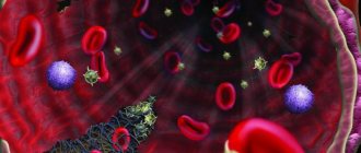
GRA is a conventional designation for one of the varieties of heterogeneous groups of polymorphonuclear cells, which is characterized by a large segmented nucleus and the presence of granulomas in the cytoplasm that glow under a microscope. The number of granular leukocytes in the blood plasma is a stable value.
Granulocytes are responsible for important functions of the human body and ensure the full functioning of the body:
- fight the root cause of inflammation in the body;
- have a negative effect on infectious agents;
- neutralize substances that cause allergic reactions in the body.
The accumulation of white cells in the blood will indicate the presence of an active inflammatory process in the human body, which will help a medical specialist determine the cause and type of pathology.
Deviations from the norm
An increase in the number of granulocytes is traditionally reflected in the results of a detailed blood test by a shift to the left side (due to the fact that immature granulocytes and their band counterparts are detected). This result indicates the occurrence of pathological processes in the body.
Factors that increase immature granulocytes can be divided into physiological and pathological.
Physiological ones include pregnancy, menstruation, stress, and high physical activity.
An increase in young neutrophils is also possible if blood is taken for analysis after the patient has eaten.
Pathologies that provoke an increase in immature granulocytes include pneumococcal infection, meningitis, pyelonephritis, septic lesions, cholecystitis, inflammation of the tonsils, otitis, thrombophlebitis, inflammation of the appendix, scarlet fever.
Video:
Purulent abscesses and the formation of phlegmon are accompanied by an increase in the percentage of all neutrophils and immature forms in particular.
Chronic skin diseases generate the activity of young granulocyte cells and, accordingly, an increase in their number in the blood.
There is an increase in immature neutrophils during intoxication of the body caused by lead poisoning, acidosis, insect attacks, uremia and other factors.
Systemic autoimmune diseases lead to changes in the entire leukocyte formula and the appearance of young neutrophil cells.
The reasons for the increase in immature granulocytes include, along with the listed pathologies, burns, manifestations of gout, myocardial and pulmonary infarction, gangrene, serum disease, the appearance of malignant neoplasms, and severe internal bleeding.
Taking androgen-containing drugs, glucocorticosteroids and lithium preparations increases the percentage of immature granulocytes.
A sharp increase in the content of band and immature forms of granulocytes should be especially alarming.
This factor often indicates myelomonocytic leukemia and extensive foci of inflammation with purulent processes occurring in them.
In these cases, granulocytes often change not only in percentage composition, but also qualitative changes in the cells occur.
Video:
An increase in immature granulocytes in childhood is often explained by reasons such as:
- infection with infectious diseases (otitis media, sore throat, acute pneumonia);
- inflammation of tissues with the appearance of suppuration;
- hemolytic anemia, severe burns;
- diabetic acidosis, the presence of trophic ulcers.
Immature granulocytes are very important because, first of all, they protect the body from the harmful effects of various pathogenic microorganisms.
Having discovered increased values of the content of immature granular leukocytes in the blood, you must definitely contact a specialist in order to identify the exact cause of the changes.
Since it is impossible to determine pathology by the number of neutrophils, it is strictly forbidden to try to restore normal values on your own and self-medicate.
The attending physician, based on the clinical picture, will prescribe an additional examination in order to make an accurate diagnosis.
Types of granulocytes in the human body and their functions
GRA (in a blood test this is an indicator of shaped white cells) have specific granules in the semi-liquid contents of the cells, which are represented by lysosomes and pyroxisomes. Granular leukocytes are the most numerous representatives of white cells of liquid connective tissue and are secreted by the organ of the hematopoietic system that carries out hematopoiesis from a single precursor cell.
Once in the hematocirculatory pathway, granular leukocytes are divided into macrophages and parietal pools. This state for granulocytes is the primary step before leaving the channel of liquid connective tissue into the organs and systems of the body, where white cells live for about 48 hours. The main feature of granular leukocytes is a reaction to normal staining during laboratory testing.
This allows granulocytes to be divided into:
- segmented neutrophils;
- eosinophilic leukocytes;
- granolocytic basophils.
| Name | Peculiarities | Functions |
| Neutrophil leukocytes |
|
|
| Segmented eosinophils |
|
|
| Granulocytic basophils | contain in large quantities:
|
|
Granulocytes: concept
All leukocytes are divided into 2 large groups. The first are agranulocytes, the second are granulocytes. The latter are the ones that are grainy. Agranulocytes, accordingly, lack it.
Granulocytes have a nucleus divided into several segments. However, it is of irregular shape. The cells are formed in the bone marrow and make up up to 70% of all leukocytes. The life cycle of granulocytes ranges from 3 to 10 days. After a few days they die. They are replaced by new cells.
Granulocytes is a collective term. These cells include eosinophils, basophils and neutrophils.
Despite the fact that they are united in one group, they are responsible for performing their specific functions. Eosinophils are cells that perform cytotoxic activities. In other words, they help destroy and eliminate parasites from the body. Eosinophils are cells that can also reduce the severity of an allergic reaction. At the same time, they can become a provoking factor in its development.
The main task of basophils is to trigger an immediate allergic reaction. They are also able to attract other granulocytes to the area of the pathological focus. Basophils also increase vascular permeability, improve blood circulation and lymphatic fluid flow, and regulate the coagulation of fluid connective tissue.
Neutrophils are cells that have the ability to move freely throughout the body. They easily leave the blood vessels and move to the pathological area. These cells are also capable of destroying fungi and bacteria. With the development of the inflammatory process, neutrophils absorb pathogenic microorganisms, but at the same time they die, releasing biologically active substances. The latter, in turn, stop the inflammatory process.
Norm
The leukogram has a certain percentage of different types of formed cells.
Standards:
The GRA norm in a blood test is shown in the photo.
| Indicator name | Children | Men | Women |
| Neutrophil leukocytes |
| 46-71% | 47-72% |
| Segmented eosinophils |
| 0-5% | 1-5% |
| Granulocytic basophilic leukocytes |
| 0,5-1% | 0-0,5% |
Granulocytes and agranulocytes
Once upon a time, in the last century, there were manual methods for counting blood cells, and there were no modern biochemical and hematological analyzers. And there was no such thing as gra in a blood test. There simply had not yet been a machine created that would issue a check with the encoded names of blood cells and their groups. There were granulocytes, and even a specialist probably could not immediately say what GRA is in a blood test.
Currently, all modern laboratories are large automated complexes, and decoding of blood tests is carried out without human intervention. And if values are obtained that differ from the reference values (normal limits), then they are rechecked manually.
Let's consider the functions of granulocytes in comparison with other blood cells, and some reasons for deviations from the norm in their total number.
Granulocytes is a collective term. All of them are leukocytes, but in addition to granulocytes, leukocytes also include monocytes and lymphocytes, which do not have granules in their cytoplasm. If we consider only granulocytes from leukocytes, then they are different - these are cells of the immune system that “live” in the blood and perform different functions. They all provide:
- recognition and destruction of foreign bacteria and foreign components in general;
- they eliminate old cells of their own body and destroy them;
- they produce immune reactions and are responsible for inflammation;
- granulocytes are the basis of the body's antibacterial defense and the substrate of allergic manifestations.
On average, a healthy adult has between 4.5 and 11 thousand white blood cells per microliter (µL) of blood.
This includes both granulocytes (basophils, eosinophils, neutrophils) and agranulocytes (monocytes and lymphocytes). The norm of lymphocytes is up to 40% of the total number of leukocytes, and monocytes up to 10% of all leukocytes. These cells are agranulocytes, that is, in their cytoplasm there are no specific inclusions or granules characteristic of granulocytes. Therefore, we can safely assume that granulocytes comprise half of all leukocytes in the human body, and their number on average is 6–7 thousand cells per microliter of blood.
There are no exact values, and this range is approximate, since the internal structure of this group is very variable, and responds by increasing or decreasing different types of blood cells to different stimuli.
Reasons for increasing and decreasing values
GRA in a blood test is a marker of a pathological process occurring in the human body. An increase or decrease in the number of polymorphonuclear cells prompts a medical specialist to identify the reasons that provoked changes in the leukocyte formula.
Reasons for the increase for all types of granulocytes:
- diseases of the body provoked by the penetration of pathogenic agents, prions, into the body;
- pathologies caused by helminths and arthropods;
- local tissue death in the body;
- poisoning with toxic substances;
- tumors consisting of malignant cells;
- recent infectious diseases;
- introduction of antigenic material to induce immunity to disease;
- systemic pharmacological therapy;
- increased sensitivity of the body to certain substances.
Increase in the percentage of blood neutrophils:
- tendency to skin hemorrhage;
- essential thrombocythemia, in which a neoplasm appears due to pathological changes in the mother cell of liquid connective tissue.
Relative increase in eosinophils:
- dysfunction of the immune system;
- disruption of the endocrine glands;
- non-parasitic infectious pathologies;
- pathological changes in the epidermis;
- disturbances in the functioning of the myocardium and blood vessels;
- connective tissue diseases.
Increased basophil levels:
- long-term inflammatory pathology of the colon mucosa;
- lymphogranulomatosis;
- dysfunction of liquid connective tissue cells;
- increased destruction of red blood cells;
- drug addiction;
- systemic use of drugs;
- inflammation of the gastrointestinal tract.
Demotion:
1. Segmented neutrophils:
- exposure to various types of ionizing radiation on the body;
- aleukemia;
- formation of connective tissue in the bone marrow with loss of hematopoietic elements;
- decrease in hemoglobin concentration per unit volume of blood;
- acute cyclical intestinal anthroponotic infection;
- pathologies provoked by non-cellular agents;
- zooanthroponotic disease with natural focality;
- damage to fibers containing collagen;
- Libman-Sachs disease;
- alcohol addiction;
- extreme exhaustion of the body;
- an increase in the volume of the unpaired parenchymal organ of the abdominal cavity;
- long-term use of potent pharmacological agents in therapy.
2. Eosinophilic granulocytes:
- intestinal infections caused by salmonella;
- prolonged increased emotional or mental stress;
- generalized inflammatory process in the body;
- operations;
- tissue damage on the body caused by high temperatures;
- multiple traumatic organ injuries;
- diseases of the hematopoietic system;
- overwork.
3. Basophils:
- inflammation of the respiratory system;
- acute infectious pathologies;
- hormonal disease with increased thyroid function;
- excess pituitary production of adrenocorticotropic hormone;
- pregnancy;
- systemic corticosteroid therapy;
- nervous overstrain.
GRA norm in AK and possible deviations
Let's consider what leads to the appearance of “lowered” granulocytes or an increase in their concentration.
Reasons for deviation of granulocytes from the norm
An increase in the number of granulocytes indicates inflammation of an infectious nature.
Differential diagnosis of an increase in different types of granulocytes allows us to specify the type of reaction:
- Allergic reactions are accompanied by an increase in the number of basophils.
- With amoebiosis, helminthic infestations, and giardiasis, the number of eosinophils increases.
A change in the leukocyte formula with a shift to the left means that there are purulent processes in the body, abscesses, and there is a possibility of myelomonocytic leukemia. With strokes, trophic ulcers, burns, neutrophils rarely increase.
An increase in substances can be caused by:
- inflammatory processes;
- infectious diseases;
- chronic diseases and endotoxicosis;
- injuries, shock, burns;
- bleeding and surgery.
Surgical intervention
There are physiological increases in the number of granulocytes:
- In women before menstruation, during pregnancy, immediately after childbirth.
- After a heavy meal.
- During physical activity.
One of the causes
of UAC with a reduced number of granulocytes indicates a hematological pathology, a viral infection. Impaired production of granulocyte cells by the bone marrow can reduce their number in the bloodstream.
Myelotoxic and hapten agranulocytosis can be provoked by medications (sulfonamides, cytostatics, antitumor drugs).
Immune agranulocytosis is caused by the destruction of granulocytes due to pathological autoimmune reactions. This condition often accompanies autoimmune connective tissue lesions (lupus, rheumatoid arthritis).
In this case, there is a high susceptibility of the skin, mucous membranes, and respiratory tract to infections. If neutrophils are low in an infant, he will suffer from constant stomatitis and frequent pneumonia.
Video of how granulocytes work:
Low levels of granulocytes or their abnormal development may be the result of a genetic pathology. Thus, in Chidiac-Higashi syndrome, due to an abnormality of lysosomes, granulocytes are not able to perform their functions, and as a result, the patient suffers from increased sensitivity to purulent infections.
Indications for the study
GRA are formed elements of liquid connective tissue that represent the body's defense system. In a blood test, granulocytes are found in a certain quantity, a deviation from which indicates a pathological process occurring in the human body.
The main indication for a clinical study of liquid connective tissue is:
- assessment of the state of the body's defense system;
- obtaining information about the cause of the disease state;
- identification of bacterial inflammatory process;
- determination of immune load;
- tracking the progress of recovery.
Position of MID in blood OCA
Formed elements are cells of biological fluid (blood). Each group of elements performs certain functions that ensure the full functioning of the body. With the development of pathological processes, the qualitative and quantitative composition of the blood changes, which is reflected in the analysis and allows one to make (assume) a diagnosis.
| Blood cells | Red blood cells | Platelets | Leukocytes |
| Functional responsibilities | Transfer of oxygen from the lungs, and transport of dioxide in the opposite direction | Ensuring coagulation (blood clotting process) | Protecting the body from invasion of foreign antigens (bacteria, viruses, parasites, allergens, etc.) |
Since leukocytes (white cells) play the role of guards of the body, their number increases when danger arises. The threat is posed by foreign agents that provoke inflammatory and allergic processes. The overall rate of leukocytes increases due to the mobilization of individual species that are responsible for the elimination of certain agents. In medicine, the process of capturing and eradicating (destructing) pathogens is called phagocytosis, which is why all leukocytes are phagocytes.
As part of the leukogram, the MID (total number of monocytes, eosinophils, basophils) can be determined or each element can be deciphered separately. The types of colorless cells and their responsibilities are discussed below. Agranulocytes (non-granular formed elements):
- lymphocytes – responsible for humoral immunity (immune response to invasion of viruses, allergens, bacterial microorganisms and activation of cancer cells)
- monocytes - provide phagocytosis of foreign substances in the peripheral blood.
Granulocytes (granular cells):
- neutrophils – capture and eliminate bacterial pathogenic microorganisms;
- eosinophils - fight parasitic infestations;
- basophils - are in correlation with immunoglobulin E, release histamine to eliminate allergic manifestations.
All colorless formed elements of blood are in a correlation relationship.
Where to get tested
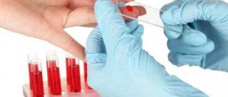
To determine the number of polymorphonuclear cells, a medical specialist prescribes a medical test. Most often, a test for the number of granulocytes is performed using the leukocyte formula, which is the percentage of white cells in the liquid connective tissue and refers to a constant value.
This type of laboratory examination can be done in any private clinic by collecting liquid connective tissue from the venous bed.
The cost of the analysis will vary from 125 rubles. up to 160 rub. depending on the level of qualifications of medical personnel and the status of the institution.
General practitioners also prescribe a clinical blood test to determine the amount of GRA. A basic study can be done in any multidisciplinary specialized treatment and preventive institution for the provision of outpatient medical care. In this case, a laboratory technician takes capillary blood from the ring finger, and the procedure is free.
Downgrade
Much depends on what type of granulocyte level is reduced.
Thus, neutrophil granulocytes decrease with the following ailments:
- Typhoid fever;
- Radiation sickness;
- Malaria;
- Primary myelofibrosis, leukemia (both chronic and acute);
- Iron deficiency and aplastic anemia;
- AIDS, hepatitis, influenza, rubella;
- Tularemia and brucellosis;
- Lupus erythematosus, collagenosis;
- Enlarged spleen;
- Cachexia, alcoholism;
- Treatment with toxic drugs.
- In an infant, a decrease in neutrophils may be due to congenital neutropenia, that is, a hereditary disease that sometimes ends in death. Most often, it manifests itself in the form of foci of chronic infection on the skin and in the body.
A decrease in the level of eosinophils is indicated by a figure less than 0.05*109/l . This phenomenon is called eosinopenia. It is observed when:
- Taking glucocorticoids;
- Acute infections, the nature of which is bacterial;
- Physical overstrain;
- Prolonged stressful situations;
- Sepsis;
- Surgical operations;
- Extensive burns;
- Polytraumas;
- Aplastic processes in the bone marrow;
- Anemia.
A decrease in the level of eosinophils in a small child indicates that the hematopoietic system and immunity are still immature. Immature granulocytes in the baby’s body are also .
A decrease in the level of basophils in the blood is indicated by a figure less than 0.01*109/l. This means that there is basopenia, which can be caused by:
- Stressful situations;
- Pneumonia;
- Acute infections;
- Diseases of the thyroid gland;
- Cushing's syndrome;
- Taking hormonal medications and problems with hormones in general;
- Ovulation and expecting a baby.
If granulocytes decrease or increase, there is a need to conduct other blood tests and more to find the cause. This is the only way to normalize their level.
Preparation
GRA in a blood test is an important indicator of the condition of the human body.
In order to obtain a reliable research result, it is necessary to follow the preparation rules:
- Blood sampling should be done in the morning;
- a few days before the procedure, you need to stop eating fatty foods;
- Before the examination, the following should be excluded from the diet 24 hours before the examination: alcoholic beverages; intense muscular loads; use of pharmacological drugs;
- in the morning, before donating blood, you can only drink water;
- It is not recommended to carry out a laboratory procedure after: physiotherapeutic treatment; Ultrasound; reflex effects on the tissues of the anterior and posterior parts of the body; fluorography;
- re-evaluation of leukocyte parameters must be done in the previous laboratory at the same time;
- liquid connective tissue must be taken for examination before starting treatment or 2 weeks after the end of therapy;
- it is necessary to inform the laboratory doctor about the presence of systemic pathologies.
The norm of neutrophils in the blood of children
A decrease in the concentration of neutrophils in a child’s blood most often indicates an infectious process. Sometimes the cause of the violation lies in an error during blood sampling or in improper preparation of the baby for analysis.
Therefore, it is necessary to understand what is causing the underestimation of the indicator and take a blood sample for repeated laboratory testing.
Why does the body need neutrophils?
There are special cells in the blood, leukocytes, that provide protection against infectious elements and cleanse the circulatory system after they are destroyed. One type of “white” blood cells are neutrophils.
These cells can be immature, rod-shaped and with a complete nucleus, and mature, in which the nucleus is visually divided into segments.
The lifespan of an individual neutrophil is several hours, so their composition is constantly updated. Normally, there should always be slightly more mature cells than young ones.
The main task of neutrophils is the destruction of bacteria, fungi and scraps of dying red blood cells. During the process of “eating” foreign elements, neutrophils also die, causing the formation of greenish pus.
Why does the concentration of neutrophils decrease?
A decrease in the concentration of mature neutrophils is observed if the bone marrow cannot cope with their timely production in sufficient quantities. Provoking factors may be:
- Taking certain pharmacological drugs,
- High physical activity
- Frequent stressful situations
- Genetic characteristics of the organism
- Deficiency of some microelements, as well as B vitamins.
This is not a complete list of diseases that lead to neutropenia; just read what symptoms of diabetes mellitus a child has to understand the prevalence of neutropenia. A decrease in the normal value can be caused by radiation or bone marrow disease.
Sometimes the analysis is considered unreliable, since the number of neutrophils changes if the child was fed or was in a state of emotional arousal before taking blood.
If in this case the characteristic symptoms inherent in a number of diseases are not revealed, the situation is a normal feature of the physiology of the body and the standard indicator is restored by the age of 2-3 years.
Normally, a child has the following neutrophil level:
- Newborn: mature – 1-17%, young – 45-79%,
- 1-12 months: mature – 0.5-4%, young – 15-45%,
- Up to 12 years: mature – 0.5-5%, young – 25-62%,
- Up to 15 years: mature – 0.5-6%, young – 40-65,
- 16 years old: mature – 1-6%, young – 47-72%.
In case of deviation from the norm, it is necessary to re-draw blood. If the picture remains unchanged, you should undergo an examination to identify the cause of the reduced neutrophil production. Only after the diagnosis is established, treatment is prescribed.
How to increase
Since a decrease in the indicator means suppression of the immune system, measures are first taken to maintain it. For this purpose, vitamin and mineral complexes are prescribed, a gentle regime is recommended, as well as enhanced nutrition.
When the pathology is accompanied by the appearance of purulent formations, periodontal disease, stomatitis, gingivitis, rinsing the mouth with chlorhexidine, bactericidal solutions and decoctions of St. John's wort or chamomile is prescribed.
In severe cases of neutropenia, they resort to measures such as blood transfusions. A decrease in the level of less than 500 in 1 ml becomes an indicator for antibacterial therapy, regardless of the cause of the disorder.
How to get a reliable blood test result
To determine the number of neutrophils, a sample of capillary blood is taken from the baby’s finger. For the analysis to be reliable, it is necessary to protect the child from eating sweets in the evening. Blood must be donated on an empty stomach, so you should not have breakfast this morning.
Children often cry in line at the treatment room. It’s worth trying to distract your child with a simple game of pats or an interesting fairy tale. Also, you should not allow the baby to rub his fingers, as this may distort the result.
If the analysis shows a decrease in neutrophils, it is imperative to find out the reason and take measures to increase the child’s immune defense.
How is the analysis carried out?
To determine the number of granulocytes, it is necessary to take capillary or venous blood.
In the first case:
- a medical specialist disinfects the extreme phalanx of the ring finger of the upper limb with wine alcohol using a cotton sponge;
- wipes the injection site with a dry cloth;
- the medical assistant takes out a scarifier needle from the sterile package, which is necessary for drawing capillary blood;
- quickly pierces the skin on the finger;
- wipe the first drops of liquid connective tissue with a cotton swab dipped in ethyl alcohol;
- using medical instruments, collects the required amount of blood to determine the number of granular leukocytes;
- Apply a sponge with an antiseptic to the damaged area.
After taking blood for testing, it is necessary to hold a piece of cotton wool at the puncture site so that the blood clots and there is no hematoma.
In the second option:
- a medical laboratory assistant disinfects the skin on the elbow with an antiseptic;
- using a vacuum syringe, pierces the epidermis and venous vessel;
- selects 10 ml of liquid connective tissue;
- The puncture is pressed down with a cotton swab dipped in wine alcohol.
Treatment
Treatment directly depends on the cause of their increase. Treatment is mainly aimed at eliminating the antigen or the inflammatory process as a whole. It is important not to try to self-medicate only based on the results of a general blood test. You can, of course, try to overcome the disease on your own if it is a common cold, but granulocytes can increase for a huge number of other reasons. Especially if they are a signal for cancer. If you are absolutely sure that you will not harm your health, then you can take a decoction of linden.
For it you will need:
Wash the linden and put it to boil, then simmer over low heat for 5 minutes, stirring constantly. For 200 ml of water you need to add 1 tablespoon of solution. Drink two hundred milliliters 2-3 times a day.
Decreased granulocytes
A decrease in this type of leukocytes does not always indicate the presence of a serious pathology, but still requires the attention of a doctor. The reason may be:
- Infection
- Recent chemotherapy
- Purulent processes
- Poisoning of the body
- A course of certain medications
- Blood disease
The decrease can only occur in neutrophils and eosinophils. And then, a decrease in these groups usually requires an additional biochemical blood test, and then a number of others, in order to determine the inflammation and the reason for such a sharp decrease in granulocytes. This may also indicate some pathologies or characteristics of the human body. In any case, this is not worth panicking and it is important to consult a doctor in time.
Treatment
Here, again, everything depends on the source of inflammation or the exact cause of the sharp decline. Treatment, as a rule, is aimed specifically at destroying the antigen or inflammation, and then at increasing the level of granulocytes. If the level has dropped after medication (for example, after chemotherapy), you can increase their level yourself, it all depends on the degree of reduction of granulocytes and the severity of this reduction. But, if you are confident that you can solve the problem without the intervention of doctors, then you can take a course of traditional medicine.
Analysis transcript
GRA in a blood test is an important diagnostic indicator. White formed elements of liquid connective tissue perform a protective function in the human body. Laboratory testing makes it possible to assess the patient’s general condition and the functioning of the immune system.
Different types of polymorphonuclear cells are responsible for specific functions in the human body.
Calculating the leukocyte formula allows the specialist doctor to see the full picture of the disease process and determine the diagnosis in order to prescribe adequate treatment in the future. From the results of scientific research, quantitative changes in leukocytes are of two types.
An increase in the number of young neutrophils, accompanied by the appearance of young granulocytes in the liquid connective tissue:
- infections with serous purulent inflammation;
- acute complex pathological process;
- release of blood beyond the vascular bed;
- loss of consciousness with fading reflexes;
- a shift in the body’s acid-base balance towards increased acidity;
- muscular fatigue;
- excessive formation of connective tissue in the bone marrow with loss of hematopoietic elements and increased extramarrow blood formation;
- acute myeloid leukemia;
- damage to the body by secondary malignant foci;
Predominance of mature neutrophils over young ones:
- malignant anemia caused by impaired hematopoiesis due to a lack of vitamin B12 in the body;
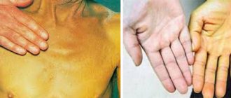
- pathologies of the hepatobiliary system;
- insufficiency of the paired bean-shaped organ that produces urine.
The interpretation of the GRA analysis is carried out by the doctor ordering the study. If there are changes in the leukocyte formula, only a medical specialist can determine the pathological process.
The following indicators and deviations are typical for a general clinical blood test:
| Indicator name | Norm in % | Promotion | Demotion |
| Eosinophilic leukocytes | from 1-5% |
|
|
| Segmented neutrophils | 41-71% |
|
|
| Granulocytic basophilic leukocytes | 0-0,5% |
|
Structure, norms and purpose of basophils
Basophilic granulocytes are a type of small white blood cell with a large nucleus and a small amount of cytoplasmic fluid. The number of nuclear segments of these bodies is even less than in neutrophils and eosinophils. When entering the blood of peripheral vessels, the circulation of basophils does not exceed 4 hours. Then, if there is an inflammatory process of allergic origin in the body, they move into the tissues.
Basophils actively participate in anaphylactic reactions, to some extent slowing down the function of lymphocytes. In pathological foci they release mediators aimed at the inflammatory process. The life cycle of basophilic granulocytes is about 12 days. Moreover, their norm in women, men and even children has approximately the same value, and is no more than 0.5 * 10 9 / l of the total number of leukocytes in the blood.
A decrease in the basophil count to less than 0.1*10 9 /l indicates the presence of an acute form of infectious pathology, for example, pneumonia, or indicates severe stress conditions. This may also be a signal of thyroid problems. This symptom can be caused by taking hormonal drugs that have an anti-inflammatory effect. And as a rule, low basophils are characteristic of pregnant women and women during the ovulation period.
Normalization of blood test parameters
If there are changes in the composition of the leukocyte formula, you must first contact a general practitioner, who, after assessing the result of the analysis, will prescribe additional examinations. Based on an additional check of the patient’s body condition, a medical specialist will decide on treatment tactics.
The main methods for normalizing white blood cell counts include:
- drug treatment of pathology that provoked quantitative changes in polymorphonuclear bodies;
- balanced diet;
- walks in the open air;
- regular light muscular exercise in accordance with age;
- sleep at least 8 hours a night;
- rejection of bad habits.
GRA is a very important indicator of ongoing processes in the human body. In order not to miss an emerging pathology, it is necessary to do a clinical blood test once a year.
Article design: Oleg Lozinsky
Normal heart rate in children by age
One of the most important indicators of a child’s heart function, along with blood pressure, is heart rate. Heart rate shows how many times per minute the heart muscle contracts. The pulse in children is constantly measured, since it is used to determine how the baby is developing and what his general condition is.
Another indicator that provides important information about the state of health and is always under the control of pediatricians is respiratory rate - respiratory rate. Using this indicator, doctors determine what kind of breathing the baby has (thoracic, abdominal), assess the functionality of the abdominal wall and chest, the rhythm and depth of breathing, and deviations from the norm.
These indicators depend on age and as the child grows, their values decrease.
Heart rate norms in children
Normal heart rate values in childhood differ significantly from those in adults. The heartbeat in children has its own characteristics and varies at different ages.
The average heart rate values for children by age are presented in the table below.
| Age | Average normal heart rate in beats per minute |
| first month of life | 110-170 |
| from 1 month to a year | 102-162 |
| from one to two years | 94-154 |
| from 2 to 4 years | 90-140 |
| from 4 to 6 years | 86-126 |
| from 6 to 8 years | 78-118 |
| from 8 to 10 years | 68-108 |
| from 10 to 12 years | 60-100 |
| from 12 to 15 years | 55-95 |
Deviations from the norm
If your pulse is too fast
If the heart rate exceeds the norm, the reasons may be as follows:
- hot weather;
- exercise stress;
- stressful situation.
In these cases, the pulse may increase three times, but this is not a pathology. A child may have a rapid heartbeat even at rest. Main reasons:
- prostration;
- overwork;
- heart diseases;
- endocrine diseases;
- respiratory diseases;
- anemia;
- infectious lesions.
If the pulse is too low
If you feel normal and no pathologies are found, a rare pulse indicates good training.
But bradycardia can be associated with pathologies and be accompanied by unpleasant symptoms. If your baby complains of dizziness, weakness, loss of strength, and has high or low blood pressure, you need to show him to a doctor as soon as possible.
What to pay attention to
If your child plays sports, you need to monitor your heart rate during exercise.
It is important that during training the heart rate does not exceed the maximum permissible values, which are calculated using the formula: 220 minus age
You should know that the heart rate should return to normal within ten minutes after stopping the exercise.
If the heart rate value is below this limit, the load can be increased.
Measurement algorithm
To carry out the test, you will need a watch with a second hand or a stopwatch. The difficulty in determining the pulse is that it is constantly changing. To measure your heart rate, you need to find an artery in your wrist, temple or neck and lightly press it with your finger. You should feel the blood pulsating under your finger. You need to count the number of shocks in ten or 15 seconds, then multiply the resulting value by six or four, respectively. This determines the pulse, which in most cases is equal to the heart rate. Now you need to compare the resulting figure with the indicators in the table, according to age. You should know that normally the pulsation should be rhythmic and clear.
Measurements must be carried out constantly and preferably at the same time. Doctors advise doing this in the morning, when the child is still in bed in a supine position. You cannot measure your heart rate after active games or emotional stress, when your heart rate increases. In this case, the result will be distorted.
If the data obtained differ significantly from the normal indicators given in the table, you need to consult a doctor to be examined and find out the cause of the deviations.
You can measure heart rate not only manually, but also using special devices that are available in pharmacies.
Finally
By constantly measuring a child's pulse, you can monitor his health and know whether he is developing correctly. Calculating heart rate makes it possible to find out about deviations in time and quickly begin treatment.
Normal limits
Regardless of a person’s age, the GRA blood indicator has its own specific normal limits.
The actual decoding takes place in the laboratory, and the doctor is provided with ready-made results, measured as a percentage. Therefore, results can be considered normal in the range from 47% to 72%. Other deviations will indicate that some disturbances are occurring in the body, which in particular led to similar results. If we consider in absolute ratios, then the decoding of the permissible norm will range from 1.2 to 6.8x(10^9)/l. In any case, you should not ignore the negative results of the study, as this can lead to significant consequences .
It is better to immediately show your blood test to a doctor and consult with further actions. You may need appropriate treatment or a repeat blood test now to confirm the results provided. Perhaps this was false information and will not be confirmed again.
Decreased levels of cells in the blood
When taking a general blood test, a specialist may detect a low level of granulocytes in a child. Based on what type of these cells is reduced, it is possible to identify the cause of this condition and begin immediate treatment. A decrease in cells in newborns is a rather dangerous pathological condition. So, with a critical decrease in neutrophils, the baby has practically no chance of survival.
The reasons for the decrease in granulocytes can be:
- blood cancer;
- lack of iron;
- liver disease;
- rubella;
- immunodeficiency;
- flu;
- intoxication.
A decrease in neutrophils in a newborn may be due to a hereditary disease. This condition requires monitoring by a specialist, as it often leads to death. Often the first manifestations of this disease include skin lesions. Focal inflammatory processes develop in the baby’s body.
When in contact with unfavorable factors, granulocytes can be destroyed or damaged, which also leads to a sharp decrease in their level in the human body.
Each such violation significantly affects the state of health, especially in young children, whose immune system has not yet matured and cannot take on such serious loads:
- Disturbances in the structure of blood cells make the body less vulnerable to aggressive infectious and bacterial environments.
- This condition can affect the baby's health. Infections and bacteria easily penetrate an unprotected body and actively begin to multiply in it.
- If the immune system is not able to cope with such problems on its own, then sooner or later this leads to death.
Therefore, if there is a decrease in granulocytes in a general blood test, it is worth immediately undergoing additional diagnostics and establishing the cause that could trigger this phenomenon. After the examination, the specialist should prescribe therapy aimed at returning cell levels to normal limits.