- October 11, 2018
- Tests and diagnostics
- Matela Victoria
What can you eat before an abdominal ultrasound? Quite often, in the process of medical examination of a patient, doctors use the ultrasound diagnostic method. During an ultrasound of the abdominal organs, their shape, size, structure and position can be assessed. A scheduled annual preventive examination will prevent the development of serious diseases. During such a study, the patient does not experience discomfort and there is no pain.
Features of the study
To obtain accurate diagnostic results, it is important to properly prepare for an ultrasound. For this, qualified specialists have developed a special diet before an ultrasound of the abdominal cavity. The main goal is nutritional adjustment. It is especially important to reduce the formation of gases in the intestines. Gases can block the image that a specialist receives on the monitor during diagnostics. Thanks to the study, it is possible to assess the condition of the organs located in the abdominal cavity:
- liver;
- gallbladder;
- spleen;
- stomach;
- abdominal aorta;
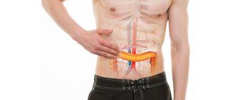
- pancreas;
- biliary tract;
- duodenum.
During the ultrasound, the doctor also examines the condition of the kidneys. This diagnosis allows you to determine the presence or absence of abscesses. Using ultrasound, you can identify a tumor, hematoma, stones, and visualize the structure of blood vessels, ducts and bile. If a person’s general health has deteriorated and stomach pain often occurs, then a specialist must prescribe an ultrasound of the abdominal cavity.
What organs can be checked with an ultrasound?
Ultrasound is very high frequency waves that the human ear cannot detect. It passes through some soft tissues - skin, muscles. Ultrasound is reflected from other tissues - internal organs, connective tissue, bones. Ultrasonic waves are absorbed by air and liquid.
It is thanks to the property of reflection that this diagnostic method became possible. The reflected waves are captured by a sensor, which transfers them to a computer. There, the sound signal is converted into a graphic signal - an image appears on the screen.
Dense tissues reflect ultrasound at a higher rate and appear white or gray in photographs. This is typical for the liver and kidneys. The bones are very dense, so they cannot be distinguished in the picture.
Softer tissues block ultrasound and appear dark gray or almost black. Air and liquid do not reflect waves at all; they appear as dark spots in the photographs.
These properties explain what an ultrasound of the abdominal organs shows:
- liver;
- gallbladder;
- pancreas;
- spleen;
- large vessels.
The stomach is a hollow organ, so it is not visible on an ultrasound; only the wall can be detected. The kidneys are also not normally visible; there is an ultrasound method for them.
The abbreviated designation OBP has a decoding - in medicine these are the abdominal organs. Therefore, when a doctor prescribes an ultrasound scan of the abdominal cavity, he assumes a disease of one of the above organs.
The types of ultrasound scans depend on what is included in the abdominal examination. All organs are rarely examined at once - this is a complex method, prescribed for serious diseases.
Ultrasound of the hepatobiliary system is most often used. It includes the liver, gall bladder and ducts. All types of examination are called abdominal - from the Latin word “abdominum”, which means stomach.
Watch a video about OBP research:
Under what conditions is it important to carry out diagnostics?
Under what circumstances should an abdominal ultrasound be performed? Causes of concern should be:
- paroxysmal pain under the ribs;
- unpleasant taste in the mouth;
- nagging pain in the abdomen;
- increased gas formation;
- systematic belching, nausea and discomfort in the abdominal area;
- organ injury;
- suspicion of cancer.
If one of the symptoms appears, it is important to immediately seek help from a doctor. This may indicate that a serious illness is developing.
Indications for use
Which doctor prescribes an examination of the abdominal cavity depends on who the person contacted and with what complaints. Usually the first specialist is a therapist. He can independently prescribe an ultrasound of the abdominal organs and kidneys, or immediately send the patient to a specialist. Among them, ultrasound diagnostics are most often prescribed by gastroenterologists who deal with diseases of the abdominal cavity.
Ultrasound of any abdominal organs is prescribed if there are indications:
- prolonged discomfort or abdominal pain;
- feeling of nausea, periodic vomiting;
- yellowing of the skin, mucous membranes;
- brown urine or yellow stool;
- persistent skin rashes;
- changes in blood biochemistry;
- abdominal organ injury with risk of internal bleeding.
In such situations, ultrasound of PD is a primary diagnostic method, that is, it helps the doctor to identify a disease that the person did not have before. They do examinations for adults and children from birth.
Ultrasound examination is also used as a monitoring method in people with chronic diseases:
- hepatitis of various origins;
- cholecystitis;
- pancreatitis;
- cirrhosis of the liver.
Ultrasound is performed annually and serves to prevent complications that may arise from irregular monitoring.
How often a person can have an ultrasound depends on the disease they have. There are no strict restrictions on this procedure, as it is safe for health. It is done as many times and as often as once a year, five years, ten years it is necessary to examine a person in order to control his disease.
Additionally, we invite you to read the article about ultrasound of the abdominal organs in children.
How to get a reliable clinical picture?
What can you eat before an abdominal ultrasound and how to properly prepare? It is important to prepare for diagnosis in advance. The accuracy of the data obtained depends on proper nutrition and drinking regime. To obtain a reliable diagnostic picture of the study, it is important to exclude the following factors:
- muscle spasm of the organ;
- presence of food masses in the stomach;
- motor activity of muscles;
- formation of gases in the intestines.
To perform an abdominal ultrasound, the patient must lie on his back. Sometimes the doctor asks you to take deep breaths or exhales, while changing position from the left side to the right. It is not recommended to drink water before an abdominal ultrasound, as you may get unreliable diagnostic results. It is forbidden to drink any liquid 2 hours before the test.
What is the procedure
Diagnosis of abdominal organs (abdominal organs) is carried out using a special sensor. Ultrasound waves are reflected from body tissues, which makes it possible to obtain a reliable diagnostic image of the organs being examined. For an accurate picture, the doctor uses a gel that does not allow air to penetrate between the body and the ultrasound machine.
Ultrasound shows:
- peritoneal organs, their shape and density;
- pathologies of anatomical structures;
- condition of blood vessels and aortas in the area under study.
Why is it important to prepare for an ultrasound?
Proper preparation for the event should ensure complete visualization of the internal organs. This will allow a thorough diagnosis of their condition. As medical practice shows, most often ultrasound is performed early in the morning, in rare cases - in the afternoon. If you properly prepare for the research process, the patient will receive the most accurate data. This will allow the doctor to make the correct diagnosis. The specialist will definitely give recommendations before an ultrasound of the abdominal cavity. You can check with your doctor for a list of foods that should be excluded from your diet. Most often, it is not recommended to eat foods that cause bloating.
Video
Abdominal ultrasound is described in detail in a video filmed by the Aesthetic Gynecology Clinic channel.
Do you have any questions? Specialists and readers of the HROMOSOMA website will help you ask a question
Was this article helpful?
Thank you for your opinion!
The article was useful. Please share the information with your friends.
Yes (100.00%)
No
X
Please write what is wrong and leave recommendations on the article
Cancel reply
Rate the benefit of the article: Rate the author ( 3 votes, average: 4.67 out of 5)
Discuss the article:
Colon cleansing and duration of fasting depending on age
The intestines should be cleansed 11 hours before the ultrasound examination of the stomach. Preparation for the examination includes the use of an enema. An enema should not be given to people who have an anal disease. In the presence of hemorrhoids, the introduction of a medical device can cause a severe inflammatory process. In the process of cleansing the intestines with an enema, you can use chamomile decoction. If the patient has not followed one of the rules for proper preparation for an ultrasound, it is important to inform the doctor about this. Otherwise, the specialist will not accurately assess the condition of the organ. It is necessary to report the medications that the person took on the eve of the study. "Dibazol", "Papaverine", "Drotaverine" may affect the accuracy of the information received.
Most common diagnoses
Among the most common diagnoses identified by UBP are the following:
- neoplasms of a malignant or benign nature;
- death of the spleen (heart attack);
- inflammation of the pancreas (pancreatitis);
- protrusion of the intestinal wall;
- cirrhosis of the liver;
- biliary dyskinesia;
- the presence of stones in the kidneys, gall bladder and bile ducts;
- inflammation of the female genital organs;
- inflammatory processes in any organ of the digestive system;
- purulent formations (abscesses);
- liver and kidney cysts;
- cholecystitis and cholecystopancreatitis;
- appendicitis;
- intestinal obstruction;
- through defects of organs (perforation).
Modern equipment allows us to conduct research with 100% reliability.
Doctors' recommendations for preparation
The specialist must give the patient not only a referral for examination, but also give recommendations on proper preparation for an abdominal ultrasound. It is important not to overeat; you should eat food in small portions. It is not recommended to drink food with food. Before the diagnosis, you are allowed to drink purified and still water. It is forbidden to drink strong and sweet tea. It is important to drink at least 2 liters of fluid throughout the day. A couple of days before diagnosis, it is important not to eat foods that can increase bloating.
Preparing for the study
General recommendations for adult patients
Please note: During a routine ultrasound examination of the abdominal organs, it is necessary to follow a diet for 2-3 days, exclude from the diet foods that contribute to stagnation of feces and gas formation .
This is especially important for elderly patients who often suffer from constipation and flatulence. Immediately before the study, if it is scheduled for the morning, then you must not have breakfast, and if it is scheduled for the afternoon, you must not have lunch. Regarding the volume of liquid, there are no special restrictions here, but it is better to drink table still water or weak black tea without sugar.
If at the same time it is necessary to conduct an examination of the organs of the urinary system (kidneys, bladder, prostate), then half an hour before starting the patient should drink 1 liter of table water .
If there is a need for an emergency ultrasound diagnosis, it is performed regardless of whether the patient has followed a diet or not.
Features of training in children
If we are talking about babies, then the last feeding should be 3 hours before the start of the procedure.
Next, as a rule, they should skip meals. For older children, the food retention time is 4 hours . Moreover, if the child complains of hunger or cries, then he can be given a small amount of water to drink .
It is very important to prepare the child psychologically for the study. It is necessary to convincingly tell that this procedure is absolutely painless and will not cause any discomfort. It should be remembered that children are often sensitive to the emotions of their parents, and therefore you need to remain calm and have a positive attitude.
How to prepare for pregnant women
Preparing for a test for pregnant women is practically no different. However, it is often much more difficult for them to cope with hunger, so they start taking it early in the morning.
Taking medications
Typically, ultrasound examination of the abdominal organs does not require special preparation. However, it is advisable that the intestines be empty before diagnosis . Therefore, patients who suffer from constipation are given a rectal suppository or a laxative tablet 12 hours in advance. If the expected result is not achieved, then a cleansing enema is performed several hours before the procedure.
Previously, before an ultrasound, activated charcoal was often prescribed (at the rate of 1 tablet per 10 kg of body weight) to cleanse the intestines. This is practiced less often now. New sorbents have also appeared (Enterosgel, Smecta, Polysorb) with higher efficiency.
What foods are not recommended to be consumed?
To obtain accurate information, after the study you need to reconsider your diet and listen carefully to the doctor’s recommendations on preparing for an abdominal ultrasound. Prohibited products include:
- chocolate products;
- cake;
- pie;
- cake;
- cookie;
- fatty meat and fish;
- sausage;
- apples;
- cabbage;
- alcohol;
- soda;
- natural juice;
- smoked products;
- mushroom dishes;
- peas;
- conservation;
Coffee and dairy products are not recommended to be included in the diet before an ultrasound. If you approach the preparation process carelessly, the specialist may postpone the study to another day due to the fact that it is impossible to fully assess the condition of the organ due to bloating and fermentation in the stomach.
Information on how a patient should prepare before an ultrasound scan
If the doctor has prescribed this diagnostic method, then it is necessary to carry out a set of preparatory actions without fail. After all, if a large amount of gas accumulates in the intestines, the doctor will not consider everything that is required. The procedure is carried out in the morning, on an empty stomach.
The recommended preparation time for ultrasound examinations is 3–4 days before the start of diagnosis. In a few days, nutrition is normalized and the intestines are cleansed. If there is constipation, then a separate diet is drawn up, medical records and documents are prepared.
The attending physician should inform you in more detail about the methods of preparation.
Authorized Products
What to do before an abdominal ultrasound? First of all, you should exclude prohibited foods from your diet. It is allowed to eat grain porridge (barley, buckwheat, oatmeal), lean meat (beef, chicken). It is allowed to include low-fat fish and boiled eggs in the diet. Food should be baked or boiled. On the evening before the diagnosis, you are allowed to have a light dinner until 19:30. It is better not to eat fatty foods. If a person often experiences constipation, before 15:00 it is necessary to clear the stomach with an enema or a laxative. On the day of the study, breakfast is prohibited. If the doctor has scheduled the procedure for 16:00, then you are allowed to eat and drink weak tea before the abdominal ultrasound before lunch. A few hours before diagnosis, it is important to drink activated charcoal. It is necessary to eat food slowly, since in this way a person will not swallow air. Many patients are concerned about what they can eat before an abdominal ultrasound, but not everyone knows that they need to eat food that does not cause fermentation in the stomach.
How is an abdominal ultrasound performed?
The test is usually carried out in the morning. This is more convenient for the patient because he does not have to fast for a long time.
The study can be carried out in a special room, both in a hospital setting and in a clinic. The diagnostic procedure is carried out primarily by a functional diagnostics doctor. Less often, a gastroenterologist who has completed specialization courses in ultrasound conducts research.
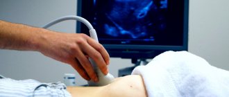
The patient lies on his back on a special couch, which is covered with a disposable towel. Next, the stomach is freed from clothes. A special gel is applied, which promotes better passage of ultrasonic waves.
The study begins with a general examination of the abdominal organs in longitudinal and cross sections. The doctor pays attention not only to the digestive system, but also to large vessels (abdominal aorta, inferior vena cava), and lymph nodes.
Then the examination of individual organs begins. For good visualization of the stomach, the probe is placed in the upper left quadrant. It is carried out slowly from top to bottom and various sections are sequentially examined - cardia, body and fundus, pylorus.
After this, if necessary, they switch to other organs. At the end of the procedure, the gel is removed using disposable dry towels.
Approximate nutrition
It is important to follow all medical recommendations before an abdominal ultrasound. This is the only way to obtain reliable data on the basis of which a specialist will make an accurate diagnosis. Preparation for abdominal diagnostics is absolutely simple. It is enough not to consume prohibited foods. It is especially necessary to approach preparation carefully for those patients who have an inflammatory process in the body. What can you eat before an abdominal ultrasound?
- For breakfast, you can boil eggs and drink green tea.
- Have a small piece of low-fat cheese.
- Dine on dietary meat, which is preferably steamed. You can drink a cup of weak tea.
- For an afternoon snack, it is recommended to prepare a portion of grain porridge.
- Dinner – low-fat fish and still mineral water.
After the examination, the doctor will give the patient a protocol containing a description of the organs. This document must be taken to the attending physician. After which the specialist will make an accurate diagnosis and prescribe treatment that will help improve the patient’s health. If there is no pathology, then the conclusion will say that the abdominal organs have no special features.
Nutrition before examination of the stomach, liver and kidneys
Plays a major role in preparing for an ultrasound. This means that the doctor at the appointment must talk in detail about the principles of nutrition , as well as give a list of allowed foods and those that need to be excluded for a while (within three days before the diagnostic procedure).
This is done so that the doctor obtains reliable results that reflect the picture of the condition of the internal organs. The quality of the resulting image is affected by the air present in the intestines , so it is necessary to exclude all gas-forming products. Feces that accumulate in the large intestine do not allow one to assess the condition of this part of the digestive tract, so if the patient suffers from constipation , it is recommended to do an enema in the evening before visiting the doctor.
Before the procedure, you should not eat any food for 12 hours. During this time, the food taken has time to pass through all parts of the gastrointestinal tract and be removed outside.
Please note: If you do not follow all the preparation rules stated by the doctor or nurse, the results of the study will be uninformative. This makes it difficult to make a correct diagnosis and prescribe treatment.
A child requires special preparation in terms of nutrition , because at any age you can withstand the required 12 hours of fasting. You can learn more about the diet in this case from this article.
What foods should you not eat?
There are a number of foods whose consumption causes excessive gas formation . Air accumulated in the intestines can seriously interfere with the visualization of organs during examination. Therefore, before an ultrasound, there is a certain list of foods that are excluded from the diet 3 days before the examination. This will reduce the amount of unnecessary air in the intestinal loops, and the signs of flatulence will be eliminated.
List of gas-forming foods that must be excluded 3 days before the procedure:
- sweet fruits;
- any confectionery products;
- bread and any other bakery products (they cause excessive gas formation and fermentation);
- strong drinks: tea, coffee, alcohol - not allowed in any form;
- milk and dairy products;
- fatty types of meat and fish (increased load on the pancreas and gall bladder);
- legumes (this includes peas, lentils, beans);
- fresh vegetables.
What is allowed?
Of course, all patients are interested in the question: what can you eat before an ultrasound? The following products are allowed to be consumed without harming the examination results:
- cereals – buckwheat, oatmeal, barley (porridge should be cooked exclusively in water);
- chicken and veal (lean meats);
- boiled eggs;
- low-fat cheese;
- low-fat varieties of fish.
Fruits and vegetables, despite the fact that they are part of a healthy diet, cannot be consumed before ultrasound diagnostics. The situation is the same with bananas, since many questions arise about this particular fruit. They contain a lot of sugar, which triggers fermentation processes in the intestines. If there are disturbances in the functioning of the digestive organs, this can adversely affect health.
Sweets also cause excess gas. This is especially true for those people who suffer from flatulence. It is best to cook dishes in the oven or steam; you can also boil foods.
How many hours before should I eat if the procedure is in the morning?
It is best if the examination is scheduled for the morning. In this case, it is most often possible to obtain reliable results about the structure and condition of the abdominal organs, since the time interval is observed - 12 hours from the last meal to the ultrasound. On the eve of the study, dinner should be eaten no later than 8 pm (for an easier bedtime). A heavy dinner is not recommended: it creates additional stress on the gastrointestinal tract. A light meal would be optimal.
If the diagnosis of the gastrointestinal tract is in the afternoon?
If a diagnostic procedure is prescribed for a patient in the afternoon or evening, then everything depends on the condition of the patient’s body. If a patient has diabetes mellitus , then it is impossible to completely prohibit him from eating before the examination . In this case, the doctor prepares the food for him, taking into account the severity of the disease. In other cases you can have a light breakfast , after which you should refrain from eating. In any case, ultrasound is performed on an empty stomach: the interval between the last meal and ultrasound should be at least 7-8 hours.
How to cleanse the intestines for people who often experience constipation?
It is important to cleanse the intestines the day before the test. To do this, you can take a drug that has a laxative effect. Guttalax is suitable for this. This product is absolutely safe for human health and rarely causes side effects. You can also use any suppositories that will facilitate the process of bowel movement. Doctors do not recommend using a strong laxative before diagnosing the abdominal cavity. Experts do not recommend taking the laxative drug Fortrans, as it can cause diarrhea.
If by morning it was not possible to cleanse the intestines with the help of a medicine, then you can use an enema. To completely cleanse the organ, you need 1.5 liters of water. If the patient does not have constipation, then there is no need to carry out such manipulation. It is not recommended to cleanse the intestines with an enema if the patient has hemorrhoids or another disease of the anus, since in the presence of pathologies, introducing an enema into the anus can cause severe pain. As a result, the hemorrhoids will become greatly enlarged and inflamed.
How to eliminate constipation in a child before an ultrasound?
If your baby has problems with bowel movements, then the day before the procedure it is important to cleanse the body. For this purpose, doctors often prescribe glycerin suppositories. The drug contains no aggressive substances, so it is absolutely safe for children. This medicine is allowed to be used even by children under 12 months. Suppositories soften feces and slightly irritate the anal mucosa. If a child has an anal fissure, hemorrhoids or inflammation of the digestive system, then it is prohibited to use any suppositories.
When inserting a candle into the baby's anus, it is important to be extremely careful so that the child does not experience any painful sensations. The suppository has a smooth structure, so during insertion it is important to hold it firmly with your fingers. It is advisable to first place them in the refrigerator before using candles. This will facilitate the process of introducing the medicinal product into the posterior opening.
How to get rid of bloating?
Many people know that before an abdominal ultrasound, you should not eat foods that cause fermentation in the stomach. But what to do if the patient often develops gas in the stomach? It is necessary to take a special medication before the ultrasound. To improve digestion, your doctor may prescribe Creon. The medicine will reduce gas formation. With the help of activated carbon, Simethicone, Espumisan, Disflatil, the functioning of the gastrointestinal tract is normalized and discomfort in the abdominal area is eliminated. Before using any medication, it is important to consult a doctor, since even harmless medications can cause side effects. If other diseases are detected in the human body or there is intolerance to one of the components of the medicine, then it should be replaced with an analogue.
Decoding the results
The standard protocol includes indicators that the doctor sees during ultrasound:
- analysis of the state of the peritoneal organs, their location, shape and density;
- description of changes in the vessels and aortas of the area under study;
- determining the presence or absence of inflammatory processes;
- exclusion of neoplasms in internal organs;
- identifying the presence or absence of free fluid in the abdominal cavity.
Norm
An ultrasound of an adult in the absence of pathology of the abdominal organs will show:
| Organ | State |
| Liver |
|
| Gallbladder |
|
| Pancreas |
|
| Spleen |
|
| Kidneys |
|
| Adrenal glands | Not enlarged |
| Abdominal aorta | Not expanded |
| Additionally | No |
| Conclusion | No structural changes were detected in the examined organs. |
Pathologies and possible diseases
In case of pathological processes, the following will be visible on ultrasound:
| Disease | Ultrasound signs |
| Fatty liver |
|
| Cirrhosis of the liver |
|
| Liver failure |
|
| Benign neoplasms in the liver |
|
| Malignant neoplasms in the liver |
|
| Chronic cholecystitis of the gallbladder |
|
| Cholelithiasis |
|
| Chronic pancreatitis of the pancreas |
|
| Pancreas cancer |
|
Factors distorting ultrasound
Points that prevent you from achieving accurate diagnostic results:
- Obesity. Organ scans may be distorted by thick deposits of fat under the skin.
- Spasm of intestinal smooth muscles. It occurs after an endoscopy performed the day before, as well as due to smoking immediately before the ultrasound.
- Excessive gas formation. The ultrasound image will be inaccurate.
- A recent X-ray examination of the abdominal cavity. In this case, the examination is postponed for 2-3 days.
- Large wound surface in the abdominal area. In this situation, it will be impossible to install the sensor.