- Preparing for a CT scan of the abdominal cavity
- Preparing for a pelvic CT scan
- Preparing for a cardiac CT scan
- General recommendations
Preparation for this is not complicated and is carried out before almost any study.
No preparation is required in cases where examination of any area of the body except the abdomen and pelvis is carried out without the use of a contrast agent. However, even in this case, it is advisable to refrain from eating for at least 4 hours before the procedure, since the doctor may see changes in native images that cannot be identified without additional contrast enhancement. This means that contrast may be needed in cases where its use was not originally planned. In some cases, the examination may require more serious preparation compared to refusing food for 4-6 hours . Let's look at these cases in more detail.
General principles of preparation
First of all, preparing for a CT scan involves determining the purpose of the study. Many patients, especially without a doctor’s referral, do not quite understand what they want to get: x-ray, tomogram, MRI, PET/CT. As a result, the research becomes meaningless, and this means radiation, time, money.
The first priority in preparation is to visit the attending physician and create a list of questions that the radiologist should answer.
First example
Patient Z (65 years old) complains of burning pain in the thigh, knee and lower leg along the back surface. He comes to a private clinic and demands to perform a “CT scan of his leg.” If the radiologist meets him halfway and performs the study, it will be meaningless.
Most likely, the patient's problems are associated with a herniated intervertebral disc, pinching the spinal nerve. He should see a neurologist and get an MRI of his spine. MSCT is not indicated.
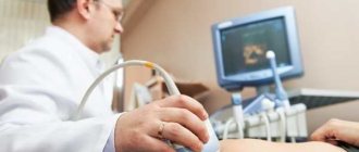
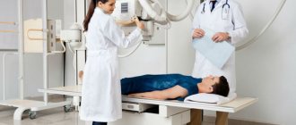
CT scan
Second example
Patient M (76 years old).
- An ultrasound revealed lesions in the liver and an enlarged pancreas.
- He was examined by an oncologist. A diagnosis of a malignant intestinal tumor with lesions in the liver was made.
- The tumor was verified pathomorphologically.
The patient came from the oncologist with a referral asking specific questions. Required:
- assess the location of the tumor, complications;
- stage the tumor using the TNM scale;
- determine the nature and cause of pancreatic enlargement.
In this case, the direction is comprehensive, nothing further is required.
Please take into account: what is much more important is not “not to eat for six hours before a tomography”, but to absolutely accurately determine the purpose of the study.
Purpose of dietary restrictions before diagnosis
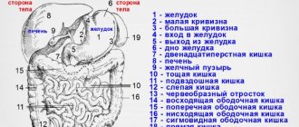
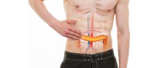
A diet before a CT scan of the abdominal cavity with contrast ensures high accuracy of examination of the condition of internal organs. In this area of the human body, all body systems are closely interconnected. When the functioning is disrupted or the size or shape of one organ changes, all others suffer.
This rule especially applies to the intestines. Before a computed tomography scan, it is important to observe some dietary restrictions, which will free the organ from feces and excess gases. As a result, the intestinal walls will make fewer perilstatic movements and the diagnosis will be more informative.
Preparing for contrast
For CT scans with contrast, iodine-based drugs are used, which have fewer complications. Iodine is in bound (non-ionic) form.
Before contrast, it is imperative to evaluate renal function. For this purpose, you must take a blood test for creatinine and urea, and calculate the glomerular filtration rate.
It is not recommended to administer contrast to patients before thyroid scintigraphy and radioactive iodine treatment. The gland tissue will accumulate iodine, which will interfere with diagnosis and treatment.
Why is a CT scan performed?
The procedure is performed to diagnose various diseases. The organs of the gastrointestinal tract, abdominal cavity, and urinary system are mainly examined. As a result, it is possible to recognize the pathology. With proper preparation, modern diagnostics helps to identify abnormalities in the functioning of the body. Namely:
- pathological changes of an inflammatory nature;
- congenital abnormalities;
- tumors of various origins (benign or malignant);
- vascular damage as a result of injury or accident;
- changes in the structure of blood vessels.
The study is carried out with or without a contrast agent. This is decided individually. Much depends on the specific disease and condition of the person.
Preparation for specific CT examinations
Research in certain areas requires specific preparation.
During CT coronary angiography, it is necessary to normalize the frequency and rhythm of heart contractions so that the ECG synchronizer can work fully and the images are as clear as possible.
Before CT colonoscopy or enterography, the following is required:
- cleanse the large (small) intestine with an enema or laxatives;
- take a contrast agent orally to “mark” remaining stool;
- stick to a diet.
Reduced peristalsis is usually not required due to the short examination time.
CT urography (kidney, urinary tract, bladder) requires that the bladder be full. Therefore, you should not urinate for 1-2 hours before the procedure.
Indications for the procedure
Diagnostics is used when the presence of malignant processes in the human body is suspected. Pathologically altered tissues are better supplied with blood compared to healthy ones. Therefore, much more contrast agent accumulates in them. As a result of the studies, doctors can find out the exact location of malignant tumors and metastases, their size and shape.
Often, contrast-enhanced CT is used to diagnose blood vessels. Using the procedure, it is possible to identify:
- cirrhosis of the liver;
- cystic formations of internal organs;
- abscess;
- anomalies in the structure of internal organs.
A similar procedure is prescribed after injuries to determine the extent of tissue damage. It is actively used before operations to determine the required volume of surgical intervention. CT scans are performed periodically during cancer treatment. This is necessary to determine the effectiveness and adjustment of the therapy used.
Eating before the procedure
For tests not related to the gastrointestinal tract, in most cases you can drink and eat as usual. Food in the stomach does not interfere with the visualization of, for example, pneumonia.
At the same time, before a CT colonoscopy or examination of the upper digestive tract, you need to follow a diet and refuse to eat for a certain time.
Before a CT colonoscopy, you should not eat in the evening, and before a CT scan of the stomach, you must stop eating at least two hours before the procedure.
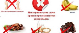
Permitted and prohibited products
Before CT enterography and colonoscopy, you should not eat “heavy” foods.
This group includes: meat, eggs, fatty fish, vegetables (cabbage, cucumbers, carrots, potatoes, turnips) and fruits. In addition, it is necessary to exclude gas-calorie-forming foods, such as sugar, peas, beans, beans, peppers, beets, milk, cheese, cottage cheese.
Diet before computed tomography of the intestine
Three days before the procedure you must:
- remove all gas and feces-forming foods from the diet;
- eat smaller meals, reducing portion sizes;
- the evening before the test you can drink a drink: water, tea, juice.
- A light dinner is possible - yogurt without sugar, semolina porridge with water.
On the morning of the test, you should not eat or drink: it is performed on an empty stomach.
Drinking regime before the procedure
Before CT enterography, it is better to refrain from drinking liquids for 6-12 hours.
- The essence of this research method is to fill the small intestine with contrast (positive or negative) through the mouth, followed by scanning.
- Immediately before the procedure, the patient drinks up to a liter of water or liquid with the addition of a contrast agent. The small intestine should be empty before this.
Before a CT colonoscopy, the drinking regime is more gentle. You may drink a small amount of water or sweet tea the evening before and the morning of the procedure.
Methods of colon cleansing
You can cleanse the colon with an enema or by taking laxatives. In the first case, a cleansing enema with a capacity of 1-1.5 liters is performed the evening before the procedure and in the morning 2-3 hours before it is performed.
Contraindications
Computed tomography with contrast is contraindicated in the presence of:
- diabetes mellitus;
- allergic reaction to injected substances;
- renal, heart failure;
- severe form of bronchial asthma;
- hyperthyroidism.
The procedure is strictly prohibited at any stage of pregnancy. During lactation, after the study, you must stop breastfeeding for two days. Performing computed tomography is impossible with a significant patient weight (>150 kg), since most devices are not designed for such a load.
Very rarely the procedure is used for patients under 14 years of age. It is performed in case of emergency for patients suffering from mental disorders and claustrophobia. If patients still need to undergo a CT scan, they are put into light medicated sleep.
Pharmaceutical preparations
Fortrans is most often used. There are a number of other drugs, but Fortrans is the most common. For adults, the contents of one package should be poured into a container of water (4 liters), mixed and taken orally in several doses. The drug intake is completed 3-4 hours before the procedure. Enemas and laxatives should be combined with diet.
Attention: be sure to read the instructions before taking the drug.
Use of pharmaceutical drugs
The need to use any medications before a CT scan with contrast is determined by the doctor. The specialist usually prescribes:
- Enterosorbents – activated carbon, Smecta, enterosgel. They help get rid of increased gas formation.
- Laxatives – Fortrans. Used when the patient is prone to constipation.
- Antispasmodics – Spazmalgon, No-shpu. Prescribed when the patient suffers from increased intestinal motility.
Drug therapy usually begins one day before diagnosis.
Possible complications
Complications may arise during preparation for a CT scan. Not all patients are able to avoid eating for several hours in a row or maintain a drinking regime. In this case, it is better to reschedule the study.
- Patients taking metformin should consult with their doctor about its temporary discontinuation, because This drug leads to increased intestinal density.
- Bismuth preparations used to treat patients with gastric ulcers lead to increased density in the intestinal lumen.
If barium contrast was previously performed, even a week before a CT scan of the intestine, the information content is reduced due to the presence of dense contents in the lumen. Intravenous contrast will delay scintigraphy or radioactive iodine treatment by at least three months.
Drug interactions
The drug increases the contrast of the image obtained during radiography due to the iodine present in its composition.
Pharmacodynamics
Trazograph is an ionic triiodinated radiocontrast agent. Increases image contrast. The effect is associated with the absorption of X-rays, which is ensured by the stably bound iodine included in the drug.
Pharmacokinetics
Trazograph is not metabolized and does not accumulate in the liver. Eliminates by glomerular filtration in a chemically unchanged form.
With the intravascular route of administration, the drug is distributed quickly in the intercellular space; connection with plasma proteins - no more than 10% of the dose. It does not penetrate the intact blood-brain barrier.
With intravenous jet injection of 1 ml/kg of Trazograph, high plasma concentrations of the drug in the blood are achieved within 5 minutes, corresponding to 2000–3000 mg of iodine/l. T1/2 (half-life) ranges from 1 to 2 hours.
Adverse reactions associated with intravascular administration of Trazograph are usually transient in nature and are classified as mild or moderate in severity, although there is information about the development of severe, life-threatening reactions, including those leading to death.
The following disorders were most often observed with intravascular administration of Trazograph: vomiting, nausea, feeling of heat, redness of the skin. By administering the substance slowly or administering it with short breaks (every 3-5 minutes), you can achieve a reduction in the sensation of fever and nausea.
Other possible disorders: dizziness, chills, sweating, fever, difficulty breathing, lacrimation, sneezing, headache, pale skin, weakness, retching and feeling of suffocation, decreased/increased blood pressure, muscle tremors, itching, urticaria, convulsions , swelling.
It is possible that these adverse reactions may be a harbinger of developing anaphylactic shock. If it occurs, the administration of Trazograph is immediately stopped and, if necessary, appropriate intravenous therapy is prescribed. The development of anaphylactoid and allergic reactions does not depend on the dose and method of administration of the solution. In patients with a predisposition to allergies, hyperergic reactions occur more often.
When using Trazograph, circulatory disorders may occur, which are expressed as dilatation of peripheral vessels and a subsequent decrease in blood pressure, reflex tachycardia, shortness of breath, cyanosis, agitation, confusion, even loss of consciousness. These side effects are severe and require emergency care.
It is necessary to take into account the likelihood of developing these complications; to provide emergency care, appropriate medications, an artificial respiration apparatus and an endotracheal tube must be ready. In isolated cases, mainly in patients with brain diseases or epilepsy, neurological complications may occur, including drowsiness, disorientation, blurred vision, temporary paresis, coma, and epileptic seizures.
In some cases, while using Trazograph, thrombosis and phlebitis may develop, and reversible renal failure rarely occurs.
It should be taken into account that Trazograph reduces the ability of thyroid tissue to accumulate radioisotope substances during diagnostic studies for a period of 14 days.
Against the background of diabetic nephropathy and in patients undergoing biguanide therapy, lactic acidosis may develop when Trazograph is administered. In order to prevent this complication, it is recommended to discontinue the use of biguanides 48 hours before the procedure. To resume therapy, it is necessary to ensure that there is no impairment of renal function.
In patients taking β-blockers, hypersensitivity reactions may be more pronounced.
In patients taking interleukin, the development of delayed reactions (including fever, itching, rash, joint pain) and the appearance of flu-like symptoms is more common.
Instructions for use
Trazograph should be administered on an empty stomach, and there are no restrictions on water intake. If there are disturbances in water-electrolyte metabolism, they must be eliminated before the procedure. This is of particular importance for patients with decompensated diabetes mellitus, generalized myeloma, polyuria, gout or oliguria.
To improve diagnosis during angiography of abdominal vessels and urography, it is recommended to thoroughly cleanse the intestines. In this regard, two days before the study, patients should avoid eating foods that cause flatulence (this mainly applies to legumes, salads, fruits, any raw vegetables, black and fresh bread).
You should eat your last meal no later than 18 hours before the start of the examination. It is considered advisable to take a laxative the night before. Long breaks in meals, as well as the use of laxatives for infants and young children are contraindicated.
To avoid the risk of developing a hypertensive crisis against the background of pheochromocytoma, it is recommended to carry out preliminary preparation with the use of α-adrenergic receptor blockers.
For patients who are afraid of the procedure, the use of drugs with a sedative effect is indicated.
The Trazograph solution should be drawn into a syringe immediately before the start of the examination.
The substance remaining after the study cannot be used. When performing angiography, it is necessary to frequently flush the catheters used with saline solution, this will minimize the possible risk of thromboembolism. Trazograph is easier to administer and better tolerated when using a solution heated to body temperature (heated immediately before the procedure). If a thermostat is used, only the number of ampoules that are intended to be used should be heated to T 37 °C.
Due to the risk of severe allergic reactions, preliminary testing of individual sensitivity using small doses of Trazograph is not recommended.
If possible, it is better to carry out intravascular administration of the substance while the patient is in a supine position. After the injection, careful monitoring of the patient's condition is required for at least 30 minutes, since the development of most complications is noted during this period. If it is necessary to use several high single doses, then the interval between administration of Trazograph should be 10–15 minutes (this allows for compensation of increased serum osmolarity due to the influx of interstitial fluid). In cases of a single injection of more than 300 ml of Trazograph, intravenous infusion of electrolyte solutions is indicated.
Trazograph is used for excretory urography, cystography and retrograde pyelography by intracavitary (into the renal pelvis, bladder) or intravenous administration.
Excretory urography:
- adults: a radiocontrast agent is administered intravenously in a stream at a rate of 0.3 ml/sec, a 60% solution of 20–50 ml or a 76% solution of 20 ml. For patients with increased body weight, it is more advisable to use Trazograph 76%;
- children: the study requires relatively high doses of the drug, which is associated with a physiologically reduced concentration ability of the still immature nephron of the kidneys; Trazograph 76% is used for research in the following doses: up to 1 year – 6 ml; 1–2 years – 8 ml; 2–6 years – 10 ml; 6–12 years – 12–14 ml; 12–15 years – 16 ml.
Infusion urography: Trazograph is administered intravenously to adults at a rate of 20–30 drops/min. For the study, a mixture of 80 ml of Trazograph (60% or 76%) and 80 ml of a 5% dextrose solution is used.
Retrograde pyelography: when carrying out intracavitary administration, it is advisable to dilute Trazograph with a 5% dextrose solution or isotonic sodium chloride solution to obtain a 30% solution. The resulting solution should be injected retrograde into the urinary tract through a catheter under low pressure, while visual x-ray control is carried out (pain in the lumbar region should not be allowed).
Cystography: a 30% solution in a dose of 100–200 ml is injected into the bladder through an epicystostomy or retrogradely (the volume should be slightly less than the capacity of the bladder), while visual X-ray control is carried out.
In order to conduct studies such as angiocardiography, aortography, arteriography, selective angiography, splenoportography, phlebography, the intravascular route is used to administer Trazograph.
Recommended dosage regimen:
- aortography: Trazograph 76% in a dose of 30–60 ml is injected into the aorta at a rate of 25–35 ml/sec;
- angiocardiography: Trazograph 76% in a dose of 60 ml is administered at a rate of 10–30 ml/sec;
- peripheral arteriography: Trazograph 60% is injected intra-arterially at a rate of 8–12 ml/sec; dose for injection into the lower limb – 20–40 ml, into the upper limb – 10–20 ml;
- selective angiography: Trazograph 60 or 76% is injected at a rate of 3–12 ml/sec in a dose that corresponds to the volume of the bed of the vessel being examined;
- phlebography: Trazograph 60% is administered intravenously at a rate of 3–5 ml/sec; dose for injection into the lower limb – 20–40 ml, into the upper limb – 10–20 ml;
- Splenoportography: Trazograph 76% is injected into the spleen at a rate of 8 ml/sec in a dose of 30–50 ml.
In case of overdose or in patients with significantly reduced kidney function, Trazograph can be removed from the body by extracorporeal dialysis.
Since the drug is used only for diagnostic purposes, reviews of Trazograph are few. It is noted that the product quickly produces the stated effect and is well tolerated.
The approximate price for Trazograph 76% (5 ampoules) is 535–652 rubles.
The method of use and dosage depend on the type of procedure.
Injections. The composition is administered slowly - 20 ml/min. To obtain a large volume - over 100 ml - the procedure lasts at least half an hour. The dosage is determined by the patient's age group.
The rate of contrast solution for adult patients (in ml): 76% - 20, 60% - 50.
| Age, months/years | Volume, ml |
| 0–12 | 7,0–10,0 |
| 1–2 | 10,0–12,0 |
| 2–6 | 12,0–15,0 |
| 6–12 | 15,0–20,0 |
| Over 12 | As for adults |
10–12.
In infants and children of primary preschool age, the study is carried out after two minutes. If the picture turns out to be uninformative, then the pictures are taken a little later.
Drip infusion. When using the system, the time for administering 100 ml of a solution of any concentration falls within the range of 5–10 minutes.
If the patient is diagnosed with heart failure, it increases to half an hour. The first image is taken immediately after the infusion is completed. Subsequent ones - after 20 minutes.
The procedure is prescribed to visualize the bladder. The drug is injected directly into its cavity using a catheter.
The duration of the procedure is half an hour, since the main reactions occur during this period. A 30% composition is used, obtained by diluting 60% Urografin with cold boiling water in a 1:1 ratio.
The dosage is determined by the following factors:
- Age.
- Weight.
- Myocardial contraction frequency.
- General health.
Preparation for the procedure involves complete cleansing of the human intestinal tract. For these purposes, two days before the expected date of the study, foods that increase the formation of gases are excluded from the menu:
- Legumes.
- Fruits.
- Black bread.
- Fresh bakery.
- Vegetables.
Dinner should take place the day before the procedure no later than 18:00. Before going to bed, take a laxative.
For confirmed pheochromocytoma, alpha-adrenergic blockers are recommended. This measure helps prevent the formation of a hypertensive crisis.
Analogs
Analogs of Trazograph are Urografin, Triombrast, Novatrizoate, etc.
There are substitutes for radiopaque contrast agents, but with slightly different compositions (contain sodium amidotriosate):
- Angiographin.
- Visitor trust.
- Urovizin.
- Urotrast.
- Verografin.
- Trazograph.
- Triombrine.
Triombrast solution is also used. In addition to sodium amidotrizoate, the drug also contains meglumine.