The organs that are located in the abdominal area are responsible for a large number of functions and processes in the body. The human abdominal cavity includes a whole complex of different organs that are responsible not only for digestion, but also the organs of the reproductive and urinary systems are located here. The abdominal organs are bounded above by the diaphragm, which separates them from the chest, and by the pelvic bones, which are located below.
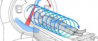
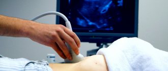
The proper functioning of all these organs is in many ways the key to good human health, so it is extremely important to strictly monitor their condition and consult a doctor if pain occurs. To more accurately identify the cause of discomfort, the doctor prescribes an ultrasound examination. This is a completely safe and painless non-invasive diagnostic method, which is based on the characteristics of the reflection of ultrasound waves from a particular type of tissue. This procedure allows you not only to see the structure of the internal parts of the body, but also to determine diseases, pathologies of the development and functioning of organs and various systems in the human body.
Abdominal walls
Muscles of the abdominal cavity
The abdominal cavity has boundaries. The top one goes below the diaphragm line. This is muscular fibrous tissue, which is located at the level of the lower ribs and delimits the chest cavity. The diaphragm takes part in ventilation of the lungs, changing the position of the dome when inhaling and returning to its original position when exhaling. It has openings for communication between the thoracic cavity and the abdominal cavity - these are the venous, esophageal and aortic openings.
In front, the peritoneum consists of several pairs of muscles:
- the outermost is the external oblique muscle;
- intermediate muscle layer – internal oblique muscle;
- the deepest is the transverse oblique muscle;
- the rectus muscle forms the abs, which is clearly visible in athletes, is involved in urination, defecation, bending over, and childbirth;
Human diaphragm - pyramidal, associated with the pubic bones (absent in 20% of the population);
- aponeurosis - tendon fibers in which the density is higher and there are few vessels.
On the side, the boundaries pass along the broad abdominal muscles, of which there are three pairs - 3 on the right and 3 on the left.
From below, the peritoneum is delimited by the pelvic diaphragm and the iliac bones. The diaphragm consists of several bundles that are woven into the prostate gland in men and the vaginal wall in women. Participates in the process of contraction of the muscles of the anus.
At the back, the abdominal cavity borders the lumbar spine.
The spleen and its role in the body
The spleen is an unpaired organ located above and to the left in the abdominal cavity. With normal physiology, the ribs completely cover it, protecting it from injury and impact.
It is in the spleen that a number of blood cells are formed, filtration and accumulation of blood occurs, and red blood cells cease their action. The organ tissue consists of red and white pulp. The spleen is adjacent to the stomach, diaphragm, part of the large intestine, and pancreas.
Aching or acute pain occurs in the spleen with the following diseases:
- splenomegaly;
- perisplenitis;
- rupture or infarction of the spleen;
- vascular thrombosis.
With diseases of the spleen, the patient is characterized by weakness, asthenia, bad mood, and low performance. What is located on the left side of the abdomen, besides the spleen, and causes discomfort to the patient, you will find out below.
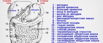
Abdominal organs and their functions
Internal organs of the human abdominal cavity
The organs in the abdomen are located in two spaces - directly abdominal and retroperitoneal. This depends on the location of the leaves - a thin serous membrane that protects the organs and delimits them from each other, and also facilitates their movement relative to each other. Thanks to the leaves, there is no friction between the organs inside the abdomen.
The abdominal cavity contains organs that belong to the digestive, hematopoietic, excretory and endocrine systems:
- Stomach. Located on the left under the diaphragm between the esophagus and the initial part of the small intestine. In the cavity, food is digested with the help of hydrochloric acid and digestive juices, as well as the absorption of vitamin B12. Here, food is decomposed into chemical components, which serve as food for all cells of the body.
- Liver. Located on the right under the diaphragm. The functions of the liver are to detoxify the blood entering its cells from throughout the body. Participates in the synthesis of bile, which digests fatty foods, regulates metabolic processes and heat transfer.
- The gallbladder is a hollow organ that stores bile. When food from the stomach enters the duodenum, bile enters the intestine and participates in digestion.
- The pancreas is an endocrine organ whose function is to control blood sugar. It produces insulin and glucagon, which break down sugar and convert it into glucose to feed the brain. It is located below the stomach on the left and is conventionally divided into three parts - head, tail and body. By releasing digestive juices, it breaks down food into small chemical components that are absorbed by the cells of the body.
Location of the spleen - The spleen is a hematopoietic organ located in the upper left, next to the stomach and pancreas. With its help, old red blood cells are recycled and new blood cells are created. Also involved in immune processes.
- The intestines are thin and thick. In it, water is absorbed and final digestion of crushed food particles occurs, and feces are formed, which move towards the exit - the anus.
- The kidneys are a paired excretory organ located in the retroperitoneum. The main function is to cleanse the blood of metabolic products. They connect to the ureters and bladder, located in the pelvis. Participate in the absorption of vitamin D and the formation of red blood cells.
All organs perform multiple functions simultaneously, such as detoxification and digestion.
Human abdominal anatomy includes the mesentery. A proposal has been put forward to consider it a separate organ of the digestive system. The mesentery is a double layer containing blood vessels, lymph nodes and nerve endings. With its help, all hollow organs are attached to the posterior wall of the abdominal cavity. It connects the intestinal loops, preventing them from twisting and keeps the organs in a certain position relative to each other.
Upper floor of the abdominal cavity
The structure of the human abdominal cavity is conventionally divided into three floors. The upper floor of the abdominal cavity is called the omental foramen. Consists of the pancreatic cleft, omental and hepatic bursa. The organs partially in contact with the pancreatic cleft are the stomach, spleen and the left lobe of the liver. The right lobe of the liver, adrenal gland and kidney border the hepatic bursa.
The omentum is 4 serous fused layers that partially cover the small intestine. In their thickness there are lymph nodes and vessels that ensure the outflow of fluid from the intestinal loops.
Average
It contains the small intestine and part of the large intestine. Bounded by the mesentery, which supports the transverse colon. There are also many depressions that are formed by peritoneal folds and the relative position of organs.
Lower
Located in the pelvis. In addition to the rectum and genitals, it includes the bladder. Men and women have different lower floor structures. In men, the peritoneum connects the rectum and testes; in women, the peritoneum connects the vagina and the posterior wall of the uterus. In this case, two depressions are formed - the uterine cavity with the rectum and the uterine cavity with the bladder.
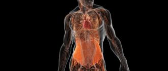
Paired organs
On both sides of the center are paired organs: lungs and bronchi
They are located symmetrically on both sides of the center. The adenoids are located in the upper back wall of the pharynx behind the nose. Palatine tonsils - behind the tongue, on both sides of the pharynx. The parathyroid glands are behind the thyroid gland (there are already 4 of them).
In the chest there are:
- Lungs. They are located behind the ribs, which protect them comprehensively.
- Bronchi. They are located between the lungs and are the connecting link between them and the trachea.
Behind the outside of the chest, on both sides, there are mammary glands. They are located at the level between the 3rd and 7th ribs in both women and men. In men, the mammary glands are practically not developed.
Abdominal organs:
- Kidneys. They are located near the back wall of the cavity, in the lumbar area. The left kidney is usually one vertebra higher than the right.
- Adrenal glands. The name itself speaks about the location - on top of the kidneys.
- Ureters. These are two tubes that connect the kidneys and bladder.
Parietal and visceral peritoneum
The serous membrane that lines the walls of the abdominal cavity and internal organs is called the peritoneum. It contains a lot of collagen elastic fibers, blood vessels, and nerve endings.
There are parietal and visceral peritoneum. The parietal peritoneum lines the walls, and the visceral peritoneum covers the organs.
In addition to its protective function, the semi-permeable membrane - the peritoneum - performs several more tasks in the body:
- Resorption. In an hour, the leaves are able to absorb an amount of exudate equal to 8% of the whole body weight. The contents of the cavity contain proteins, decay products, bacteria, and remains of necrotic tissue.
- Exudation or secretion of fluid. The upper part of the abdominal cavity is most active in this regard; in the lower direction, the intensity of the discharge decreases.
- Barrier. The greater omentum provides mechanical protection to organs and protects against infections by delimiting areas of inflammation.
The total area of the peritoneum is approximately equal to the area of human skin.
Peritoneal hernia
A peritoneal hernia occurs when there is a weak point in the abdominal wall, causing part of the intestine to protrude out of the abdominal cavity. An abdominal hernia is an exit or protrusion of the small or large intestine or parts thereof from the cavity in which they are located through a congenital or acquired opening in the peritoneum.
An abdominal hernia can occur as a result of prolonged pressure of internal organs on the walls of the abdominal cavity or weakening of a certain point - for example, as a result of pregnancy, obesity, constant physical activity, etc. A peritoneal hernia protrudes when part of the abdominal cavity protrudes and forms a hernial sac, which sometimes contains part of the small or large intestine. The only effective treatment for hernia is surgery.
Retroperitoneal space
The retroperitoneal or retroperitoneal space also refers to the abdominal cavity, but it is limited by the parietal peritoneum. This includes:
- kidneys, adrenal glands and ureters;
- pancreas;
- parts of the duodenum;
- lymphatic vessels and nodes;
- inferior vena cava, abdominal aorta.
The organs of the retroperitoneal space are in a fatty membrane for safety.
Structure and arrangement of human organs
They are arranged into two categories: hollow or tubular (for example, the intestines or stomach) and parenchymal or solid (for example, the pancreas or liver).
The former have several layers in their tubes, which are also called shells. The inside is lined with a mucous membrane, which plays a mainly protective function. Most organs have folds on it with projections and depressions. But there are also completely smooth mucous membranes.
Next comes the submucosa, which consists of connective tissue and is mobile.
In addition to them, there is a muscular layer with circular and longitudinal layers separated by connective tissue.
The human body contains smooth and striated muscles. Smooth - prevail in the respiratory tube and genitourinary organs. In the digestive tube, striated muscles are located in the upper and lower sections.
In some groups of organs there is another membrane where blood vessels and nerves pass.
All components of the digestive system and lungs have a serous membrane, which is formed by connective tissue. It is smooth, allowing the insides to slide easily against each other.
Parenchymal organs, unlike the previous ones, do not have a cavity. They contain functional (parenchyma) and connective (stroma) tissue. The cells that perform the main tasks form the parenchyma, and the soft skeleton of the organ is formed by the stroma.
Abdominal diseases
Inflammatory bowel diseases
Abdominal diseases include:
- Injuries – punctures, cuts, tissue ruptures with subsequent bleeding. They occur due to mechanical damage and are accompanied by heavy blood loss.
- Inflammation – acute or chronic. The pancreas, gallbladder, and bladder are most often affected. The reason is infectious agents.
- Chronic diseases of organs with periodic exacerbation. May be accompanied by organic lesions and tissue changes.
- Tumors – malignant and benign. They can develop in any organ of the abdominal cavity and spread to nearby tissues through metastasis.
- Intestinal diseases are autoimmune or acquired as a result of long-term poor lifestyle choices.
- Infectious diseases - hepatitis, enteritis and others.
The most dangerous disease is peritonitis. It can be caused by several problems - ruptured appendix, perforation of organs, complications after surgery, tuberculosis, intestinal obstruction. In the case of peritonitis, inflammation of the layers of the peritoneum occurs - parietal or visceral. This condition is life-threatening and requires immediate surgical intervention.
Which organs belong to the same system?
- stomach;
- Pancreas;
- Intestines;
- Liver;
- Kidneys;
- Spleen;
- Gallbladder and bile ducts;
- Bladder.
There are also gender differences in the number of organs in this part of the body and how they are located: women have the uterus and ovaries in this area, while men have mostly external genitals.
Most often, to find out the causes and discomfort in the stomach, the doctor prescribes an ultrasound of all abdominal organs in order to obtain complete information about the state of a person’s health and the structural features of his internal organs.
In addition to the above organs, the abdominal area contains a large number of important blood vessels and lymph nodes. All these parts of the body are very important for human health, so it is very important to consult a doctor at the first signs of pain for timely diagnosis and treatment.
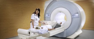
Methods for examining the abdominal cavity
There are several ways to examine organs located in the abdominal cavity. The simplest and most accessible is ultrasound. It is prescribed when a person complains of abdominal pain. MRI is done when it is necessary to confirm or clarify the diagnosis. An abdominal CT scan is performed for people who cannot undergo an MRI.
There are also invasive methods in which instruments are inserted into the cavity of organs - intestines, stomach, ureters and kidneys, gall bladder. These are gastroduodenoscopy and laparoscopy.
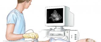
Ultrasound examination of the abdominal cavity
Ultrasound is performed to detect hidden diseases
This is an absolutely painless examination, which is based on the reflection of sound waves from the diseased and healthy organs. Depending on the condition, the sensor transmits a different signal, and the doctor makes a conclusion about the patient’s health.
Ultrasound is indicated for minor ailments and painful sensations. Usually a full examination of the internal organs is prescribed, because the localization of pain does not always coincide with the diseased organ.
Indications for examination are general ailments - increased gas formation, pain, examination is carried out for pregnant women. Using ultrasound, you can detect tumors, tissue ruptures, structural anomalies of internal organs, and inflammatory processes.
CT and MRI
Abdominal CT
MRI is used to perform surveys, angiography, and contrast examinations. You can see the relationship between damage to certain organs and their effect on healthy tissue. An MRI cannot be done if the patient has an artificial heart valve or titanium pins in the bones, since the method is based on the influence of a magnet.
The CT method is based on x-rays. In this case, a layer-by-layer image of an organ or its section is obtained. CT scanning is allowed for people with artificial valves and metal inserts in bone tissue.
Laparoscopic method
Laparoscopy procedure
This is a minimally invasive diagnostic method. It is also used to perform simple surgical operations. Through punctures in the skin, the doctor inserts an instrument into the abdominal cavity, at the end of which a camera is attached. Through it the image is transmitted to the screen.
Using a laparoscope, you can examine every organ of the abdominal cavity - the stomach, pancreas, liver, gallbladder, intestines and others.
The advantage of laparoscopic examination is the accuracy of diagnosis, as well as rapid recovery after the intervention and the absence of complications. The patient can be discharged from the hospital after 1–2 days.
Gastroduodenoscopy
Gastroduodenoscopy
Gastroduodenoscopic examination is carried out to examine the mucous membrane of the stomach, esophagus and duodenum. A rubber tube is inserted through the mouth, with a small chamber at the end. With its help, the doctor sees the condition of the mucous membrane on a computer monitor. The examination is prescribed after ultrasound diagnostics in order to better examine tissue areas and make an accurate conclusion about the diagnosis. Most often, duodenoscopy is prescribed for gastritis, stomach ulcers, or suspected internal bleeding due to perforation of the stomach.
How to prepare for an ultrasound examination
An abdominal ultrasound is a procedure that you should prepare for in advance to ensure accurate results. For example, if a woman on the eve of the examination consumed foods that caused flatulence, then after an ultrasound, this fact will cause difficulties in visualizing the spleen, pancreas, liver or biliary structure. Or if the patient is taking medications, then you need to stop taking them or warn the specialist who will conduct the ultrasound examination.
It is especially necessary to take the diagnosis of the pelvis seriously: before undergoing an ultrasound, you need to cleanse the intestines, and a few days before you start drinking herbs and medications that improve digestion and reduce gas formation: tea made from lemon balm, mint, chamomile, ginger. If the examination is required for a child, it is advisable to also put him on a diet on the eve of the ultrasound. A couple of days before the appointed date, give him enzymes (festal, activated carbon) to avoid flatulence during the ultrasound examination.
How many days in advance should you start the diet?
Patients always ask whether it is possible to eat before an abdominal ultrasound? Yes, but doctors warn that for three days before the procedure you need to follow a special balanced diet. It is advisable to eat every three to four hours, and there should be at least 4 meals. It is recommended to eat low-fat cheese, meat, and fish. Your daily diet must include grain porridges: buckwheat, oatmeal, barley. 1 boiled egg per day will harmoniously complement your diet.
- Is it possible to dye your hair during your period?
- Numbness in the fingers of the left hand
- Technical passport for the apartment
What not to eat before the test
On the eve of the ultrasound, a diet is prescribed so that the research conclusions are correct, because ultrasound waves will not be able to pass through the air in the stomach. Before undergoing the procedure, you should avoid any products that contribute to gas formation: dairy and fermented milk, baked goods, raw vegetables, sweets, carbonated drinks. You also need to give up too salty, spicy and fatty foods, and immediately before the procedure - from alcohol, smoking, chewing gum, and candy, so as not to provoke stomach cramps.
How far in advance can you eat on the day of an ultrasound?
The cleaner the body is on the day of the ultrasound, the more accurate the diagnosis will be, and, as a result, the more effective the treatment and faster the recovery. A short-term diet before an abdominal ultrasound will help improve the condition of the whole body, which is important for both men and women. The day before the procedure, you need to have dinner no later than 19:00, and you cannot eat anything on the day of the ultrasound.
Should I drink before an abdominal ultrasound?
During the diet, 2-3 days before the ultrasound, doctors recommend drinking herbal infusions, weak tea or non-carbonated water, but not more than 1.5 liters per day. You should not drink anything on the day of the ultrasound. It is recommended not to drink for several hours before the procedure so that the digestive system is completely empty. But this does not create much inconvenience for patients, since most doctors prescribe an ultrasound in the morning, and after the examination they are allowed to drink and eat as much as they want.
If an ultrasound of the kidneys or bladder is planned, preparation for the procedure includes the use of water for the acoustic window, so the patient is instructed to drink plenty of fluids. But we must take into account that you need to drink non-carbonated drinks slowly, without swallowing a lot of air, so that during the examination it does not form a space in the stomach that will not allow the device to correctly read the information.
Small and large intestine
The longest part of the gastrointestinal tract is the small intestine. This is where food is completely broken down. Here, almost all nutrients are absorbed into the blood - amino acids, vitamins, minerals. Remains of food and fiber continue their movement through the intestines. Loops of the small intestine occupy the posterior lower part of the abdominal cavity, predominantly the left side.
The diameter of the large intestine is much larger than that of the small intestine. The main function of this area is the formation of feces. A lot depends on the microflora of the colon.
What is located on the left side of the abdomen in men and women and can cause nagging pain and discomfort? Most likely, this is the large intestine making itself known. This may be colitis, proctitis, irritable bowel syndrome, left-sided ulcerative colitis, spastic constipation. For an accurate diagnosis, you need to contact a gastroenterologist.
Stomach as a source of pain on the left side of the body
This organ is located in the center of the abdominal cavity, but most of it is located on the left. It is the second organ of the gastrointestinal tract after the esophagus. At the entrance to the stomach there is a ring-shaped muscular sphincter. A similar, but smaller size is also available at the outlet. The stomach is necessary for the normal functioning of the gastrointestinal tract; it is the “battlefield” of enzymes and acids produced by the pancreas, gall bladder and liver. With stomach diseases, as if by a “domino effect,” all human life and health collapses.
Patients often wonder what organ is located in the left lower abdomen, while the stomach is the source of discomfort. Pain from an ulcer can radiate through nerve fibers to the upper and lower parts of the abdominal cavity. Gastritis pain is easy to distinguish, since it is most often associated with food intake - it is aggravated by hunger and overeating. Erosion of the lower part of the esophagus and the mucous membrane of the stomach walls can also cause serious discomfort for the patient.
Stomach polyps are also a common reason for patient questions about what is on the left side of the abdomen and hurts. Gastric polyps are benign tumor formations. Most often, they develop gradually, over many years, due to the growth of the mucous membrane, which is characterized by an inflammatory process.
Left kidney and ureter
These organs are a common cause of pain. The left kidney is located below, just below the level of the lower back on the left side. The left ureter is located next to the anterior abdominal wall. This is a thin tube about twenty centimeters long, which is directed from the hilum of the kidney to the bladder.
What is located in the lower left abdomen in men and is the source of pain? Most likely it's a kidney. Pain can occur with nephroptosis, hydronephrosis, pyelonephritis, glomerulonephritis, and urolithiasis.
Patients often ask the question: “What hurts and what organ is located in the left lower abdomen?” Depending on the nature and severity of pain, we can conclude that the cause is in the organs of the urinary system.
If a stone comes out, the pain will be sharp and unbearable. You need to contact an ambulance. If the patient has a fever, feels feverish and nauseous, and the lower back ache on the left side, most likely he has pyelonephritis or glomerulonephritis. In this case, it is also forbidden to let the disease take its course. Without medical intervention, even death is possible.
What is located in the lower left abdomen and causes throbbing pain that occurs from time to time? This is probably sand passing through the ureter. 65% of men and women have a tendency to form kidney stones (this is also facilitated by the quality of drinking water). But more often than not, the stones never form and come out in the form of tiny sand through the ureter.