The volume of amniotic fluid becomes larger, and the placenta begins to actively develop. By week 40 she will weigh approximately 800 grams. The “growth” of the fetus is about 9 millimeters, and the weight reaches 1.3 grams. Because the baby is so small, the mother cannot yet feel him, but meanwhile he is moving very actively.
Obstetric 6th week of pregnancy is actually 4th week from conception. The baby’s body shape at this age resembles the letter “C” or looks like a curved oval. The amniotic sac is formed around it, the main function of which is protective. Until birth, it will protect the child from mechanical influences. The baby reacts to them because the development of muscle tissue has already begun.
An important stage of intrauterine development is the formation of two chambers of the heart - this is happening right now. Germinal red blood cells are produced, and the first blood vessels, ventricle and atrium grow rapidly. After a while, a septum will appear that will divide the heart into 4 chambers.
Starting from the 6th week, the fetal heartbeat can be heard on ultrasound. Its contraction frequency is 110-130 beats per minute.
Every day the fetus becomes more and more human-like. The rudiments of legs and arms have formed, and in a week tiny fingers will begin to form. Ears appear in the form of bulges on the sides of the head. The eye sockets and mouth are already visible.
Why is an ultrasound done at this stage?
There are several reasons why your doctor may recommend an ultrasound:
- a woman complains of feeling unwell and has a stomach ache;
- signs of toxicosis suddenly disappeared;
- there is a risk of developing a frozen or ectopic pregnancy;
- An ultrasound is performed as an emergency when the patient develops bloody discharge;
- if there were unsuccessful pregnancies (miscarriage or frozen pregnancy).
At six weeks, research is carried out in order to obtain as much information as possible. Determine whether the fetus is developing and exclude the possibility of a frozen pregnancy. The study also helps to exclude the possibility of developing an ectopic pregnancy.
The results of blood tests for progesterone and hCG can also be considered an indication for an ultrasound. If there are any deviations in them, the gynecologist can send the woman for an examination to make sure that everything is okay with the expectant mother and her child.
It may be recommended to undergo an ultrasound examination when a woman becomes pregnant as a result of IVF or during stimulation. In such a situation, the entire period of bearing a child will be under the careful supervision of specialists.
Reference! If pregnancy is marked by pronounced toxicosis with all its unpleasant signs, then the disappearance of symptoms by the doctor can be perceived as an indication for an ultrasound.
Why is pregnancy testing carried out?
First of all, in the sixth week we can clarify how the pregnancy is developing and is it developing? There are also 3D ultrasound and 4D ultrasound. This is a study that provides a more detailed understanding of the organs and systems of the fetus. It is prescribed by doctors if there are any doubts about the diagnosis or unclear visualization of the defect. And to confirm the diagnosis, an expert 3D ultrasound examination is prescribed.
Ultrasound also evaluates the health of the expectant mother. Assess risks. During an ultrasound examination, attention is paid to whether there are any pathological space-occupying formations in the ovaries or myomatous nodes. There are myomatous nodes that interfere with the development of pregnancy and lead to miscarriages. In this case, it is necessary to resolve the issue with fibroids first and then plan the pregnancy. Also, in addition to large and multiple nodes, there is another risk - the growth of fibroids themselves. Some fibroids grow during pregnancy due to the influence of the hormone progesterone. They increase many times over. In this case, such conditions require dynamic monitoring.
Myomas
It is also, of course, important to examine all adjacent organs. These are the kidneys, liver and pancreas. If there are problems in these organs in the form of conglomerates (stones) or cysts, then in this case, consultation with related specialists is necessary to determine management tactics before pregnancy. Determine which medications can be taken and in what doses so that it does not affect the fetus. Of course, if these are large formations, it is advisable to remove them, because compression (squeezing) during pregnancy of these formations can lead to their rupture, which will subsequently lead the patient to the operating room.
Any surgical intervention can lead to premature birth of the patient, which can negatively affect the condition of the fetus and can even lead to death. Of course, all this should be explained to the patient by the antenatal clinic doctor at the time when she began preparing for pregnancy, in the so-called pre-gravid period.
If the pregnancy is spontaneous, it is very difficult for the patient to decide what to do and who to contact. All risks must be assessed. And if they exceed, then it is worth deciding on surgical intervention, especially when it threatens the life of the mother. First of all, doctors act in the interests of the mother and her life. In order to avoid such difficult choices and situations, high-quality diagnostics and timely resolution of issues with chronic diseases are necessary.
Mother's condition and how pregnancy develops
Week 6 is considered almost the very beginning of pregnancy; at this stage, a woman can guess her situation by the following symptoms:
- Increased breast sensitivity. Not only the mammary glands, but also the nipples become extremely sensitive. When you touch your chest, you can feel a strong, bursting pain.
- Changes in the quality of the skin. A woman develops acne; it can be localized on the face; the skin becomes looser and more sensitive to allergens, chemicals and cosmetics.
- The expectant mother’s well-being changes, she becomes more capricious. The woman is worried about weakness, she experiences irritation, and she may have problems sleeping. And the reason for everything is unstable hormonal levels.
- The taste of food changes, a sharp reaction to the smell appears. Attacks of toxicosis may bother you, and strange taste preferences may arise.
At this stage, the pregnancy is developing with all its might. Hormonal changes and corresponding changes occur in the body. A woman's uterus may put slight pressure on the bladder, and there is a frequent urge to urinate, which is not associated with kidney or bladder disease.
In the sixth week, the baby’s muscle tissue begins to form, and blood begins to circulate throughout the baby’s body. The brain and spinal cord of the embryo are formed from the fetal neural tube.
Pain
Take abdominal pain very seriously, it can mean a possible miscarriage, especially if accompanied by bloody discharge. Also, abdominal pain may be less noticeable and cramping, which is quite normal in this position. Contact your doctor to rule out any possible threats to you and your baby.
Often expectant mothers suffer from lower back pain; they, as in the first case, can be safe, but can indicate possible infections that have a detrimental effect on the future health and development of the child.
In any case, the cause of the pain should be determined by a specialist; do not risk your health and the life of your baby.
What will the diagnostics show?
When conducting early pregnancy research, many mothers think that an ultrasound will help them find out the sex of the child. But that's not true.
The doctor will pay attention to:
- possible pathologies in fetal development;
- the fetus may already have a heartbeat;
- embryo imaging;
- the presence of one or more fertile eggs in the uterus;
- the exact location of the fertilized egg in the uterus.
Looking at the monitor, the expectant mother will see her baby. It will resemble a pea or a small dark spot. In this case, you can consider the legs and arms. The baby's eyes are poorly developed at this stage of pregnancy, for this reason they will be tightly closed.
The specialist will study the fetus, its internal organs, and will also help eliminate possible defects in the functioning of the heart, liver and other organs.
And at this time, a happy mother can find out that she is expecting twins.
Hydatidiform mole
Hydatidiform mole or trophoblastic disease is a pathological degeneration of the chorionic villi, intended to form the placenta. As the disease progresses, complete or partial transformation of capillary villi into vesicular formations occurs. The disease is detected in the early stages, mainly at 5–6 weeks, and is characterized by an extremely aggressive course.
A typical echo picture shows the accumulation of bubbles in the uterine cavity, increased echogenicity, indicating the presence of blood in them. The body of the uterus is enlarged; a round formation is clearly visible in the thickness of the myometrium, surrounded by a dense rim and having a hypoechoic middle. Even minor trophoblastic processes affecting single villi completely exclude the possibility of the development of a normal embryo.
Ultrasound image: hydatidiform mole
How do they do it?
Research at this time can be carried out in two ways:
- Ultrasound through the anterior abdominal wall. This procedure is considered less informative, but safer. In addition, not all medical institutions are equipped with the appropriate equipment to conduct research using another method.
- Transvaginal ultrasound with the insertion of a particularly sensitive sensor into the woman’s vagina. It has high accuracy and is used in the absence of contraindications. It helps not only to most accurately determine the duration of pregnancy, but also the presence of pathologies in the fetus, which is extremely important.
If we talk about the examination that is carried out through the anterior abdominal wall. It is not much different from any other ultrasound procedure. The woman is placed on a couch, a highly sensitive gel is applied to the abdominal area and ultrasound diagnostics begin. The specialist will talk with the patient, report on the condition of the fetus, and issue a conclusion upon completion of the procedure.
Ultrasound using a transvaginal sensor follows a slightly different pattern. The woman lies down on the couch, the doctor treats the sensor with a special antiseptic, puts a disposable condom on it and inserts it into the woman’s vagina.
An image appears on the monitor screen, the doctor determines the fact of pregnancy, examines the fetus for the presence or absence of developmental pathologies. Upon completion of the procedure, he issues a conclusion to the woman.
When going for an examination, you must take with you:
- Referral for ultrasound (if available).
- Medical card of the pregnant woman.
- A clean diaper or large towel.
- Slippers or shoe covers.
You also need to reserve a certain amount of time, since the procedure will take from 10 to 15 minutes.
Yoga
The 6-week-old embryo, the photo of which was already shown above, is now actively developing; for proper development and positioning, you need to perform a special set of exercises. You should forget about regular exercises; now yoga is gentle, aimed more at relaxation and breathing. Exercises such as figure eight and circular movements of the hips, cat stance (good, evil) and other light exercises will help.
Be sure to rest between exercises, breathe deeply and evenly, if after exercise you feel unwell, shortness of breath or pain, then immediately consult your doctor; exercise should be postponed.
Now it is very important to master breathing techniques, they will help you both during childbirth and calm down in a stressful situation. How does breathing help during childbirth? Breathing is one of the best pain relievers, as attention is concentrated on the technique of the exercise, as a result of which the pain is dulled. Breathing exercises also saturate your baby’s body with oxygen, which is also very important and useful.
You can attend special yoga courses for pregnant women or do the exercises yourself. For this, there are special video courses or articles with photographs for correct implementation. Please note that the exercises should be appropriate to your stage of pregnancy so as not to harm the baby. Involve your spouse in these exercises.
Pay more attention to walks in the fresh air, eat right, follow the recommendations of your doctor, then the pregnancy period will become an unforgettable period of your life, in the truest sense of the word. Now there are a lot of pleasant worries and troubles, there is no time for frustration or illness. Be healthy and patient. Good luck to you and your kids!
How to prepare?
Preparation for the procedure depends on the method of ultrasound and takes place in several stages:
- preparing the intestines for the examination;
- compliance with hygiene procedures before visiting a doctor;
- filling the bladder with water to obtain accurate results.
2-3 days before the ultrasound diagnosis, you should stop eating foods that lead to excessive gas formation in the stomach. It is worth excluding spices, legumes, sparkling water, etc. from your diet.
It is recommended to take a shower before going to see the doctor. Carry out hygienic procedures for the genitals.
This is especially true for those patients who are undergoing transvaginal ultrasound.
In the 1st trimester, the study is recommended to be carried out with a full bladder. Before the procedure, it is advised to drink about 500 ml of clean water. This will help you see the fetus and uterus better, but in some cases it is not necessary to drink water. For this reason, it is worth checking with your doctor or nurse about this.
Fetal development
The 6th week of pregnancy (fetal development during this period is very active) is characterized by the following changes:
- The structure of the heart becomes more complex. After some time, it will divide into the atria. On ultrasound you can already detect contractions.
- The neural tube is becoming more perfect. It is completely covered by connective tissue. In the brain, the formation of depressions and convolutions occurs; the organ already regulates the activity of the muscles and heart.
- The tail of the embryo disappears, and the intestinal tube is formed. Its upper part will later form the larynx and pharynx, the anterior part will form the esophagus, the middle part will form the basis for the intestines, and the posterior part will form the excretory system.
- The digestive system is being formed.
- The thymus gland appears, which is responsible for the immune system. The respiratory system is separated from the rest, but its full functioning will begin only after the baby is born.
- Skeletal bones, cartilage, muscles, and tendons are formed.
6th week of pregnancy, fetal development. - Active division of nerve cells occurs, the nervous system is rapidly developing, so the unborn child is already able to respond to external stimuli.
- You can already recognize the face of the future baby; the ears and mouth are visible. Baby teeth are being laid.
- Hands and feet are formed, elbows and knees are clearly visible, and there are rudiments of fingers.
- The size of the ovum during this period is 10-18 mm, the size of the fetus is approximately 4-5 mm. The amount of amniotic fluid increases, which allows the fetus to move freely in the uterus, but its size is not yet sufficient to feel movement.
Who is recommended for research at this time?
Some women independently sign up for an ultrasound at 6 weeks, trying to find out about their “interesting situation” in this way. But it is better to go for research after the doctor’s recommendation.
Ultrasound examination is carried out:
- in case of IVF;
- to register a woman for pregnancy;
- if there is a suspicion of a frozen or ectopic pregnancy;
- urgently if there is a threat of miscarriage.
Reference! Examinations are carried out not only to detect pathology in the development of the fetus, but also to verify the fact of pregnancy and accurately determine the period.
Despite the fact that the procedure is not included in the list of planned ones, its implementation should not be considered something out of the ordinary. A doctor may recommend an ultrasound scan for a patient if there is confusion about the timing or the woman independently insists on having the study performed.
Hematometer
Gravidar hematometra is a small hematoma localized in close proximity to the site of implantation of the fertilized egg. The cause of hemorrhage is blood vessels damaged during the implantation of a fertilized egg (implantation hemorrhage). Detection of hematometra using ultrasound, on the one hand, is unambiguous evidence of intrauterine pregnancy, and on the other hand, it may mean the possibility of spontaneous termination of pregnancy.
The main task of ultrasound when detecting a hematoma is to determine its volume, structure (liquid or coagulated blood), dynamics of progress or regression. All these indicators can characterize the duration of the process and, accordingly, possible risks, since the connection between the fertilized egg and the uterus at 6 weeks is quite weak and an increase in pressure from a growing hematoma can disrupt this connection.
Ultrasound shows an anechoic zone located outside the amniotic sac, wrapping around the fundus of the membranes. To assess the dynamics of changes in the hematoma, a repeat study is required after 1–2 weeks. The appearance of a hematoma is noted in more than 50% of pregnant women and in the vast majority of cases, the hemorrhage completely resolves by 14–15 weeks.
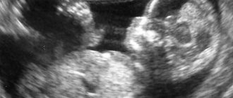
What can be seen in an ultrasound photo at 6 weeks of pregnancy?
Having received the treasured photograph of your future baby, you should not try to see in it who the child looks like - this is impossible.
If you look closely at the photo, you will be able to notice two small spots, one of them is the yolk sac, the second is your baby.
Photo of the fetus
In general, the future parents will not see anything significant in the photo yet, since the baby is still too small. But this has its own charm - since all the pleasant moments are yet to come.
Photo of twins
What changes occur in a pregnant woman's body?
At week 6, many pregnant women begin to suffer from a headache, which may appear due to a decrease in vascular tone and a decrease in blood pressure. To bear a child, changes occur in the mother's body. So, to make it easier to bear the baby’s weight, the ligaments that support the sacrum are softened in the abdomen. In this case, nagging pain in the lumbar region is a sign of pregnancy, not illness. Such pain can last up to 20 weeks.
If pain in the back or lower back becomes intense and radiates to the stomach, you should urgently seek medical help. They can be a sign of serious pathological changes and an incipient miscarriage. You should not take medications without consulting your doctor.
Not only can this harm the baby, but it can also make diagnosis difficult, which can lead to inappropriate treatment.
Back pain, excluding the lumbar region, is not typical for the 6th week of pregnancy; your doctor will tell you the cause and treatment. Low-intensity chest pain serves as an indicator that the mammary gland has begun preparing for feeding. This is a completely normal process. It starts and ends spontaneously. But the absence of such pain should not be a cause for concern.
Important!
If the pain is significant, you should consult a doctor. Particular concern during pregnancy is the appearance of abdominal pain accompanied by bloody discharge. With such a combination of complaints or if pain of moderate and high intensity occurs, you need to lie down, call an ambulance and undergo hospital treatment, if the visiting doctor insists on it.
Diagnostic results and norms
If we talk about normal indicators, then the weight of an embryo at 6 weeks does not exceed 1 gram. The size of the fertilized egg is about 2.5 cm, the heartbeat is from 110 to 150 beats per minute.
Indicator table
| CTE (embryo length) | 4–6 mm |
| Yolk sac diameter | 3.01 m3 |
| Amniotic fluid | 2-3 ml |
Certain deviations from the norm are allowed, but they will be assessed by a doctor. Changes in indicators are not always perceived as pathology. Because there are acceptable limits.
Alcohol and smoking
Any mother does not want her behavior to become the reason for the abnormal development of the baby. The 6-week embryo is just beginning to develop, important vital organs are being formed, all toxins (alcohol, drugs, smoking) can cause pathologies and mental retardation. You should categorically refuse all harmful factors, because we are talking about the future of your baby, do not ruin his life in the womb.
Contraindications
Possible contraindications depend on the method of ultrasound. If the procedure is carried out using a highly sensitive sensor, then the contraindications may be as follows:
- threat of miscarriage or increased uterine tone;
- gynecological diseases.
The study performed through the anterior abdominal wall has no obvious contraindications. It is not performed only if the patient has extensive damage to the skin at the site where the gel was applied.
Determining the sex of the child
At this stage of gestation, germ cells are laid down, which determine the sex of the unborn baby. Boys develop testicles. But the sex of the child cannot yet be determined using ultrasound, because the development of the genital organs has just begun. However, thanks to the advances of modern medicine, this can be done using a blood test.
A DNA test will show whether there is a Y chromosome in the baby’s genome. If so, it means a boy will be born. Otherwise, you should wait for a girl. But such research is quite expensive, and there is also a possibility of error.
Where do they make it and how much does it cost?
The examination is carried out in the following medical institutions:
- perinatal center;
- gynecology;
- maternity hospital at the place of registration.
You can also do the examination in a private clinic; you don’t even need a referral for this, but you will need to pay.
The cost of the procedure depends on the method of implementation; it varies from 1 to 5 thousand rubles.
If you want to conduct a 3D examination, then its price is higher and can reach 9 thousand rubles.
Signs of pregnancy
This stage of pregnancy is characterized by a clear manifestation of signs of the development of new life inside the body. Some of these unpleasant symptoms can make the expectant mother feel anxious. Such signs include, for example, small amounts of brownish discharge. But if they are not accompanied by nagging pain in the lower abdomen, then there is no cause for concern. This is the body's reaction to hormonal changes.
The most common pregnancy symptoms that appear by week 6 include:
- Increased fatigue;
- Vomiting that occurs in the morning;
- Increased salivation;
- Frequent urination;
- Unregulated mood swings (rapid);
- Lethargy felt in the muscles;
- Pain in the lower abdomen (not in all cases);
- Increase in the volume of the mammary glands.
Due to the characteristics of each organism, not all women can detect the manifestations of some pregnancy symptoms. Until the 6th week, some women do not notice any changes within themselves at all.
Also, the 6th week of pregnancy is characterized by the appearance of minor painful sensations in the breasts, as well as darkening of the nipples. The discomfort that occurs in the lower abdomen is the result of the changes that occur in the uterus during this period.
New sensations in the expectant mother are caused by hormones. In addition to increased sensitivity to various odors, irritation may occur, as well as unpredictable food desires. By the 6th week of pregnancy, the following are also noted:
- Dizziness;
- General weakness;
- Temperature increase.
Typically, all symptoms that arise on a hormonal basis disappear by 10-14 weeks.
Questions - answers
What threat does alcohol pose to the baby in the sixth week of pregnancy?
Alcohol consumption is contraindicated at all stages of pregnancy. Drinking alcohol during the development of fetal systems and organs is most destructive. It can cause the development of various pathologies and deformities. Large amounts of alcohol consumed can cause miscarriage.
I have severe toxicosis, I am six weeks pregnant. How can you alleviate your condition?
First, remove all strong-smelling items: perfumes, creams, deodorants. Try not to come into contact with household chemicals: washing powders, dishwashing detergent and others. Let your family take on all the household responsibilities during this time. Identify foods that also make you feel nauseous and eliminate them. Try to switch to frequent, small meals. Choose foods that you enjoy eating, but do not forget about your child’s needs for vitamins and microelements.
Drink fresh, home-squeezed juices. If you vomit frequently, drink as much fluid as possible to avoid dehydration. Give yourself a break as often as possible. If you feel very bad, take a vacation for this period. This will allow you to relax a little and pay more attention to yourself. If you have toxicosis, do not eat fatty foods and smoked foods - this will only worsen your condition. Try to experience positive emotions - stress increases toxicosis. Be patient, soon the toxicosis will end, and you will enjoy your position as a future mother. Don’t forget, taking good care of your health will allow you to bear and give birth to a healthy baby.
During the six weeks of pregnancy, I gained 5 kg. What weight gain in the sixth week of pregnancy is considered normal?
There is a table from which you can calculate the rate of weight gain taking into account your build. You need to divide your weight in kilograms by your height in square meters. This will give you the coefficient of your physique. A coefficient up to 19.8 is a thin build, from 19.8 to 26 is an average build, and over 26 is a plump woman.
Then, after studying the data in the table, you can see the rate of weight gain for your body type and stage of pregnancy. You should have gained between 0.6 and 1.4 kg. According to this table, your weight is above the norm. You need to reconsider your diet, limit carbohydrates, especially simple ones, consume more fiber, vegetables, and sweet fruits with caution. Large weight gain can lead to obesity.
I'm six weeks pregnant, but I can't quit smoking. Is smoking during the sixth week of pregnancy dangerous for the fetus?
The composition of cigarettes itself is poisonous. Nicotine has a negative effect on blood vessels, narrowing them; hydrogen cyanide is part of the rat poison; carbon monoxide reduces oxygen levels in the blood; benzene is one of the carcinogens; formaldehyde is toxic, an aqueous solution is used to create anatomical materials. This is the complex composition of poisons you offer your baby. You are not only destroying your own body, you are also killing your baby. Studies have shown that the placenta is not a barrier to toxins that reach the child from a smoking mother.
Toxic substances entering the mother's blood narrow the vessels of the uterus, reducing blood flow to the baby. The baby is experiencing oxygen starvation. The child is underweight. Mothers who smoked during pregnancy give birth to babies who are underweight, weak and require special care. Maternal smoking also harms the fetal brain. If both parents smoke, the risk of sudden death syndrome increases.
Is flying in the sixth week of pregnancy dangerous for the baby?
Air travel is restricted at 37 weeks. At this stage, during the flight, labor may begin. For periods longer than 14 weeks, you can afford to travel by plane or train. There are certain restrictions for women with increased uterine tone or the threat of miscarriage. The most dangerous period for traveling is considered to be the first trimester of pregnancy. The first trimester of pregnancy is a difficult period of adaptation for the female body. Hormonal changes and toxicosis are not the best time to travel.
There are contraindications for traveling by plane: toxicosis, pregnancy after IVF, threat of miscarriage, anemia of 1st and 2nd degrees, inflammatory processes or exacerbation of chronic diseases. Climate change plays a big role. A woman’s body already experiences increased stress associated with pregnancy, and climatic adaptation is also imposed. Before you get ready to travel, think about whether this will harm your unborn child?
Why do you need an hCG test in the sixth week of pregnancy?
This is an analysis that determines pregnancy through the presence of chorionic tissue and human chorionic gonadotropin in a woman’s body. Using an hCG test, you can determine (after just 6 days) whether a woman is pregnant or not. At the beginning of pregnancy, this hormone stimulates the production of estrogen and progesterone for the normal course of pregnancy. This process is then controlled by the placenta. HCG is prescribed to women to exclude ectopic pregnancy, to determine pregnancy at the earliest stages, if there is a threat of miscarriage or suspicion that the fetus has frozen.