Mother's condition at 37 weeks
According to statistics, childbirth at 37 weeks occurs quite often, so the expectant mother should actively prepare for the upcoming event. It is possible that already from this period the “first bells” or harbingers of labor will appear - a lowering of the abdomen, a slight slowdown in the child’s movements, an increase in discharge, and sometimes the release of a mucous plug. It is recommended to urgently decide on a maternity hospital, if this has not already been done.
Preparatory stage
In the last weeks there is no need for special preparation. There is enough amniotic fluid to avoid filling the bladder before the study. There is also no need to diet or fast.
An exception can only be if a woman is prone to increased gas formation. A pregnant woman will first need to take a prescribed drug to suppress gases or review her diet. For 2 days the following are excluded from the menu:
- beans;
- soda;
- cabbage;
- sauces;
- radish;
- radish;
- apples and pears.
2 days before the examination, a woman should stop eating radishes.
You should take a change of shoes, a diaper and napkins with you to the examination. It is important to bring an exchange card.
Children born at 37 weeks of pregnancy - how they feel
There is no need to be afraid that the baby will be born right now. Many children literally strive to be born at 37-38 weeks, because all body systems are ready to begin active work. The hormone cortisone, which helps the lungs mature, has already been formed in the child’s body, and meconium, the first feces, has accumulated in the intestines.
The only difference between the baby's appearance at 37 weeks and at 40 is his height and weight. Usually, after 36 weeks, babies add a centimeter every 7 days, therefore, a baby born to the end of pregnancy looks larger. After 37 weeks, the development of the fetus is almost complete, and it is a full-fledged human being, so it is ready to be born!
Presentation
Screening at 37 weeks is carried out to determine the location of the baby. During this period, the fetus should lie head down, it is preparing to emerge. If it is positioned correctly, then the woman can give birth on her own.
If the fetal presentation is transverse, an experienced doctor can turn the baby head down before birth. If the attempt fails, a caesarean section is performed.
Also, the baby's presentation can be breech. This diagnosis occurs in 5% of cases. With a breech presentation, it is quite possible to give birth on your own. The reasons for breech presentation are the same as for transverse presentation. It can be mixed, knee, gluteal or leg.
What happens to the baby at 37 weeks
At the beginning of the week, the child is still developing: the nerves are covered with myelin sheaths, but this process is not completed even after birth and then lasts about a year. The cartilages on the face (nose, ears) harden. There is still lanugo (fluff) on the body. The fetus at the 37th week of pregnancy is already able to accept and digest food, because the mucous membranes of the gastrointestinal tract are fully formed, and intestinal motility has started.
The fetus at 37 weeks can maintain its own body heat, its metabolic and thermoregulatory processes proceed normally. The liver stores iron, which is needed immediately after birth to create blood cells. The baby’s cranial bones are still soft, the fontanelles are open, because otherwise the head will not pass through the birth canal.
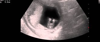
What does a baby look like at 37 weeks of pregnancy? From the ultrasound image you can see the following parameters:
- The size of the fruit is 48-50 cm.
- Child weight – up to 3 kg.
- The hairs on the head are up to 2-4 cm long.
- The skin folds have almost smoothed out.
- Individual facial features have been formed.
- The head and tummy have an equal circumference.
What happens to the baby when an ultrasound is performed at 37 weeks of pregnancy? He can sleep while sucking his thumb, he can grab the umbilical cord or stroke his tummy. His head is already lowering towards the pelvis. By the way, determining the sex of the child from an ultrasound photo at this stage can be difficult: he no longer moves due to lack of space, and the genitals are usually not visible for viewing.
Fetal development at the thirty-seventh week of pregnancy
Thirty-seven weeks of pregnancy corresponds to the 9th obstetric month. From this moment on, the birth is not considered premature. By this time, the child, as a rule, is already ready for life without the mother’s body. The child can already do the following:
- can accept, digest and assimilate incoming food;
- suck on your own;
- inhale and exhale independently;
- maintain body temperature at the same level.
But, despite a certain level of fetal maturity, organs and systems continue their further development.
In the stomach, the processes of absorption of necessary substances begin, peristaltic movements of the intestines are activated, and original feces - meconium - are formed.
By the 37th week of the child’s development in the womb, intensive growth of the nervous system and connections between neurons of the brain continues, coordinated movements appear, he holds on to the umbilical cord, touches himself, sucks his finger. The circumference of the head of the skull and abdomen are compared.
Subcutaneous fat accumulates daily, which smooths out skin folds. The baby's weight by this time can reach 3500 grams. It becomes possible to determine individual facial contours. The bones of the skull are still soft and mobile due to the presence of fontanelles, which facilitates the birth process. The cartilage of the nose and ears thickens. There is already hair, nails, and an individual papillary pattern on the skin. The fluff and birth lubricant practically disappear from the skin.
Lung tissue continues to produce surfactant, a surfactant that prevents the pulmonary alveoli from sticking together. Thanks to the alveoli and surfactant straightened after the first breath, the child will be able to breathe independently in the future. In the liver, there is an intensive accumulation of iron and trace elements necessary for the production of red blood cells.
The adrenal glands enlarge significantly and begin to produce hormones that adapt the newborn to extrauterine life. Already thanks to this degree of maturity of the systems and organs of the fetus, it is more prepared for childbirth than before.
What are the indications for ultrasound at this stage?
Usually the third and last scheduled ultrasound is performed at 30-32 weeks. Most women have already completed it, and the expected due date has been adjusted. But for a number of reasons, another examination will be required immediately before birth. Most often it is recommended to pregnant women who have not had their last scheduled ultrasound. Other possible indications for this diagnosis:
- Low weight of the child and other developmental abnormalities in order to control the quality of treatment and diet.
- Twins to assess the position of babies before birth.
- Pelvic, transverse position of the fetus to determine delivery tactics.
- The need to correlate the size of the pelvis with the weight and growth of the fetus.
- Low water or, on the contrary, a large amount of amniotic fluid.
- Suspicion of hypoxia or umbilical cord entanglement based on other studies.
- Placenta previa is dangerous for the baby.
Ultrasounds are also performed at this stage for mothers who are planning to have a caesarean section. According to statistics, most indications for ultrasound examination before childbirth are related to studying the position of the baby. Not all babies lie head down - some are in a breech position or even positioned transversely. The first version of presentation is not an unambiguous indication for a cesarean section, the second is, although sometimes the baby literally lies down before birth as intended by nature.
Objectives
The use of an ultrasound method for diagnosing the fetus at the thirty-seventh week is necessary to determine its degree of maturity and assess the compliance of the child’s physiological development with all relevant normative values, as well as determine the approximate date of birth. An ultrasound diagnostic doctor provides invaluable assistance in the management of a pregnant woman and answers a number of the following questions:
- fetal heart rate and its compliance with the norm;
- location of the baby in the uterine cavity;
- degree of maturity of the placenta;
- the state and level of maturity of the child in the womb;
- the total amount and volume of amniotic fluid, its condition;
- cervical condition;
- correspondence between the size of the pelvis and the size of the child;
- the location of the umbilical cord and the presence of its entanglement around the fetal neck.
When ultrasound is supplemented with color Doppler mapping, it becomes possible to visualize blood flow in the vessels. This determines the blood flow in the arteries of the umbilical cord, the vessels of the uterus and the ratio of utero-placental blood flow.
Ultrasound of the fetus at 37 weeks - how it is done
Usually no preparation is required for examination of a pregnant woman. There are no special dietary requirements before an ultrasound; of course, there is no need to fast either. The only point: if you are prone to increased gas formation, you can take a drug that reduces flatulence (for example, Espumisan) a couple of hours before the procedure.
You should also not drink water while filling your bladder before the examination. The amount of amniotic fluid at this stage is large, and they provide an excellent opportunity to thoroughly study the fetus and its development, as well as examine the placenta, uterus, and cervix.
Ultrasound at the specified period is performed only in a transabdominal manner. You need to lie on your back and relax. A cushion is often placed under the right side, especially when the inferior vena cava is compressed by the uterus. After applying a special gel to the skin, a sensor will be moved across it - from the very top of the abdomen to the pubic joint. There will be no unpleasant sensations.
The price of an ultrasound at 37 weeks is practically the same as the cost of a routine examination five weeks earlier. Depending on the type of clinic, it will range from 1000-1500 rubles to 3000 rubles and more.
The need for ultrasound and CTG
The main purpose of an ultrasound at 37 weeks is to diagnose the full maturity of the fetus. Normally, at this stage the baby is fully formed and ready to be born. A scheduled third ultrasound usually occurs at 30–32 weeks. Late examination is possible only if there are certain indications.
The main goals of ultrasound and CTG at the 37th week are presented in the table.
| Purposes of ultrasound | First of all, the fact of fetal maturity and readiness for independent life support is established. The child already has a certain coordination of movements, which can be tracked thanks to ultrasound. This is especially clearly visible during 3D and 4D examinations. With an ultrasound, a woman can see the baby grasping the umbilical cord with his hands. This indicates the formation of the grasping reflex. During the examination, you can notice how the baby moves his arms and explores his body. If amniotic fluid leaks, an ultrasound can diagnose slight oligohydramnios. It is important to make sure that this factor is not caused by pathological processes. During the examination, the mother may see the child hiccup. At 37–38 weeks, the doctor diagnoses that the vellus hair has already fallen off. The main objectives of the examination include: • determining the presentation of the child; • determination of heart rate; • establishing the baby’s maturity; • determination of the condition and amount of amniotic fluid; • determination of placenta maturity. If desired, you can take photos and videos of the study. In addition, an ultrasound can be used to see whether the umbilical cord is wrapped around the neck. The fetometry of the child is measured. |
| Goals of CTG | CTG at the 37th week is necessary to determine: • heart rate; • heart rate variability; • acceleration; • uterine activity; • deceleration. The study demonstrates the functionality of the fetus. The examination shows whether the child has formed correctly. CTG allows us to exclude some serious abnormalities, such as, for example, hypoxia. If necessary, a woman can be referred for emergency childbirth. Based on the survey results, a certain score is assigned that will characterize the current condition. |
In the last weeks, ultrasound and CTG are prescribed only when indicated. During a normal pregnancy, there is no need for procedures.
Read also: cardiotocography of pregnant women.
Ultrasound at the 37th week allows you to assess the volume and condition of the amniotic fluid


