It is a well-known fact that in the initial stages any disease can be cured easier and faster, so you should not refuse regular preventive examinations. This is especially true for the female half of humanity - most pathological processes of the mammary glands occur without pronounced symptoms, in no way stimulating a visit to a specialist.
Thanks to breast ultrasound, you can quickly and painlessly diagnose many diseases in their initial stages. This allows you to prescribe appropriate therapy on time and save the woman from serious complications and even premature death. In addition to prevention and detection of pathologies, ultrasound examination of the mammary glands is indispensable for the postoperative and recovery period.
Indications for diagnostics
You need to go for an ultrasound examination of breast tissue if you have the following indications:
- compaction;
- soreness;
- change in the shape of the nipple or mammary gland;
- discharge;
- roughening of the skin or loss of mobility;
- injury;
- polycystic ovary syndrome;
- pregnancy planning;
- enlargement of the mammary gland and regional lymph nodes;
- hormonal disorders;
- breast growth in men (gynecomastia);
- clarification of the diagnosis after mammography.
Ultrasound is also prescribed after operations, installation of implants, and in the presence of pathologies in the past. In this case, the examination is routine and is done within the time frame established by the attending physician.
If women have no complaints, a routine examination is done once every three years. After 30 years, the procedure is performed annually.
At what age is it permissible to do an ultrasound of the mammary glands, depends on the presence of alarming symptoms in the patient and the recommendations of the attending physician.
When an ultrasound is prescribed and what it helps to determine is described in the video below. Taken from the channel “Clinic of Aesthetic Gynecology”.
Decoding the testimony
During the examination, the ultrasound specialist examines all corners of both the right and left breasts of the woman.
After completing the ultrasound examination, the patient is given a protocol with the results in a few minutes:
- It is considered normal if the thickness of the glandular tissue is in the range of up to 14 mm . If the patient’s age is 40 years or older, then this indicator has a normal thickness of up to 20 mm.
- The specialist also examines the milk flows, and the normal condition that determines the norm is their visibility without deformations, as well as the absence of expansion in the areolar segment.
Deviations from the norm may indicate the beginning of the development of breast disease.
When to do an ultrasound
The period of the procedure is influenced by the patient’s age, the purpose of the examination, and the use of certain medications. To make an accurate diagnosis, an examination must be done in the first half of the menstrual cycle. When it is best to perform an ultrasound of the mammary glands in each specific case, the attending physician will tell you.
What day of the cycle
A gynecologist or mammologist refers the patient to the procedure from the 5th to the 10th day of the menstrual cycle. It is not advisable to undergo examination a few days before the onset of menstruation. At this time, changes occur in the hormonal background that can significantly affect the diagnostic result.
During menopause or taking hormonal medications, the examination is scheduled on any convenient day.
An inconsistent menstrual cycle makes its own adjustments. If there is a tendency to take a long break - more than 35 days, an ultrasound scan is prescribed on the 10th day after the start of menstruation. When the cycle is 21 days, the examination is done on the first day or the first 5. The exact date in such cases is selected by the gynecologist.
Pregnant and nursing mothers
When breastfeeding, an ultrasound is done to identify the cause of milk stagnation and to understand the overall picture of the pathology. An ultrasound examination is also recommended upon the onset of lactation during the prenatal period.
Mastitis often occurs during breastfeeding, which can only be detected using ultrasound.
Women during menopause
During menopause, ultrasound is prescribed at least once a year. The procedure can be done in conjunction with a mammogram or separately. If the mammologist has not prescribed a mammography examination, ultrasound is done twice a year.
How often can the study be carried out?
When frequent monitoring of the condition of the glands is required, for example on different days of the same cycle, there is no need to worry. The procedure will not cause any harm to the body. Ultrasound scanning should be done as often as necessary in a particular situation.
Which is better: mammography or ultrasound?
It is very difficult to say unequivocally which of these methods is better to choose. Generally, women under 40 years of age are recommended to undergo an ultrasound scan, and after 40 years of age, a mammogram is recommended.
Advantages of two diagnostic methods:
- The main advantage of ultrasound over mammography is that ultrasound shows the most accurate picture of the disease. It can detect even small tumors. Mammography shows neoplasms in the mammary glands, but is unable to distinguish whether they are benign or malignant.
- The advantage of mammography is its price; compared to ultrasound, it is several times smaller and more affordable . An ultrasound scan costs 2,000 rubles or more on average.
The world is full of diseases that are unexpected for us and we should never neglect any specialists in their field.
A timely ultrasound examination is a guarantee of maintaining your health and a warning about an upcoming disease that can be prevented in a timely manner. Having prescribed the appropriate and correct treatment for you. Don't skimp on your health, always choose the best.
Research methodology
Ultrasound of the mammary glands is done in the supine and lateral position. The doctor will tell you when to change your position. The patient is required to undress to the waist and relax.
Operating principle of an ultrasound machine
An ultrasound machine consists of a sensor and a monitor. Ultrasound is sent through the sensor to the organ being examined. It “bounces” off tissues, creating a picture on the monitor like an echo.
How is it performed and how long does the procedure last?
The ultrasound procedure is carried out as follows:
- The doctor applies a gel to the area being examined. Often, an ultrasound of the mammary glands involves examining the nearest lymph nodes.
- The doctor moves a special sensor from the patient's nipple to the side, returning and repeating for each area of the breast. It takes 5-10 minutes. The examination takes place in several positions for each gland.
- The monitor displays the structure of the mammary gland - tissue and ducts. If it is necessary to take a picture, the doctor records the image. On the echogram, the found formation can be highlighted with arrows or lines. The digital coordinates of the location of the suspicious area are indicated on the side.
- At the end of the procedure, an ultrasound examination protocol is drawn up.
How is diagnosis carried out?
- The patient is in a horizontal position on her back, with her arms thrown back behind her head.
- The doctor begins to treat the area of study with the prepared gel in order to improve contact of the sensor with the skin.
- Pressing the sensor tightly to the chest, he continues the study, moving the sensor along the mammary gland.
- Ultrasound, thanks to the penetration of its waves at different angles, displays a complete picture on a computer monitor, where a specialist already assesses the condition of the patient’s mammary glands.
- The procedure takes about 30 minutes.
- The examination itself is completely painless; discomfort may be present from pressing the sensor on the chest.
- If you are in an uncomfortable position, you can inform the doctor about this, he will show you the most comfortable position and change it without compromising the examination.
Interpretation of results
The resulting images are usually called an echogram. It will show the exact location and size of the formation if one is detected. The obtained ultrasound results and their interpretation are given to the patient or pasted into his card. This form is called a survey protocol.
What indicators are reflected in the ultrasound protocol
The following information is entered into the form:
- description of the structure of the predominant tissues, their density and echogenicity;
- the condition of the lymph nodes and the size of the ducts;
- information about the presence of pathology - location, size and description with photo.
Doctor's report
The doctor’s conclusion always includes an analysis of the factors in their totality:
- ultrasound diagnostic data;
- gender and age of the patient;
- day of the menstrual cycle;
- taking hormonal contraceptives or other hormone-containing drugs;
- patient complaints;
- presence of pathologies in the past.
After analyzing all available information, the doctor makes a diagnosis.
No pathology detected
The normal interpretation of breast ultrasound means that the ducts are not dilated. The tissue structure is defined as an echogenic zone 2 mm thick. Thickening is observed closer to the nipple area. Adipose tissue with low-echoic ellipsoidal lobules predominates. There should be no formations or deformations of the lobules or ducts inside the breast. An important point is the correspondence of the structure and condition of the glands to the day of the menstrual cycle in women and the age of the patient.
Mastitis
Mastitis is an inflammation caused by stagnation of milk (lactostasis). Therefore, it is defined as swelling inside the mammary gland. Swelling is noticeable on the surface of the chest. Mastitis is accompanied by an increase in temperature.
Signs of a benign tumor
Benign breast formations:
- Fibroadenomatosis. Fibroadenomatosis, or mastopathy, can be of several types - diffuse, nodular, fibrous. Small uniform compactions inside the gland should be defined as diffuse mastopathy. If the location of tiny areas is limited to one place, we are talking about nodular mastopathy. Dense small areas in the connective tissue - fibrous mastopathy. It is accompanied by the release of transparent discharge from the nipples, premenstrual pain and engorgement of the mammary glands.
- Fibroadenoma. This is a benign tumor, one of the forms of nodular mastopathy. The leaf-shaped form of fibroadenoma is located inside the ducts.
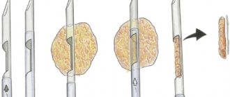
- Cyst. These are cavities with fluid that can occur alone or accompany a fibroadenoma. It may be palpable or go unnoticed. These cavities are formed in places where the ducts grow. This happens due to hormonal imbalances. Painful sensations occur when it grows and resemble a burning sensation. A sample can be obtained by performing a biopsy. This will help you learn more about the pathology.
- Lipoma. It is a wen - a voluminous growth of adipose tissue.
- Adenosis of the mammary glands. Pathology is the growth of glandular cells. Precancerous form of mastopathy.
Signs of a malignant tumor
If an ultrasound shows how a lump grows into the connective tissue, causing deformation of the neighboring area, and does not have clear boundaries, this lump should be defined as cancer. In this case, a puncture is performed.
The degree can be determined by assessing the accompanying signs:
- from the lymph nodes;
- appearance of one and/or both mammary glands;
- size and location of the formation.
Ultrasound examination of the breast
Ultrasound (sonography) of the mammary glands and its lymph nodes is scanning of breast tissue with ultrasound at a frequency of up to 19 MHz (megahertz). During the examination, the sensor is applied to the skin. The echo signal passes through the liquid unhindered and is reflected from the ribs and hard tumors. Ultrasound waves are absorbed differently by healthy and altered glandular tissue.
The echogenicity of the structures appears on the sonographer’s monitor in the form of a black and white picture. The denser the fabric, the lighter the area. The accumulation of liquid is colored black. Inhomogeneous (heterogeneous) lesions contain two colors, shades of gray.
Types of ultrasound for examining the mammary glands:
| Types of sonography | Features of the method |
| Doppler ultrasound, sonography with color Doppler mapping | In Doppler ultrasound, standard ultrasound is combined with Doppler: it allows you to assess the condition of the mammary glands and blood vessels, the nature of the tumor |
| Ultrasonic elastometry | The sonoelastography method helps to identify the boundaries of benign or malignant formations and perform a targeted biopsy. On the monitor, hard areas are highlighted in blue, healthy tissue is colored green, and changed tissue is colored red, orange, yellow |
| Duplex (UZDS) | Combines Doppler and AB ultrasound mode. The image on the monitor is two-dimensional |
| Triplex (UZTS) | This type of ultrasound combines the capabilities of duplex scanning and color mapping (CDC). The image is 3-dimensional, allows you to see differences in the direction and speed of blood flow |
There are portable models of sonographs: a doctor can be called to your home. This medical service is inexpensive and is useful for examining the mammary glands of people with disabilities.
Ultrasound diagnostics (ultrasound) of the breast is done in the late follicular phase of the ovary, and in emergency cases - on any day of the menstrual cycle. Sonography of the mammary glands is performed for the purpose of preventive research, determining the type of pathology, and monitoring the effectiveness of treatment.
Ultrasound is prescribed before medical or plastic surgery. In the first case, the severity and localization of changes are clarified; in the second, they are used more often for the selection of female breast implants. It is mandatory to evaluate the mammary glands using an ultrasound at the stage of preparation for IVF.
To watch a video of how ultrasound diagnostics are performed before mammoplasty:
Features of the procedure for children
Children (boys, girls) have the same diseases as adult women, plus the likelihood of birth defects and chromosomal disorders. A child can have a breast ultrasound at any age, including the neonatal period. There are no differences between the preparation and procedure for performing the procedure for children and adults.
Which doctor should I contact to prescribe an ultrasound, where is the best place to do it and how much does it cost?
An ultrasound of the mammary glands is prescribed by a mammologist, gynecologist or therapist, but you can contact specialists without a referral. Especially when it comes to a private clinic. In public hospitals, upon referral, the examination will be done free of charge. The cost of breast ultrasound in private clinics varies depending on the city and the characteristics of the examination. If screening includes examination of lymph nodes, it will be more expensive. It is possible to use a Doppler sensor to study blood vessels, which will also affect the cost.
The approximate cost of ultrasound screening in private institutions in Russia is:
- 450-1500 rubles - standard procedure;
- 950-2500 rubles - ultrasound of the mammary glands and lymph nodes;
- 1800-3500 rubles - examination of the mammary glands, lymph nodes and blood vessels using Doppler.
Breast ultrasound during menopause
With menopause, ultrasound examinations should be performed more often due to the fact that many women, during the period of absence of menstruation, take hormonal drugs to smooth out the symptoms of menopause, which can provoke the growth of malignant cells. With all this, you can undergo the study at absolutely any time, because the state of the organs against the general hormonal background is quite stable. It is worth mentioning that doctors recommend mammography for women after a certain age for a clearer and more detailed examination. It is not entirely correct to separate ultrasound and mammograms from each other when deciding which is the best choice. After all, it is in the combination of both methods that one can obtain an unambiguous answer in making a diagnosis. Therefore, both methods complement each other rather than exclude each other.
Photo gallery
Structure of the mammary gland
Absence of breast pathologies on ultrasound image
In the photo - breast cyst
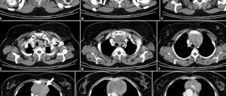
This is what cancer looks like on an ultrasound
Who is the procedure indicated for?
The main indications for doing an ultrasound are:
- Painful sensations.
- Doubtful lumps and neoplasms identified by palpation.
- Monitoring the condition after implantation.
- Age from 30 years.
- Previously diagnosed pathology of the mammary gland.
In addition to the above, the need for examination is determined by the following factors:
- Coarsening of the skin.
- The presence of secretion of a dubious nature.
- Obvious breast or nipple deformity.
It is important to remember that pathological formations can occur not only in women, but also in children. About 40-60% of men, depending on age, are also susceptible to gynecomastia. When the volume of glandular tissue increases, a procedure for examining the mammary glands in men is necessary.
Video
Below is a video about how a breast ultrasound is performed and what can be revealed during the examination. Taken from the GrudExpert channel.
Do you have any questions? Specialists and readers of the HROMOSOMA website will help you ask a question
Was this article helpful?
Thank you for your opinion!
The article was useful. Please share the information with your friends.
Yes (100.00%)
No
X
Please write what is wrong and leave recommendations on the article
Cancel reply
Rate the benefit of the article: Rate the author ( 2 votes, average: 5.00 out of 5)
Discuss the article:
Ultrasound of the mammary glands during menstruation
The timing of a breast examination depends largely on the current situation. Sometimes diagnosis needs to be carried out as soon as possible, without waiting for favorable days in a woman’s monthly cycle. As practice has shown, it is advisable to immediately perform an ultrasound examination of the mammary glands during menstruation in the following cases:
- if there is severe swelling or areas of hyperemia on the skin of the mammary glands;
- presumptive formation of a cyst in order to study its contents, whether it contains fluid or a cyst with a dense, homogeneous filling;
- in order to monitor the dynamics of the increase or decrease of the cyst;
- determination of the nature and nature of papillomas located on the chest;
- if the mammogram results showed an atypical condition of the mammary gland, there is a need to determine the nature of the detected neoplasm (cyst or tumor).
Also, the procedure for examining the mammary glands during menstruation using ultrasound is often done in atypical situations that exclude changes directly in them, namely:
- after surgery with the installation of breast implants;
- suspicion of the development of mastitis after childbirth;
- when choosing a hormonal contraceptive;
- after trauma to the chest in the chest location;
- in the event of gynecological disorders caused by dysfunction in the ovaries.
It is not advisable to conduct a breast examination to detect a tumor in the second phase of the menstrual cycle, since at this time the ducts in it are dilated and do not allow obtaining true results. Therefore, as a rule, if there is a suspicion of existing tumors in the mammary gland, whether they are malignant or benign, an ultrasound is performed at the very beginning of the menstrual cycle, preferably on the first day of menstruation.
Elastography method and its advantages
Separately, ultrasound with elastography in mammology should be considered. The tissues of the human body have rigidity (elasticity). Healthy areas and tissues of benign neoplasms are more elastic than cancer cells (they have a stiffness index 28 times higher than normal). Therefore, the level of elasticity can determine the presence of oncology in the body, even at an early stage of the disease. Let's look at how ultrasound of the mammary glands with elastography is done. A special sensor generates a beam that allows you to evaluate tissue stiffness. The information received is processed in a special program, on the basis of which a conclusion is drawn. The organ being analyzed is projected on the screen; healthy and diseased cells are highlighted in different colors.
The accuracy of detecting cancer cells using ultrasound elastography is 95%.
Elastography allows you to determine the smallest neoplasms that are not noticeable upon palpation. Thanks to this procedure, you can completely eliminate the need for a biopsy or more accurately select the coordinates for tissue sampling.
Interpretation of ultrasound of the mammary glands is carried out immediately after examining the patient. In this case, the quality of the device and the experience of the specialist play a significant role. When choosing where it is better to have an ultrasound of the mammary glands, you should give preference to a clinic with modern equipment.