Cardiological diseases are multifaceted in nature. Conventionally, they can be divided into functional and structural (organic).
- For the former, the development of changes in work is typical, without visible deviations in the anatomy of the organ. This is the most common option and occurs in the initial stages of a large number of diseases.
- For the latter, a typical feature is the presence of both structural changes and functional disorders.
Ultrasound of the heart shows deviations in the anatomical structure of the organ and partly its functional features, and is used as the main instrumental method for examining patients and suspected cardiac patients.
The question of whether it is worth doing the study usually does not arise - the ultrasound technique is considered mandatory in all cases, including a preventive examination to exclude organic disorders of the myocardium and other structures.
Ultrasound of the heart, also known as Echo CG, is a universal instrumental way of studying the organ and local circulatory system, despite its “routineness” it provides a lot of information.
What diseases does ultrasound detect?
What disorders can be detected based on the results of the diagnostics? An ultrasound of the heart will show a group of abnormalities:
- Vices. Mostly congenital. Caused by hereditary causes or previous diseases. Up to a heart attack and other changes. Based on the results, doctors can prepare for treatment and work out treatment tactics.
- Thrombosis of different localization. Blockage of blood vessels with blood clots. Adequate medical correction requires an assessment of the position of the abnormal object, its structure and size.
- Inflammatory processes of the heart: endo-, myocarditis and others can be diagnosed using ultrasound. They have a specific clinical picture and are clearly visible from the results of the study. On the other hand, in some cases verification through additional measures is required.
- Cardiac ischemia. Accompanied by chronic malnutrition and gas exchange in myocardial structures. It is not as rare as it might seem. Including young patients. It is considered a harbinger of emergency conditions. Therefore, ultrasound is used as a preventive examination measure.
- Heart attack. Acute cardiac nutritional disorder. Accompanied by the death of large volumes of muscle tissue. It is eventually replaced by scar structures. They differ in density from the normal functional lining, therefore the changes are clearly visible on the screen of an ultrasound machine. Read more about the symptoms of a pre-infarction condition here.
- Arrhythmias. Echocardiography is a method for identifying not only organic disorders. Thanks to the study of contraction frequency and other aspects, it is possible to determine the state of the myocardium, contractility and pumping abilities, and the evenness of the rhythm. Of course, not in the same way as an ECG machine does, but it is quite possible to obtain basic information. Usually both events: ultrasound and electrocardiography are carried out in parallel. One almost cannot exist without the other.
- Cardiomyopathies. A wide group of diseases for which the development of changes in the muscle layer is typical. Usually growth, transformation of structure, structure. There can be many diagnosed variants. The interpretation is carried out by a cardiologist and diagnostic specialist on site.
- Arterial hypertension. Secondary signs of the pathological process are noticeable even according to ultrasound results. Basically, there is hypertrophy of individual chambers of the organ. The left ventricle suffers most often, since it bears the main burden of pumping blood under high pressure conditions. This is an informative indicator.
- Tumors. Benign (for example myxoma) or malignant. Unfortunately, we can only state the fact of the presence of neoplasia. But it’s impossible to say exactly what it is. A biopsy will be required and a tissue sample will be tested in a laboratory.
- Chronic heart failure. Variation of ischemic disease with a decrease in myocardial contractility.
- Pathologies of vessels of the local circulatory network. Aneurysms, other abnormalities. Also atherosclerotic changes with the deposition of cholesterol plaques on the walls of the arteries and their calcification.
There are other finds, but these are the key ones.
What is an echo kg (echocardiogram) of the heart?
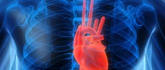
Echocardioscopy, echocardiography of the heart is an ultrasound method of examining the heart, allowing one to diagnose pathological changes in the structure of the heart (both congenital and acquired), valves, and vessels coming to and leaving the heart.
In addition, cardiac echography allows you to track indicators of the filling of the heart chambers—atria and ventricles with blood—during the full cardiac cycle. Echo CS is a diagnostic that needs to be carried out regularly.
Diseases that can be diagnosed using ECHO-CG:
- cardiomyopathy;
- narrowing of the lumen between the atria and ventricles of the heart, regulated by valves (in medical terminology - stenosis);
- deviation from the norm of the structure and density of the muscle and integumentary tissue of the aortic wall and aortic arch (aneurysm, hematoma);
- insufficiency of nutrition of the left or right ventricle or atrium;
- defect of the septum between the left and right parts of the central circulatory organ;
- the presence of blood clots in the heart and its vessels;
- violation of the thickness of the heart muscle in different parts of the heart;
- inflammation of the pericardium (its increase, change in density, appearance of excess fluid in the pericardial cavity).
Echocardioscopy allows you to diagnose diseases at the initial stages of their development. Correct treatment prescribed by a cardiologist after a heart echogram allows one to count on a complete cure and recovery of the patient.
Indications for the study
There are many reasons for diagnosis. In general, we are talking about suspicions of a particular process.
- Pain in or behind the sternum. Most are of cardiac origin. But not always. The task of doctors is to assess the nature of the discomfort and draw conclusions.
- Shortness of breath for no apparent reason. Reduced tolerance to mechanical load. Tolerance usually falls as a result of poor blood circulation in the tissues of the muscular organ and low contractility. The development of insufficiency is clearly visible from the diagnostic results.
- Disorders of physical development in children. Slow or low weight gain, slow growth, stunting and general weakness. He speaks about pathological processes almost unambiguously. Mainly about birth defects.
- Professional sports. Just a high level of daily physical activity and exertion. Individuals in this group are at risk of developing cardiomegaly. Changes in the size of a muscular organ. Usually it becomes significantly larger than normal, but this does not make it work any better. On the contrary, changes require correction as soon as they are found.
- Heart murmurs. You won't be able to feel them subjectively. This is an objective sign, it is detected by the results of auscultation. Listening to the chest by a doctor.
- Feelings of improper functioning of a muscle organ. Interruptions. False stops, changes in rhythm on the go and other uncomfortable phenomena.
- Weakness. Asthenic processes. Inability to perform daily duties at work and at home.
- Paroxysms of loss of consciousness. Syncope is considered to be the result of a sharp decrease in blood circulation to the brain. This is usually a consequence of a disorder in the vertebral arteries, but not always. The heart may also be to blame. The question requires careful instrumental clarification.
- Chilliness in the limbs.
- Pale or bluish skin, cyanosis of the nasolabial triangle.
- Pregnancy. As part of gestation, the study is prescribed as a screening technique for disorders in the body of the expectant mother. Ultrasound of the heart is done for preventive reasons, so as not to miss possible disorders.
- Childhood.
- Preparation for surgical treatment. It is necessary to study the functional and structural features of the muscular organ, to decide on tactics, access, volumes and scales of measures. The above-mentioned techniques, including ECHO-CG and electrocardiography, are used for these purposes. Other, more detailed ones as needed.
- Study of the effectiveness of the therapy. Including medication.
Assessing the parameters of the heart, its structural and organic features allows us to draw conclusions about the presence or absence of diseases. Clarify the nature of the problems and prescribe the correct treatment.
Indications and contraindications
Echocardiography is indicated for patients with shortness of breath, rapid heartbeat, pain in the left side of the chest, swelling in the legs, as well as for patients with rheumatism and high blood pressure. All people with chronic lung diseases are referred for this study. Newborns are also required to be examined if a congenital heart defect is suspected.
There are no contraindications to this procedure, as it is an absolutely harmless method for diagnosing diseases. An ultrasound of the heart can be performed as many times and as often as required in each specific case.
Preparation
Special preparation for ultrasound examination of the heart is not required: just go to a medical facility at the designated time and provide access to the chest.
On the day of the study, it is forbidden to engage in physical activity, especially intense. It is also worth maintaining emotional peace so as not to artificially distort the results.
During the procedure, you must follow the recommendations and instructions of a diagnostic specialist. This will allow you to get accurate results. Upon completion, the patient can return to standard, familiar daily activities.
You need to take disposable towels or other fabric with you to remove the gel. It is also a good idea to take a diaper to ensure your own hygiene while on the couch.
Preparatory activities depending on the method of carrying out the procedure
In some cases, transesophageal cardiac scanning or stress echocardiography is prescribed. Both research methods require more thorough and specific preparation from the patient.
Preparation for examination of a cardiac echo through the esophagus consists of the following rules:
- the last meal should be no later than 7.5-8 hours before the procedure;
- You can drink water and take medications only 2 hours before the scan, no later;
- It is important on the eve of the examination to limit the intake of coffee, tea, eliminate alcohol, cigarettes and heavy physical activity.
Before transesophageal echocardiography, it is necessary to honestly answer all the ultrasound doctor’s questions: have there been any allergic reactions to any medications, are there any diseases of the esophagus, pharynx, or is swallowing impaired. If you have dentures, they should be removed before the procedure.
On average, the procedure lasts 2-3 hours, of which 25-30 minutes are allocated for the scanning itself, and the rest of the time is spent on preparation (administration of local or general anesthesia) and analysis of the results obtained. The cost of transesophageal echocardiography is 2500-8000 rubles.
Stress echocardiography consists of ultrasound scanning of the heart before and after special physical activity. The study may use a bicycle ergometer, a treadmill (like a treadmill), medications, or electrical stimuli, and its goal is to identify hidden cardiac pathology.
Features of preparation for the examination:
- 2 days before stress echocardiography, you should avoid taking medications (strictly after consulting your doctor);
- stop drinking caffeine-containing drinks, alcoholic beverages, and cigarettes;
- physical activity on the eve of the study is minimized;
- The last meal should not be later than 3 hours before the appointed time.
Doctors also recommend wearing comfortable clothes and shoes, taking a towel and water with you. The procedure can take from 20 to 40 minutes, the price of the service ranges from 2000-9000 rubles.
Progress of the study
The process is extremely simple for the patient.
- At the designated time, the person arrives in place and takes off his clothes in the office to provide access to the chest.
- Next, the specialist applies a special gel to the skin, which ensures better conductivity of ultrasonic waves. This is the standard start.
- Already on the couch, the patient should lie quietly. Usually in positions on the back, on the side. Postures may change during the diagnostic process. You must follow all instructions from the specialist. Holding your breath, accelerating it, and other options are possible. This is necessary for survey purposes.
The whole procedure takes about 15-20 minutes. Plus or minus. Depending on the findings, the experience of the medical staff and the complexity of the clinical case.
At the end of the event, the patient awaits the results. He receives a diagnostic protocol and a specialist’s opinion on the presence and nature of violations.
It is recommended that you contact your cardiologist or other doctor who referred you for this procedure with documentation.
Interpretation of ultrasound indicators
After the examination, the sonologist writes a conclusion.
In the conclusion that the patient receives in his hands, certain indicators are included that require decoding. When deciphering the indicators determines any discrepancies with the norm, then after the ultrasound examination you need to visit a cardiologist.
If you have a conclusion in hand, then an adult can decipher it yourself, knowing some medical terms. Below are the terms that are indicated under normal conditions in an adult. When deciphering, you will need to build on them, determining your norm or pathology.
- The normal value of the right atrium is 2.7–4.5 cm.
- Fractional ejection 55–60%.
- The normal thickness of the right atrium is 1.9–4.0 cm.
- Stroke volume from 60 to 100 ml.
- The systolic size should be between 3.1-4.3 cm.
- The normal size of the aorta should be 2.1–4.1 cm.
It should be noted that the test results are influenced by the age of the child or adult, gender, and health status. An accurate diagnosis can only be made by the attending physician using a series of tests.
Video about diagnostics
Results standards
Among them:
| Index | Normal values |
| Stroke volume. The amount of blood that the heart pumps per unit time. Demonstrates left ventricular function (LVF). | 60-110 ml on average. |
| Left ventricular volume at rest. That is, in diastole (end diastolic size, abbreviated as EDD). | 3.3-5.5 cm. |
| Identical sizes, but in systole, at the time of contraction (end systolic size, ESD). | 2.3-4.1 cm. |
| The area of the fully active, open mitral valve. | About 4 sq. cm. |
| Opening of the aortic valve. | From 1.5 cm and above. |
| Exudate, transudate. | Absent. |
| Ejection fraction. | From 50% to 85%. |
| Thickness of the posterior border of the LV (wall). | 0.8-1.2 cm. |
| Foreign objects. | No. |
| Mass of the LV muscle layer. | 130-147 grams. Depending on the gender of the patient. |
There are many other indicators. They are not always described in a single protocol. It all depends on the scale of the research conducted.
Also, usually the results are presented not in the form of a summary table, but in the form of a free conclusion. Interpretation of cardiac ultrasound is the scope of activity of a cardiology specialist.
You also need to keep in mind that approximate levels are named. In fact, deviations are possible in both directions. This does not always indicate pathology. Just as in not all cases, the absence of obvious changes guarantees the elimination of problems.
Normal ultrasound readings of the heart muscle are determined in the context of the situation, taking into account the individual characteristics of the individual patient.
Types of heart ultrasound and how they are done
Echocardioscopy of cardiac condition/function comes in three types.
- One-dimensional, or the so-called M-mode. The ultrasonic wave is sent along a single axis; as a result, the image transmitted to the monitor looks like a projection from above. Information about other parts of the studied area can be obtained by moving the sensor along the body.
- Two-dimensional. When performing this type of ultrasound, two views of the area being studied are obtained. The beams are sent at a frequency of approximately 30 times per second perpendicular to each other. A three-dimensional image is obtained demonstrating the order of movement of the analyzed structures.
- Doppler. The method is based on estimating the speed of an object based on changes in the speed of the reflected signal. When an ultrasound wave encounters moving red blood cells, it slows down or speeds up, which is called a Doppler shift. Blood flow velocity, turbulence, and ventricular filling characteristics can be measured.
Based on how echocardiography is performed, transthoracic, transesophageal, stress (regular and with load), and intravascular forms are distinguished. Let's take a closer look at them.
Transthoracic
Transthoracic echocardiography is the correct name for standard ultrasound of the heart. It involves studying the structure of the heart non-invasively through the chest. To undergo it, the patient undresses from the waist up and lies down on the couch, turning on his left side. This position brings the apex of the heart closer to the outside of the sternum, which reduces the distance the waves need to travel and improves visualization of the four chambers.
The ultrasound specialist applies a special gel to the surface of the skin that will come into contact with the sensor. Subsequently, he moves it along the study area, choosing positions that clearly demonstrate the target areas. The patient does not experience pain or discomfort. The duration of the procedure is 10-15 minutes.
Transesophageal
In situations where the transthoracic form of diagnosis is unavailable for some reason, a transesophageal, or transesophageal, echo is performed. Such reasons may be a thickened layer of subcutaneous fat, anatomical obstacles (muscles, joints, bones, etc.), a prosthetic valve that provokes acoustic interference. The sensor is attached to the end of the swallowing tube, which is inserted into the esophagus. The receiving device is located in close proximity to the left atrium, allowing detailed examination of the smallest structures of the heart. The duration of the invasive probe stay is about 12 minutes. Contraindications to transesophageal ultrasound are bleeding, varicose veins of the esophagus, inflammatory lung diseases and others.
Stress with stress test
This type of Echo-CG involves applying a load to the cardiovascular system, but not through physical exercise, but using medications - dobutamine or adenosine. This method is chosen in cases where an exercise bike or treadmill is not available, and there is a need to evaluate the functioning of the organ in stressful situations. It allows you to assess the body’s response to stress, its tolerance, the likelihood of ischemia, and the effectiveness of prescribed therapy.
Stress
This is another type of stress test, performed while the patient is playing sports (on any cardio machine). It is prescribed because ultrasound performed at rest is not able to detect hidden disorders of myocardial functionality.
During the procedure, the sonologist takes readings about the behavior of the heart muscle and the nature of blood flow. The load level is selected individually, taking into account the age and diagnosis of the patient. Session duration is approximately 45 minutes.
Intravascular
Intravascular echo is prescribed for cardiac catheterization. From the name it is clear that the microsensor is inserted into the cavity of the blood vessel. The choice of method is determined by the need to diagnose stenoses or occlusions.
How a heart ultrasound is performed for women
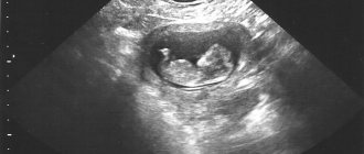
Echo-CG in women and men follows the same pattern. Since the study is absolutely safe, it is prescribed to pregnant women and young mothers during breastfeeding. The procedure is mandatory when registering for pregnancy and childbirth. A scan of the mother’s heart is mandatory (especially if there is a history of epilepsy, diabetes mellitus and other endocrine diseases), and if there are suspicions of abnormalities in the development of the unborn child, an ultrasound of the fetal heart is also recommended. The optimal period is 11-13 weeks. At this time, you can check the presence of cameras, measure the rhythm of heart contractions and other parameters.
Indications for additional studies
Among these are:
- Regular pain presumably of noncardiac origin. Shooting, strong stabbing sensations. Changes in the intensity of discomfort when standing up, physical activity. Usually these are just grounds for excluding cardiovascular disorders.
- The absence of any deviations according to the results of the ultrasound examination. Probably the essence of the violation lies in something else.
- Atypical symptoms. For example, hemoptysis, a feeling of fullness in the sternum, cough, shortness of breath, sputum production. The development of pulmonary problems is likely. Falsely assessing them as cardiac diseases.
- The presence in the blood of certain markers indicating possible tumors. There are also suspicions of neoplastic processes. Tomography, biopsy and histological analysis of tissue samples will be required.
The list is not complete, but these are the variations that doctors encounter most often in everyday practice.
Ultrasound of the heart is an indispensable and simple way to urgently diagnose the condition of a muscular organ; it allows you to see structural abnormalities in the myocardium and partially assess functional features. It is carried out in all cases without exception.
It is highly accessible, preparation for ultrasound examination of the heart is not required, there are no contraindications, which makes the method one of the main ones.
What not to do before the examination
The level of preparation for cardiac echography depends on the method of diagnosis itself. What not to do before a cardiac ultrasound:
- worry, be exposed to emotional stress, as this can lead to incorrect results;
- on the eve of the manipulation, visit the pool, gym, run;
- drink alcohol, large amounts of coffee, smoke before the procedure;
- even before standard echocardiography, it is not recommended to overeat and drink a lot of water;
- The use of any homeopathic medicines must be agreed upon with the doctor who referred the patient for examination (generalist/pediatrician/cardiologist).
In some cases, visualization of blood vessels and the heart may be difficult due to the large size of the mammary glands in women, severe curvature of the chest bones, and pronounced hair on the chest in men.
What is revealed by ultrasound?
During the procedure, diseases or their signs are identified. The most common include:
- rheumatism;
- myocarditis;
- pericarditis;
- hypertension;
- myocardial infarction or a previous condition;
- disturbance of heartbeat rhythms;
- disease of the aorta or arteries;
- heart defects of any etiology;
- hypotension;
- cardiomyopathy;
- intracardiac formations;
- ischemia;
- vegetative-vascular dystonia;
- heart failure.
The appearance of neoplasms and expansion of cavities can also be detected. Hypo- and hypertrophy is indicated by thinning or thickening of the heart walls.
