What is EchoCG
This simple examination provides information about the general condition of the heart muscle.
Due to complete safety, it is performed on both adults and children from the first days of life. In the process of short diagnostics, even the smallest violations are determined. As a result of this, the doctor can make the correct diagnosis. The method helps to identify deviations that do not bother the patient in any way, but are no less dangerous.
Purpose of the study
Due to the high level of versatility, there are many indications for the study. These include pain in the chest area of unknown etiology. Patients cannot always accurately assess their well-being.
Unpleasant sensations can be observed with pathologies of the gastrointestinal tract, intercostal neuralgia and other disorders in the body. The pain is easily relieved with medications, but it is necessary to assess the condition of the heart muscle.
Unstable blood pressure readings play a big role. Hypertension does not occur out of the blue. Primary provoking factors are formed in cardiac diseases. Secondary manifestations of the disease are explained by kidney pathologies and hormonal changes.
Thanks to the ultrasound method, it is possible to identify organic disorders that are accompanied by an abnormal rhythm. Due to the violation, doctors additionally prescribe an ECG.
Pronounced clinical signs of heart pathologies are direct indicators for diagnosis.
These include cyanosis of the nasolabial triangle and discoloration of the fingers. A similar study is carried out if the patient has atherosclerosis or cancer. Echocardiography is indicated if the presence of neoplasms is suspected. The development of the neoplastic process is said to be:
- dyspnea;
- general weakness;
- dark circles under the eyes;
- rhythm disturbance.
Ultrasound diagnostics can give a rough idea of the cause of the pathological condition. An accurate diagnosis can be made through MRI.
Echocardiography is used to determine complications and possible adverse reactions to the treatment process. It evaluates the effectiveness of therapeutic measures, including surgery.
During pregnancy, an ultrasound examination is prescribed to assess the general condition and functioning of the organ. Ultrasound makes it possible to identify the presence of contraindications for natural childbirth. Based on the results, the doctor determines the method of delivery.
Interpretation of ultrasound scan of the heart
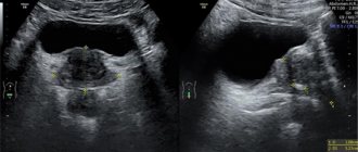
Regardless of heart disease, there are two main methods of instrumental diagnostics, which are quite informative and accessible to the population. An ECG allows you to assess the presence of pathologies in the conduction of impulses and create a general idea of the state of the organ. Using an ultrasound of the heart, you can evaluate its structure, the size of its components (walls, valves, septa), track the movement of blood through the departments and detect any space-occupying formations (tumors, abscesses, fibrinous deposits, and so on).
The quality of ultrasound depends not only on the technique, but also on the interpretation of the results. If the indicators are interpreted incorrectly, it is possible to make an incorrect diagnosis and choose inadequate treatment tactics. Despite the fact that if anyone knows the norms, they can determine the presence of abnormalities, only a specialist can predict a certain disease based on these data. Therefore, it is important that only a qualified doctor interprets the diagnostic results.
Indications for echocardiography in children
Echocardiography is prescribed for children if abnormal development is suspected and for diseases identified during the perinatal period. The main indications for prescribing ultrasound for children are the following cases:
- loss of consciousness for a short time;
- shortness of breath without signs of infectious and viral diseases;
- freezing extremities on a systematic basis;
- cyanosis;
- fatigue, tearfulness;
- some abnormalities in the development of the heart;
- presence of noise when listening with a phonendoscope.
Children during puberty are recommended to undergo a full examination. Diagnosis of abnormalities using echocardiography will give a complete assessment of the condition.
Study parameters and possible diagnoses
Using the study, several parameters are determined.
- General dimensions.
- The thickness of the walls of the organ and atria.
- Dynamics of rhythm.
Using an ultrasound image, a cardiologist can detect the presence of scars, tumors, and blood clots. Echocardiography provides complete information about the condition:
- hearts;
- adjacent tissues;
- valves located between the ventricle and the left atrium.
A Doppler examination allows the doctor to fully examine the condition of the vascular tissue, the level of blockage, the intensity and volume of blood flow.
The information obtained during the study allows us to make correct diagnoses and prescribe appropriate treatment. Ultrasound of the heart reveals the following pathologies:
- ischemic disease;
- pre-infarction condition;
- staging of hypotension and hypertension;
- structural abnormalities;
- cardiac decompensation;
- valve malfunction;
- abnormal heart rhythm;
- development of rheumatism;
- organ damage due to inflammatory processes;
- reduction of lumen in large vessels.
Pathological lesions in case of untimely diagnosis and lack of therapy lead to serious complications, including the death of the patient. Therefore, this procedure is the main method for identifying even minor violations.
Interpretation of ultrasound of the heart
7 minutes Author: Irina Bredikhina 83895
A modern method of hardware diagnostics - echocardiography or ultrasound of the heart, is based on the use of oscillations of high-frequency sound waves. Through ultrasound examination, a medical specialist determines the cause of functional failures in the organ, identifies changes in the anatomical structure and histological structure of tissues, and determines abnormalities in the vessels and valves of the heart.
The prerogative aspects of ultrasound diagnostics are:
- absence of damage to the skin and penetration into the patient’s body (non-invasive);
- harmlessness. Ultrasonic waves are safe for health;
- information content. Clear visualization of the heart allows you to accurately determine the pathology;
- no contraindications to the use of the method;
- the ability to observe dynamic processes;
- relatively low cost of research;
- insignificant time costs for the procedure.
Ultrasound of the heart is performed by a doctor from the radiology department based on the direction and recommendation of a cardiologist. If you wish, you can go through the procedure yourself.
Decoding results
Thanks to echocardiography, doctors are able to make a clear analysis of heart performance indicators for the entire cycle. This is a period consisting of one contraction and one relaxation. When the rhythm is not more than 75 beats per minute, the duration of one such period should be 0.8 seconds.
The analysis of indicators is done sequentially. Each individual unit is analyzed by the doctor in the examination report. An accurate conclusion is made by a cardiologist after a detailed analysis of the data obtained.
Parameters and standards for pediatric echocardiography
Analysis of the results of echocardiography of the organ and the functioning of the child’s circulatory system is calculated based on deviations from the norm.
Norms.
| Name | Indicators (mm) |
| max opening of AK flaps | 15-27 |
| LV/RV EDR | 37-56 / 10-18 |
| LV ESR | 26-27 |
| Thickness of the interventricular septum | 7-12 |
| Thickness of the posterior wall of the left ventricle of the heart | 7-11 |
| Anteroposterior LA size | 20-40 |
| LA/RA size in apical position | <40-48 / <38-46 |
| Thickness of the heart muscle on the right in diastole | <5 |
| Aircraft diameter | 15-28 |
| Bottom PV diameter | 12-25 |
An important point when deciphering the data obtained is the ratio of the size of the heart to the total surface area of the patient’s body.
When is the examination scheduled?
Ultrasound diagnostics is done if the patient suffers from cardiac pathologies, lung diseases, rheumatism, pulmonary regurgitation, or problems with pressure in the pulmonary artery. Other indications for echocardiography: lack of air, heart pain, dizziness, swelling of the legs.
The examination is performed when the patient is recovering from heart surgery, after a heart attack, with thrombophlebitis and varicose veins. Ultrasound examinations are also performed on infants if they have signs of a congenital defect: underweight, bluish skin tone, heart murmurs.
Ultrasound allows the doctor to identify disturbances in the functioning of an organ, determine its size, heart rate, blood circulation speed inside the organ and other important indicators. When interpreting the results of echocardiography, it is possible to analyze the condition of the heart vessels and pathological changes in them. Cardiac echocardiography is performed in conjunction with Doppler ultrasound to assess blood flow characteristics.
Ultrasound of the CS is a safe procedure that is prescribed to patients of any age. It is usually performed if there are changes after an ECG of the heart. There are no restrictions on such diagnostics. The only thing is that it can be difficult to examine the heart if a woman has large mammary glands, or the patient has a deformed chest, or has attacks of bronchial asthma.
Arrhythmia is one of the indications for research
Standards for adults
The conclusion is not made only on the basis of the results. The attending physician compares the obtained normal cardiac ultrasound readings with other information about the patient - laboratory tests, clinical manifestations. EchoCG should not be evaluated as an independent method for identifying pathologies.
Normal valve values
The aortic opening under normal conditions is 25-30 mm2. Reduced values indicate a narrowing of the vessel. An ultrasound scan should not reveal tumors or thrombosis in the valves.
Analysis of the functioning of the valves is carried out by comparing the normal sizes of the heart and deviations from them at 4 levels:
- I – 2–3;
- II – 3–6;
- II – 6–9;
- IV – over 9.
The given indicators make it clear how much the valve sags when the valves contract.
Normal values for the whole organ
The normal range of indicators and normal cardiac ejection fraction in adults are presented in the table:
| Name of parameters | Women | Men |
| Total LV mass | 95-141 | 135-142 |
| Left ventricular myocardial mass index is normal | 71-89 | 71-94 |
| End systolic size | 46-57 | 31-43 |
| LV during relaxation | 1.1 | |
| Release of blood during contraction | 50-55% | |
| The number of plasma ejected into the vessel | 60-1000 ml | |
| pancreas | From 0.75 to 1.25 cm/m2 | |
| RV wall | up to ½ cm | |
| End diastolic volume | from 20 ml to 1/10 l | |
| LP | 18,5–33 | |
| CDR pancreas | 0.95 cm–2.05 cm | |
The attending physician will tell you what the ESD (end-diastolic size) of each ventricle is and what indicators are a deviation when examining the diagnostic results.
Heart ultrasound indicators
Until recently, the ECG was considered the “gold standard” for diagnosing cardiovascular diseases. However, it allows you to evaluate only a small number of indicators, such as heart rate, heart rhythm, the position of the electrical axis of the heart, the presence of metabolic disorders in the myocardium, the presence of acute or chronic myocardial damage.
In the second half of the 20th century. With the beginning of the use of ultrasound in medicine, a procedure such as echocardiography (ultrasound of the heart) became available. Interpretation of the results of this study helps to assess both structural and functional changes in the heart, as well as assess the condition of the valves and blood vessels.
Interpretation of cardiac ultrasound is a complex process and involves the study of a number of indicators. And therefore, it should be carried out only by an experienced sonologist, since the correct diagnosis and prescription of the correct treatment by a cardiologist depend on the correct interpretation of the examination results.
Further in the article we will tell you how to decipher an ultrasound of the heart, what indicators can be assessed by echocardiography, what is the norm for ultrasound of the heart in adults and children, and also what deviations from the norm may indicate.
Advantages of the technique
Echocardiography has many advantages over other diagnostic methods.
The first is a painless procedure that does not cause any discomfort to the patient. The examination is carried out as a typical ultrasound. Before the examination, the patient does not require injections or oral administration of medications or other types of medical procedures. Also, the procedure is completely harmless even for newborn babies. Often performed on pregnant and elderly patients.
EchoCG is an accessible and simple examination method, since equipment for this procedure is available in every medical institution. The cost of cardiac ultrasound is much lower compared to MRI. The main feature of this type of survey is its information content.
Echocardiography determines the electrostatic frequency of the rhythm and structures of the heart. The diagnostic method is reliable in identifying abnormalities and making an accurate diagnosis.
How is CDR done?
To make the image as clear as possible, and also to avoid various types of interference, a special substance is applied to the sensor, which is intended for sound examination of the CDR. It prevents substances from getting between the patient and the transducer, thereby improving the performance of the ultrasound beams.
Study of the heart by CDR, based on the analysis of the structures of ultrasound signals reflected from it. A special sensor both emits and receives ultrasound pulses reflected from the structures of the heart, which are called echoes. With sectoral cardiography, the ultrasound beam is scanned in the form of a sector with an angle from 30 to 90 degrees.
A smaller sector angle provides higher quality display by increasing the clarity of the frame. For an ultrasound examination of the heart, you have to choose points where it is directly adjacent to the chest, that is, look for the so-called echo window. It should be noted that bones, cartilage and air prevent the passage of ultrasound.