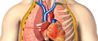
The inside of the human abdominal cavity is lined with a thin membrane called the peritoneum, which provides the secretion and absorption of a small amount of fluid for the better functioning of all organs. However, there are organs that this membrane does not affect: they are located behind the peritoneum. That is why the space limited in front by the peritoneum, and behind by the lumbar muscles and spine, is called retroperitoneal, or retroperitoneal. Its examination using ultrasound is often included in the standard protocol and is carried out together with ultrasound of the abdominal organs.
What is included in a retroperitoneal ultrasound?
In medicine, the peritoneum is the serous tissue that covers the walls of the abdomen from the inside with one sheet, and the internal organs with another. Both tissue segments are connected to each other: in men completely, in women partially.
Ultrasound of the retroperitoneal organs is performed on the internal parts of the body where the peritoneum is located. Typically, the study involves the lumbar area of the back. The organs of the retroperitoneal space include:
- kidneys;
- adrenal glands;
- aorta;
- ureters;
- the inferior vena cava, which runs along the line of the spine.
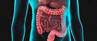
Some organs are partially covered by the peritoneum and enter the retroperitoneal cavity:
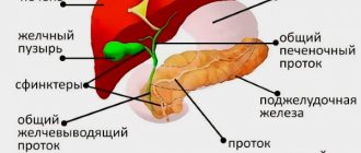
- duodenum;
- ascending, descending colon;
- pancreas.
Ultrasound of the retroperitoneal space not only shows the condition of the organs, but also evaluates the fatty tissue responsible for the supporting function. It transmits ultrasonic waves to the maximum. Fatty tissue tightly envelops the organs, which helps to accurately determine the diagnosis.
What is the essence of the procedure
MRI is a non-invasive diagnostic method. The survey is considered innovative. Internal organs can be visualized qualitatively.
The procedure is based on the action of magnetic fields. In response to them, organs and other structures send signals that are processed by special software. Subsequently, a detailed cross-sectional image is formed from the obtained data.
The examination does not generate ionizing radiation.
The operating principle of MRI is based on the signal sent by the nucleus of a hydrogen atom in a magnetic field. In this way, the entire body is scanned. Diagnosis is made due to the saturation of organs and tissues with hydrogen.
Modern medical equipment has a power of 1.5–3 Tesla. This helps to significantly reduce the duration of the procedure.
The MRI procedure is quite simple.
Indications and contraindications
The doctor interviews the person before prescribing an ultrasound scan of the retroperitoneal organs. Indications for examination are:
- Previous abdominal injuries.
- Kidney failure.
- Presence of stones in the ureters and kidneys.
- Disturbances in the functioning of the genitourinary system: pain when urinating, detection of blood and other impurities in the urine.
- Diabetes.
- Changes in body temperature without any particular reason or additional symptoms.
- Glomerulonephritis.
- Suspicions of the presence of malignant or benign neoplasms.
- Hydronephrosis.
- An infectious disease in a chronic form.
- Pyelonephritis.
- Atherosclerosis of the abdominal aorta.
- Periodic pain in the pelvic area down to the lower back.
The advantage of ultrasound of the retroperitoneum (retroperitoneal space) over other methods is the small number of contraindications. A person is not prescribed a study if there is an extensive wound on the abdomen or the integrity of the skin over a large area is compromised. Ultrasound diagnostics is not performed for the formation of ulcers on the abdomen and other inflammatory processes.
The procedure has no age restrictions. The study is prescribed for both adults and young children.
In what cases should an MRI be performed?
Tomography is performed only if indicated. Diagnostics is recommended for developmental defects and abnormal changes in the organ. The procedure is required if an inflammatory process is suspected.
You must get a doctor's order for an MRI.
An examination is the only way to determine the type and stage of advanced cancer. Manipulation allows you to accurately determine the nature of the neoplasm.
The main indications are presented in the table.
| Indications | MRI is prescribed to patients with: • pain in the lumbar region; • hormonal imbalances; • cystic formations; • neoplasms of a benign or malignant nature; • nodular hyperplasia; • adrenal adenoma; • abscesses; • post-traumatic changes; • improper development of the kidneys. |
| What does it visualize? | Tomography helps to visualize: • infectious processes; • neoplasms at any stage of development; • various damages in the tissue structure of the organ; • inflammatory processes. |
Diagnostics helps to study the condition of soft tissues, bones, vascular system and organs.
During the examination, there is no need to introduce equipment into the structure of the body. The examination method is non-invasive.
MRI is used to examine the liver
MRI of the retroperitoneum shows the condition:
- liver;
- spleen;
- kidney;
- perirenal fiber;
- gallbladder;
- adrenal glands;
- ureters;
- stomach;
- intestines.
The existing pathology will be detected regardless of the stage of the disease. Diagnostics may also be recommended in order to ensure the effectiveness of the prescribed treatment.
How to prepare for research
Preparation for an ultrasound scan is always individual. This depends on the organ being examined. General requirements include the presence of a disposable diaper, which is needed as a bedding on the couch, and a napkin for wiping off gel residues after an ultrasound. But usually paid medical institutions provide diapers, shoe covers and other similar supplies.
The nuances of preparing for examination with an ultrasound sensor, depending on the organ or system being examined:
- Genitourinary system. No special preparation is required. Before diagnostics, reduce water consumption. If the requirement is ignored, the ultrasound results may be inaccurate: due to the active work of the kidneys, one of the pelvis expands. During the examination, this is interpreted as normal or pathological. For an ultrasound of the bladder, on the contrary, it will need to be filled.
- Adrenal glands. On ultrasound diagnostics, adrenal tissue is practically undetectable. Therefore, the doctor visually examines the area where the organs are located. This is enough to identify formations, if any. The procedure is carried out on an empty stomach: it is forbidden to eat, drink any liquid or water 8–9 hours before the test.
- Aorta with inferior vena cava. Before performing an ultrasound of the retroperitoneal space, food is excluded from these vessels, which leads to gas formation in the intestines. You cannot take medications: enterosorbents, enzyme medications and carminatives.
What are the advantages of MRI
MRI has a number of advantages that make the procedure universal and in demand. The main advantage is the absence of radiation exposure. Thanks to this, the manipulation has almost no contraindications, and it can be repeated many times.
During diagnostics, you can obtain images with a high level of detail. That is why MRI is often sufficient to establish a final diagnosis and further selection of therapeutic therapy. The manipulation is not accompanied by any painful sensations.
Magnetic tomography helps to detect a tumor and determine the nature of its formation. Manipulation allows you to monitor the effectiveness of the selected treatment. Unlike CT, MRI is prescribed for children. However, despite this, it is important to consult a doctor first.
Diagnosis will be 100% safe only after consultation with a doctor. It is highly discouraged to resort to research at your own discretion.
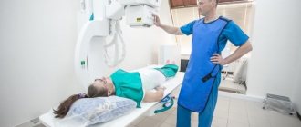
How is ultrasound done?
Ultrasound of the retroperitoneal space is practically no different from examination of other organs by this method. The person exposes his stomach and lower back and sits on the couch, having previously laid down a disposable sheet. The sonologist spreads the gel on the skin, which helps the sensor with built-in ultrasound move freely.
Ultrasound is performed in different positions: the sonologist may ask the person to turn on his side, stomach or back. But scans of the kidneys are often done in a standing position, since lying down cannot be determined whether they are prolapsed or enlarged. Prolapse or increase in the size of the kidneys is considered a pathology.
To study large vessels, Doppler ultrasound is used. The purpose of the program is to assess the state of blood flow. The additional procedure does not cause any discomfort in a person.
The signals from the ultrasound sensor transmit the reflection of waves from the organs to the scanner, where everything turns into a three-dimensional image on the screen of the ultrasound machine. If necessary, photographs and video recordings of diagnostics of the retroperitoneal space are taken.
Watch a detailed video about ultrasound of the BP and retroperitoneal space:
Progress of the study
Ultrasound can be performed using several methods, but they all differ little from each other. Actions during the procedure do not cause pain or other discomfort to the patient.
- Initially, he is asked to remove clothing from the entire anatomical area of the abdomen and lie down on a horizontal couch.
- The projection site of the kidneys or other retroperitoneal organs is covered with a gel that improves the contact between the skin and the sensor.
- The latter delivers an ultrasonic wave, which is reflected from structures of various densities and forms an ultrasound picture on the screen of special equipment.
- The shown figure is analyzed by a specialist doctor and gives a conclusion based on it.
Decoding the results
After the ultrasound, the sonologist gives the person a research protocol. It indicates the parameters of the kidneys: shape, size, position and condition of the parenchyma enveloping the tissue organ. Indicators of the adrenal glands, lymph nodes, and pancreas are also taken into account: size, location.
The protocol indicates the nature of echogenicity of each organ. It can be normal, increased or decreased. The last 2 indicators indicate the presence of diseases. When examining veins, the thickness of their walls, the speed of blood flow and the width of the lumen are determined.
Ultrasound of the retroperitoneal space accurately diagnoses:
- Kidney diseases. During the examination, the doctor first of all pays attention to the location of the paired organ. In normal condition, the left kidney is slightly higher than the right. When breathing deeply, a maximum displacement of 2 centimeters is allowed. With increased echogenicity, the development of neoplasms is diagnosed.
- Anomalies of the pancreas. When diagnosing, shape, size and structure are taken into account. Ultrasound diagnoses pancreatitis, cysts, inflammation, tumors of a malignant or benign nature.
- Disturbances in the blood flow of the aorta and inferior vena cava. It is possible to detect aneurysms, abdominal ischemic syndrome and vasorenal arterial hypertension.
- Adrenal diseases. Normally, the longitudinal size of the adrenal gland does not exceed 3 centimeters. If during diagnosis the organ is visually enlarged, this may indicate an anomaly. In this case, an additional study is prescribed - multislice tomography.
- Presence of stones in the ureters. Usually, the ureters are poorly visible on ultrasound of the retroperitoneum. Small stones and neoplasms are detected only during a minimally invasive research method. The essence of the method is to insert a miniature sensor upward through the lower urinary tract.
See what the retroperitoneal space looks like on the monitor:
The video shows an example of a formation in the kidney:
Normal indicators and the most common pathology
A qualitative study of the retroperitoneal space using ultrasound is impossible without determining the norm.
Kidneys
The shape of a normal kidney is oval or bean-shaped, the contour is clear and even, sometimes wavy. The longitudinal size should not exceed 12 cm and be less than 10 cm. However, the size of the kidneys depends on the constitutional characteristics of a person and the type of his activity, for example, professional athletes may have larger kidneys.
The echostructure should be homogeneous, the echogenicity is average or normal, that is, the kidney parenchyma is slightly darker than the liver on ultrasound. The center of the bud, on the contrary, looks white.
Diffuse kidney changes
There is a change in the echostructure and echogenicity of the parenchyma of one or both kidneys.
- Pyelonephritis. In an acute process, the kidney will be enlarged, it will become darker, which indicates inflammatory edema of its tissues. An expansion of the pelvis with cloudy contents may be observed. This process can spread to the fiber of the entire retroperitoneal space, which can lead to a deplorable condition. The chronic process does not have specific signs on ultrasound.
- Kidney failure. Acute failure will lead to an enlargement of the kidney, but its parenchyma will become much lighter. At the same time, the abdominal cavity system of the kidney also expands, that is, the calyces, pelvis, and ureter. In chronic renal failure, the kidney gradually becomes smaller in size, its parenchyma becomes thinner, and fibrous fibers appear in it. The result may be a shriveled kidney.
- Arterial hypertension. Kidney damage due to high blood pressure is similar to chronic renal failure, but at the initial stage of development it may be indicated by triangular retractions of the parenchyma - these are scars.
- Nephrosclerosis. This is a general concept that includes a decrease in the size of the kidney, thinning of its parenchyma, uneven contours and a decrease in basic functions (as indicated by other studies).
Focal pathology
The most common formations detected by ultrasound of the kidneys are cysts. They can be single or multiple, small and giant, round and irregular in shape. Small cysts need to be monitored, that is, examined once a year. Very large sizes - removed.
How much does the examination cost?
The cost of ultrasound diagnostics of the retroperitoneal space is determined by region. Price – 1000–2000 rubles. An additional procedure, Dopplerography of the retroperitoneal vessels, costs 1100–2200 rubles.
Ultrasound of the retroperitoneal space is considered the most informative diagnosis. No special preparation is required.
Have you undergone such an examination? Tell us about it in the comments. Share the article with your friends on social networks. Be healthy.
How is an MRI performed?
A classic tomograph is a large cylinder surrounded by a magnet. The patient is placed on the movable table of the apparatus. The couch is pushed inside the device capsule.
Open types of tomographs have also been created. It is recommended that people with a weight of 120 kg or more and severe claustrophobia undergo diagnostics in such a device. The duration of the examination is on average 20–40 minutes.
The examination is completely painless. The indicators obtained during the examination are transferred to a computer. There, the data is processed by special software and forms a slice-by-section detailed image.
Advantages and disadvantages of the MRI method
The MRI method has many advantages:
- the research time is relatively short (on average up to 30 - 40 minutes, less often - an hour), the results are ready almost immediately after it, a detailed description is prepared in no more than two days;
- contrasting, informative images obtained in different sections, the possibility of computer modeling of internal organs and vessels up to a three-dimensional model;
- To obtain images, it is not necessary to use X-rays, which are harmful and have an adverse effect on the body. The tomograph creates a magnetic field that is used to scan the human body and does not harm it, despite the fact that its magnitude exceeds the Earth’s own magnetic field by tens of times. To put it simply, an MRI is a big magnet;
- this is a non-invasive method, that is, there is no need to make incisions on the patient’s body and put him into a state of artificial sleep, which is harmful to the heart;
- MRI can be done in many public and private clinics;
- the widespread distribution of MRI machines leads to a reduction in the cost of research and a reduction in the waiting time for scanning in line;
- There are no restrictions on the number of scans performed on one patient, especially if surgical intervention and follow-up are required.
The MRI method also has a number of serious limitations. If the patient ignores these restrictions or deliberately hides at least one of the factors listed below from the doctor, the consequences can be dire, even fatal. Here are the main points to consider:
- Patients who have pacemakers, artificial valves, metal prostheses and bone rods are not allowed to scan. Like a magnet, the MRI machine will attract any metal object to itself, even if it has to be torn out of the human body;
- tattoos with a ferromagnetic component will leave a burn on the skin;
- you need to lie still for at least half an hour to get clear pictures;
- psychological discomfort from the belts on the couch, which slides into the tomograph, closed space and the noise from the operating device;
- children most often undergo an MRI under anesthesia in the presence of an anesthesiologist due to the fact that they cannot lie still;
- It is uncomfortable for pregnant women to lie on their back for a long time;
- the effectiveness of lung examination is reduced due to their movement during the patient’s breathing process;
- the room in which the scanning takes place must be protected from any interference;
- the size of the device itself sometimes becomes an obstacle: patients weighing more than 120 - 130 kg will not fit in the device;
- Only highly qualified specialists can work with MRI.
MRI should be performed with caution in patients with claustrophobia, those suffering from psychological disorders, people who cannot control themselves, and pregnant women, although pregnancy itself is not a contraindication. Often pregnant women are prescribed an MRI instead of an X-ray.
Pathology
Damage
Retroperitoneal hematomas most often occur with fractures of the pelvic bones and spine; in addition, they can be the result of damage to an organ located in the third section or in the abdominal cavity (kidney, pancreas, duodenum, stomach, etc.). In more rare cases, the source of retroperitoneal hematomas can be large vessels (aorta, inferior vena cava, renal vessels). Retroperitoneal hematomas can also occur in hemophilia.
Typically, retroperitoneal hematomas are formed as a result of direct trauma without or with damage to bones or internal organs. They can also be the result of a blow to the anterior abdominal wall, while injury to internal organs not only significantly worsens the condition of patients, but also complicates the diagnosis of hematomas.
The clinical picture of retroperitoneal hematomas depends primarily on the nature of damage to the bones, 3. organs, abdomen, as well as on the size of the hemorrhage. As studies by P. P. Kiselev, V. G. Tsuman and Yu. A. Muromsky have shown, up to 2 liters of blood or more can accumulate in the third section. Small hematomas may be asymptomatic. The symptomatology of extensive retroperitoneal hematomas is similar to the clinic of damage to internal organs, especially soon after injury, and is accompanied by shock (see), blood loss (see) and a picture of an acute abdomen (see). Shock to one degree or another almost always accompanies a retroperitoneal hematoma associated with damage to bones or internal organs. Most patients experience abdominal pain, often on the side of the injury. With massive hematomas, the Shchetkin-Blumberg symptom and flatulence are observed.
Hemorrhagic syndrome with small hematomas is not expressed. However, with significant hemorrhages, anemia is observed with a drop in blood pressure up to collapse (see).
Diagnosis of retroperitoneal hematomas is based primarily on anamnesis (the nature of the injury, hemorrhages in the past with hemophilia) and local manifestations - swelling, pain in the lumbar region or in certain parts of the abdominal cavity, rapidly developing flatulence. Symptoms of dysfunction of the urinary system are of great importance. A characteristic symptom is the forced position of the patient with the thigh adducted to the stomach on the side of the hematoma. With massive bleeding, there is a progressive decrease in hemoglobin and the number of red blood cells in the peripheral blood, as well as blood pressure.
Differential diagnosis should be carried out primarily with damage to the abdominal organs and urinary system.
A retroperitoneal hematoma on radiographs is manifested by blurred contours and displacement of the kidney, psoas muscle, bladder or retroperitoneal parts of the intestine.
Treatment of isolated retroperitoneal hematomas should be conservative. The set of measures includes measures to combat shock, blood loss, and intestinal paresis. If there is a suspicion of injury to the organs of the 3. p., they should be inspected using retroperitoneal access on the corresponding side.
In some cases, laparocentesis is performed and, as a last resort, if it is impossible to exclude damage to internal organs, laparotomy is performed. Moreover, a retroperitoneal hematoma cannot be emptied into the abdominal cavity due to the risk of infection. For isolated retroperitoneal hematomas, the prognosis is usually favorable. Infection of the hematoma leads to the development of an inflammatory-purulent process in the 3rd section.
Diseases
Inflammatory and purulent processes in 3. p. occur with direct injury (primary infection), metastatically and per continuitatem. The causative agent of infection can penetrate hematogenously from peripheral purulent foci and lymphogenously during inflammatory processes located in the pelvis, lower extremities, chest, intestines, etc. The most common causative agents of inflammation of cellulose 3. p. are staphylococcus, streptococcus and intestinal wand. About a quarter of patients have a mixed flora.
There are serous (catarrhal), purulent and putrefactive inflammatory and purulent processes in the retroperitoneal tissue.
According to the anatomical and topographic structure, inflammatory-purulent processes are divided into paranephritis (see), paracolitis (see) and inflammation of the retroperitoneal tissue itself. At the beginning of development, the inflammatory process is localized in any one section of the 3rd section. As the integrity of the fascial sheets is damaged, the process spreads to neighboring areas of the 3rd section and beyond, forming extensive purulent leaks.
Wedge, the picture of inflammatory-purulent processes in 3. p. is characterized by an increase in temperature, changes in the peripheral blood. In severe cases, significant disorders of the cardiovascular system, breathing, urination, etc. are observed.
The onset of the disease can be gradual, with a slow development of symptoms, or sudden, with a rise in temperature and severe intoxication. As the disease progresses, nausea, vomiting, flatulence, and symptoms of peritoneal irritation are observed. Often, inflammatory and purulent processes in the third area are accompanied by the development of exudative pleurisy, often on the affected side, which sometimes takes on a purulent character. In the blood there is pronounced leukocytosis, a band shift in the blood formula, and later accelerated ROE. Changes in urine are possible.
As the purulent process develops in the tissue, local symptoms appear more clearly: pain, muscle tension in the lumbar region, the formation of an infiltrate, changes in the contours of the lumbar region. Each anatomical localization of the inflammatory-purulent process in the third area is characterized by its own form and location of the infiltrate. When located in the retroperitoneal tissue itself, the infiltrate is located in the area of the outer part of the iliac fossa in the form of a compaction running obliquely from top to bottom and inwards. In lumbar localization, the infiltrate does not have sharply defined boundaries; a compaction of elastic consistency is determined above the wing of the ilium. For retroperitoneal localization of the abscess, a characteristic symptom is flexion contracture of the hip on the affected side.
When the inflammatory process spreads to the surrounding cellular spaces, local symptoms change accordingly, along with this, general reactions intensify. When an abscess breaks into the abdominal cavity, symptoms of local or general peritonitis appear (see). Retroperitoneal phlegmon can spread to the mediastinum, to the thoracic and abdominal organs, leading to the formation of secondary osteomyelitis, external intestinal fistulas, and inflammation of the perirectal tissue. There have been cases of swelling spreading to the gluteal region and even to the thigh.
Difficulties in diagnosis are determined by both the latent onset of the disease, the wide variety of wedges, manifestations, and the commonality of symptoms with other diseases.
Of great importance for diagnosis, along with the wedge, assessment of general and local manifestations, is rentgenol. study. If there are symptoms of purulent inflammation, a diagnostic puncture 3. p. is sometimes necessary.
Differential diagnosis of the inflammatory-purulent process in 3. p. is often difficult. If general reactions predominate, it is necessary to exclude infectious diseases. With more pronounced local reactions, the purulent-inflammatory process in the 3rd point should be differentiated from diseases of retroperitoneal organs, as well as abdominal organs (cholecystitis, appendicitis).
Treatment of inflammatory-purulent processes in 3. p. depends on the stage of the process. In the absence of signs of suppuration, anti-inflammatory therapy (broad-spectrum antibiotics, sulfonamides) and restorative treatment are indicated. Cellulitis and abscesses must be opened and drained. For this purpose, the best is the Pirogov incision above the wing of the ilium. Existing leaks are opened through additional incisions and counter-apertures. Untimely and inadequate treatment can lead to severe septic and thromboembolic complications.
The most common outcome of acute inflammatory diseases or injuries can be retroperitoneal fibrosis. Wedge, its picture is manifested by pain and disorders of the urinary system. Roentgenol, the study reveals narrowing of the ureter (usually at the level of the middle third), expansion of the renal pelvis and calyces, as well as delayed release of the contrast agent during excretory urography. Retroperitoneal fibrosis is differentiated from sarcoma 3. p., teratoid tumors, neuromas, lymphoblastomas, malignant tumors of the ureter and pancreas, as well as metastases.
What is MRI?
MRI (magnetic resonance imaging) is one of the most effective methods for non-invasive diagnosis of the condition of blood vessels and internal organs of a person. The American chemist Paul Lauterbur was the first to think about using a magnetic field to obtain images. He wrote an article about this in the journal Nature in 1973. There he explained in detail how, using the effect of nuclear magnetic resonance (NMR), one can obtain an image of a slice (projection in one direction) of the human body.
The MRI device was developed later through the joint work of Lauterbur and British physicist Peter Mansfield. For this medical breakthrough they were awarded the Nobel Prize in 2003.