The X-ray method of examining the kidneys using a contrast agent is the most accurate and informative diagnostic method known today. Thanks to its capabilities, it is possible to thoroughly study most pathologies of the urinary system.
Recently, several types of contrast-enhanced X-ray techniques have been developed, which allows the doctor to select the most suitable one, based on the patient’s symptoms. This approach helps the specialist obtain comprehensive information and prescribe adequate treatment.
What is research
An examination using radiation exposure to the human body helps to obtain an image of an organ, evaluate its size, location, structure, and see pathological formations, for example, tumors.
If a contrast agent is used during an x-ray examination, the patency of the natural pathways to and from the organ can be assessed. The results obtained help to draw a conclusion about the functionality of the organ and violations of its activity.
The examination is carried out in specialized diagnostic centers or hospitals. The result of the study is given to the patient or forwarded to the attending physician if it was done in the same institution where the patient is observed. The results come the next day.
How is urography performed?
Before entering the X-ray room, the patient must take off his clothes, putting on a special robe and metal jewelry. Pictures are taken in a lying or standing position, as determined by the doctor. The examination time depends on the method, the use of contrast agents and the characteristics of the kidneys. The procedure is completely painless, and the main thing that is required of the patient is not to move for several seconds while the image is being taken.
The result of an x-ray is one or a series of images of the examination area. The images are saved on a disk (or other storage medium), and based on the images, radiologists will draw up a conclusion. It describes in detail the structure and condition of the kidneys, bladder and ureters, detected pathologies and their features.
Types of X-ray examination
Medical practice uses several types of x-rays that are used to diagnose kidneys. The choice of technique remains with the doctor, who knows what results this or that procedure will show. This is taken into account when it is advisable to confirm a particular diagnosis.
Survey radiography
Among the simple types of examination is digital x-ray. This is a common type of procedure, it is performed quickly and is available in almost every medical institution.
The irradiation area is placed in front of an X-ray screen, and the rest of the body is protected by a lead apron. The procedure is painless.
A survey X-ray examination makes it possible to see the organ as a whole and determine deviations. The disadvantage of such an image is that not all pathologies can be examined and identified.
CT scan
A modern examination for kidney diagnostics is computed tomography. The research is carried out using special high-precision equipment. With a CT scan of the kidneys, the image is displayed on a computer monitor. Using a special program, they work with images that are of high quality.
Therefore, when an X-ray image is enlarged, the doctor sees those pathologies that are hidden from the view of a specialist with a regular X-ray. Layer-by-layer imaging is extremely effective for identifying pathologies - tumors, cysts, stones.
The doctor receives not only the image itself with the localization of the pathology, but also data on the size of the tumors, their structure and other characteristics.
X-ray with contrast
Contrast x-ray of the kidneys
Another type of examination is an x-ray of the kidneys with a contrast agent. The patient is specially injected with a substance that transmits rays through itself differently than tissues and organs.
Once in the cavities and vessels, the contrast agent is distributed into them and displayed on an x-ray. This helps the doctor to see pathologically enlarged cavities, places of protrusion of vascular walls, thrombosed vessels and other results.
For each type of examination, with the exception of computed tomography, preparation is made for radiography of the kidneys. The doctor tells the patient about this when he gives a referral for diagnostics.
Types of diagnostic methods
Modern types of studying the state of the urinary system provide the doctor with almost all the necessary data about the structure of its organs - the bladder, ureters and urethra (urinary canal). The main methods that are widely used in medicine and have proven themselves in making a diagnosis are:
- overview urogram (image);
- intravenous urography;
- retrograde pyelography;
- antegrade pyelography;
- urostereoradiography;
- contrast pyeloureterography.
Almost all of the listed methods involve the introduction of a contrast agent - urografin intravenously or using a urinary catheter. Despite their general similarity in the form of studying the urinary system, they differ quite significantly in their essence and features.
Review urogram
This method does not require the use of a contrast agent and is considered the simplest and most reliable of the other x-ray methods. It is prescribed when the doctor is confident that such a study will be sufficient or if the patient has an allergic reaction to the contrast agent. Survey urography involves the creation of images of the organs of the urinary system.
An overview image of the kidneys and other organs of the urinary system, allowing you to assess your health status
The image allows you to recognize pathological processes or changes in the structure of organs, such as:
- calculi (stones) in the renal pelvis and urethra;
- displacement or prolapse of the kidneys;
- hypoplasia (underdevelopment) or doubling of the kidneys;
- bladder abnormalities;
- atypical course of the urinary canal.
Survey images can detect the presence of gas in the peritoneum, which is a life-threatening symptom for the patient. This sign indicates a perforation (destruction) of the intestinal wall, and the patient needs emergency surgical care as soon as possible.
The use of this method helps specialists quickly make a decision about the need for surgical intervention when stone formations are detected in the kidneys or the possibility of using conservative therapy. In other words, the method allows us to understand the causes of pathological manifestations without the use of contrast.
Intravenous urography with contrast
Of course, the introduction of contrast during urography provides much more opportunities for establishing a reliable diagnosis. Thus, the so-called intravenous (IV) urography is performed using Urografin or Omnipaque, which is injected into the cubital vein and serves as a contrast stain for the entire urinary system. Due to the gradual removal of the drug from the body and its entry into the urinary system, the procedure takes place over different time periods.
So, the first image is created at 7 minutes after drug administration, the second at 15, and the third at 21 minutes. These intervals are necessary to study the excretory (urinary) activity of the kidneys. Normally, the urinary system eliminates (removes) the contrast into the bladder after half an hour, and at 7 minutes the drug enters the renal pelvis. At 15, the pelvis and urethra already reach almost dense filling, which ensures not only their detailed examination, but also the position and course of the urethra.
Urography on control time periods with varying degrees of contrast staining
As a result, the radiologist receives highly informative data that is easy to read and shows not only the anatomical structure of organs and pathways, but also the movement of Urografin. At 21 minutes, an X-ray of the kidneys with contrast reflects the current state of the bladder. This method received another name among specialists - intravenous excretory x-ray.
Contrast-enhanced pyeloureterography
Contrast pyeloureterography is an X-ray method that makes it possible to assess the condition of the urethra and renal pelvis when using a contrast agent. To introduce the substance into the organs being examined, urological catheters of various calibers No. 4, 5, 6 according to the Charrière scale are used. It is most preferable to use a catheter number 5 - its caliber is sufficient to ensure normal outflow of urine in case of overflow of the pelvis.
Before inserting Omnipaque or Urografin, a survey image of the paired organ being studied - the kidneys - is taken to clarify the location of the distal part of the catheter. This will become a control point for confirming or rejecting an x-ray of the kidneys with contrast. Urografin is administered exclusively in its pure form, which prevents the occurrence of spasms of the pelvicalyceal sections.
This examination has certain features, strict adherence to which will ensure a reliable and least physiologically costly result for the patient. These include the use of low concentrated Urografin, since at high concentrations, which increases the likelihood of diagnostic inaccuracies.
When performing the procedure, a 20% solution is used, but it is ideal if it is possible to carry out diagnostics using liquid or gaseous contrast agents - Sergozin, Cardiotrast or Triyotrast. Modern preparations containing three or more iodine groups form clear shadows due to their polyatomic structure.
Pyelography
Pyelography, also known as ureteropyelography, is an X-ray examination of the renal pelvis and calyces using contrast agents. The introduction of a substance to indicate organs in the image is carried out in two ways, depending on the symptoms present - along the flow of urine or against its movement.
A contrast-enhanced test in which a substance is injected or catheterized directly into the kidney and then the doctor watches it pass through the urine is called antegrade pyelography. The entry of the drug first into the calyxes, then into the pelvis and the rest of the urinary tract, makes it possible to monitor the violation of urinary function at its various stages.
To carry out such a diagnosis, a kidney puncture is necessary.
The second method must be used if the patient has a certain number of disorders that prevent the passage of urine in the usual way, or a decrease in kidney function, leading to urine retention in the vessels and parenchyma. Then the study is carried out with the introduction of contrast against the flow of urine, and for this study it is called retrograde pyelography.
A contrast agent is injected into the urethral canal through its external opening using a catheter, and the drug, rising, stains the urinary tract, which makes it possible to examine existing pathologies. The urethra, bladder, then the ureters and renal pelvis with cups take turns. And after 30 seconds, X-rays are taken.
Such a short time is quite enough for the substance to fill the ureters, and if the exposure time increases, then due to the influence of the substance the diagnostic value of the study is significantly reduced.
Diagnostics makes it possible to accurately determine strictures (narrowing) of the tracts, the presence of diverticulosis, neoplasms or injuries of various types. Since this type of procedure poses a risk of infection, it is not performed on patients with hematuria (blood in the urine) and inflammation of the urinary system. Retrograde, as well as underground pyelography, allows much better visualization of the cups and pelvis of the kidneys than urography. Therefore, if the patient has no contraindications to the use of these methods, the doctor will prescribe one of them to obtain more information.
Urostereoradiography
This method of using X-rays is used quite rarely - it consists of creating a whole series of successive photographic images offset by 6–7 cm from the previous one. As a result, during exposure, the doctor has the opportunity to study the entire animated image using stereo binoculars. Obtaining material of ideal quality using this method is very difficult due to the constant movement of urine through the urinary tract, which does not give it any advantages over other diagnostics. But at the same time, it is able to detect urolithiasis, dilation of the pelvis and calyces, neoplasms and kidney tuberculosis.
Indications for x-ray examination of the kidneys
X-rays are performed for clear indications when the patient has symptoms of pathology of the urinary system. Such pathologies arise both during the process of intrauterine development and are considered congenital, but they can also be acquired.
Therefore, according to indications, x-ray examinations are carried out for both adults and children, and for young children - in cases of extreme necessity, if other methods fail to establish a diagnosis.
If a person’s kidney function is impaired, serious consequences that can be fatal develop, so you should never refuse the procedure if the doctor insists on performing it. The referral is issued by a general practitioner, nephrologist, surgeon, or oncologist. Data on the condition of the kidneys is valuable for cardiologists and gynecologists, so they can also refer the patient for an X-ray of the kidneys.
Indications for this procedure are:
- signs of renal pathology;
- abnormalities in urine test results indicating improper kidney function;
- the appearance of blood in the urine;
- pathological changes in the urinary organs detected using other studies, for example, ultrasound;
- kidney injuries;
- urolithiasis disease;
- visualization using ultrasound of tumors, cysts;
- renal colic;
- frequent exacerbations of infectious pathologies of the urinary tract;
- control after surgery.
Pathologies can occur without pronounced symptoms. If there are no signs of pain, patients rarely go to doctors. If the patient’s well-being worsens, a urine test is prescribed.
The following symptoms may alert a doctor when visiting a patient:
- patient complaints of pain in the lower back, cutting or aching pain;
- the patient experiences swelling in the morning - on the face, in the lower extremities;
- change in the amount of urine excreted per day;
- the acquisition of uncharacteristic signs by urine - a brown or dirty yellow color, the appearance of sediment or flakes in the urine, an unpleasant sweetish odor or, conversely, a pungent odor;
- patient complaints of pain when urinating;
- general weakness - increased fatigue, headaches, insomnia, increased body temperature.
With all these signs, careful diagnosis is required, since kidney pathologies affect the condition of the body. If the pathology is not detected in a timely manner, you can miss the acute period, during which it is best to treat the disease, but it is difficult to deal with chronic pathologies.
Methodology of the procedure
Having familiarized yourself with all the indications and contraindications, as well as preliminary preparation, it’s time to understand how intravenous urography of the kidneys is done.
Equipment for urography
The procedure is carried out in several stages. The patient lies down on the X-ray table, after which several standard images are taken. After the first stage, the patient is administered a contrast agent intravenously.
It is usually inserted into a vein in the elbow. A contrast agent is a medicinal composition that, when conducting radiological studies, allows you to visualize the area being examined as accurately as possible and significantly increases the accuracy of the data.
The contrast is completely harmless and cannot cause negative consequences (such as an allergic reaction).
However, in some cases, a person who receives intravenous contrast may experience some discomfort such as headache, dizziness, nausea, and vomiting. This is quite rare and is of an exclusively individual nature.
One of the most important points when performing intravenous urography of the kidneys is that the medical professional very slowly injects the contrast agent into the patient (the duration of administration takes about two minutes). This technique allows to minimize the occurrence of discomfort and unpleasant sensations in the patient.
Some time after the administration of the drug (within 5-10 minutes), the X-ray procedure begins.
Several new images are taken at different time intervals, which are determined by an experienced urologist individually for each patient. The injected contrast agent helps doctors monitor how long it will take for it to be excreted by the kidneys, and also makes it possible to most accurately determine the state of the renal and urinary systems, and to detect oncological tumors and kidney stones at an early stage.
In some cases, another stage of examination may be required at a later date after the administration of the contrast agent (on average an hour later). The doctor can also refer the patient for an x-ray in a standing position.
This will allow you to observe the work of the kidneys in dynamics and track their mobility, and in addition, detect pathology or anomaly regarding the location of the kidneys.
The procedure is absolutely painless; you may only experience slight discomfort when inserting a needle with a contrast agent. However, since intravenous procedures are quite common in medical practice and are familiar to almost every person, intravenous administration of the drug should not cause any concern.
Intravenous urography of the kidneys is a fairly safe procedure, especially if performed by experienced medical specialists. However, it is a prerequisite that the radiography room contains all the necessary equipment to provide first aid if the patient feels unwell when the drug is injected into a vein.
What will a kidney x-ray show?
X-ray examination of the kidneys helps to identify the following pathologies:
- stones and sand;
- compression of the urinary tract by stones or tumors;
- incorrect position of the organ (more often – prolapse);
- cystic formations;
- benign or malignant tumors;
- abnormalities in the structure of organs (for example, the presence of only one kidney);
- polycystic disease;
- hydronephrosis;
- pyelitis;
- pyeelectasia;
- glomerulonephritis;
- kidney injuries (ruptures);
- pyelonephritis;
- tuberculous kidney damage;
- ureteral rupture.
Based on the data obtained, doctors make a diagnosis and begin treatment of the patient. This is extremely important, because when kidneys fail, patients experience severe intoxication - poisoning with products of their own production, which will inevitably lead to death without qualified medical assistance.
X-ray of the kidneys and urinary tract in Moscow
We invite you to urography and other examinations at our clinic. Our priorities in work:
- Caring for the patient and his health.
- The quality of services provided.
- High diagnostic accuracy thanks to advanced equipment.
We are confident in the professionalism of our doctors - each of them has extensive work experience, regular advanced training in courses and trainings, and specialized training in the use of the clinic’s diagnostic equipment.
All this allows you to quickly and accurately make a diagnosis and determine the optimal treatment method for a wide variety of pathologies.
Want to learn more about kidney x-rays? We are ready to answer your questions and make an appointment at a convenient time by phone: +7(495) 478-10-03.
Contraindications for the study
If it is necessary to conduct a study, doctors may refuse the patient and offer alternative diagnostic methods if there are contraindications to the procedure.
Such contraindications may be:
- pregnancy;
- patient's age;
- presence of obesity;
- ascites;
- acute infectious diseases occurring in the body;
- exceeding the radiation dose recommended for the year.
If the patient does not currently have the opportunity to resort to alternative diagnostics, and the condition is critical and requires immediate decision-making, then doctors, even in the presence of the above contraindications, will perform fluoroscopy of the kidneys.
If it is necessary to take an x-ray with a contrast agent, the situation is different. Contraindications to contrast X-ray are an allergic reaction to the injected substance, renal failure, tendency to bleeding, tumor formations of the adrenal glands, acute period of glomerulonephritis, decompensated cardiac pathologies.
In this case, kidney urography is undesirable, but if there is an urgent need, doctors take responsibility for conducting the study and do everything necessary to minimize the risks of complications. In this case, data on the patient’s condition is recorded extremely carefully in the accompanying medical documentation, and the decision on diagnosis can be made collectively.
How is a kidney x-ray done with contrast?
During the procedure, the patient should not wear any jewelry or metal objects. You must first visit the restroom and empty your bladder. The patient is asked to put on a special medical gown, which will simplify the task of the medical staff during the examination.
The patient is warned in advance about the possibility of unpleasant sensations, for example, burning, nausea, slight redness of the skin. In some cases, the patient is given sedatives or analgesics to drink.
A widespread practice is to organize a survey x-ray, followed by an x-ray with a contrast agent. After a series of photographs taken at regular intervals, one photograph is taken in a standing position.
Before the procedure, you should empty your bowels and bladder.
The examination lasts on average about 45 minutes. The exact time cannot be predicted, as it depends on the individual characteristics of the patient, his state of health and the advanced stage of the disease. If stones have been identified in the organs of the urinary system, then most of the time is spent determining their size, as well as establishing their exact location.
Sometimes the images obtained after the administration of a contrast agent are not sufficiently informative, resulting in the need for a repeat examination. Images are considered low-quality in the following case: the upper part of the urinary tract is clearly visible due to the filling with a contrast agent, and in the lower part there is no contrast agent, which is why there is no information about the condition of the organs of this department. In this situation, a re-examination is required after an hour and after two hours. It is late X-ray images that are the most informative for diagnosing some renal pathologies, as well as for determining the normal functioning of the paired organ.
Preparation rules
The doctor will instruct the patient in advance about preparing for a kidney x-ray.
To obtain clear images, it is necessary to ensure that the kidneys are not covered by the shadow of a crowded intestine. Therefore, it is important for doctors that when performing an x-ray, the intestines are freed from digested food and gases.
It is recommended to carry out preparation two to three days before the x-ray. The patient needs to reduce the amount of heavy foods that cause gas formation and take a long time to digest in the intestines. These are beans, cabbage, baked goods, cucumbers, and dairy products.
To reduce bloating, it is recommended to take sorbents - “Simethicone”, “Enterosgel”, “Polysorb”. A day before the x-ray, it is necessary to cleanse the intestines with Fortrans, and before the examination, either have a light dinner or not eat at all.
In the morning before the procedure, you can drink some water and take a walk in the fresh air. For patients with constipation or those who ate before the x-ray as necessary, a cleansing enema is performed.
What are the features of examining children?
X-ray of the kidneys in adults has significant differences from a similar examination in children. Before deciding on the need for an x-ray examination, the child is first sent for an ultrasound examination. Only after an ultrasound, according to the doctor’s indications, can a decision be made to send a small patient for an x-ray.
The child’s parents must be present during the procedure.
To avoid unnecessary anxiety on the part of the child, he is given sedatives or light anesthesia is used. During the examination, the presence of a doctor and an anesthesiologist is mandatory. To secure the child in a certain position, the procedure also requires the presence of parents who wear special protective aprons.
If a repeat examination is necessary, it is done after a decent break, so you should try to get everything right the first time. To do this, parents are advised to give their children a cleansing enema before the procedure. It would also be a good idea to take Espumisan a couple of days before the x-ray.
To make the child less worried, you can take with you a pacifier or a toy that does not contain metal parts. With familiar objects, any child feels more comfortable and calm.
Carrying out urography
Urography is performed in a horizontal position - the patient lies either on his back or on his stomach. To determine the mobility of the organ, the patient will need to stand up. If there is a need to localize a tumor, stones or cyst, urography is performed in the patient’s lateral position.
The X-ray emitter is installed approximately one meter from the area being examined. The procedure of a survey x-ray along with an excretory study is informative.
At the first stage, the patient takes a regular X-ray, and at the second stage, a radiopaque substance is injected into the blood. A series of pictures of the kidneys are taken at certain intervals so that the contrast has time to fill the cavities.
Types of urographs
Generally speaking, there are only two types of x-ray diagnostics of the kidneys - a survey examination and with the use of a contrast agent. The first serves as a complement to urine and blood tests, and can also be prescribed after surgery to monitor the patient’s condition. The most informative method is infusion urography. It allows you to identify the most severe pathologies.
Intravenous urography
This diagnostic method involves the injection of a contrast agent into the patient’s blood, which is captured by the kidneys and allows one to examine the anatomical structure of the organ being examined. This type of study is especially effective when it is necessary to monitor excretory function. A normally functioning urinary system completely eliminates the contrast agent in about 30 minutes.
Direct pyelography
This procedure is based on direct injection of contrast into the upper lumen of the urinary tract. This examination is effective for diagnosing changes in the mucous membrane, diverticula of the calyces and pelvis. The technique is contraindicated for inflammation in the urinary system and macrohematuria. Contrast x-rays of the kidneys are used for a detailed study of the condition of the vascular network and ducts.
Urography in children
Urography is performed for vital indications, so even in childhood, the study can be performed at any time, regardless of the patient’s age. For young children there are some special features when performing x-rays:
- Before the study, a gas tube is placed; enemas are recommended for older children;
- X-rays are performed using digital machines to minimize radiation exposure;
- during carrying out unnecessary areas are covered with protective aprons;
- Before an x-ray of the kidneys with contrast is taken, small children are given anesthesia, the administration of drugs is calculated according to the weight and age of the baby;
- Older children can only be given sedatives and, if necessary, antiallergic drugs.
Process description
The procedure is permitted only after the feasibility of the method has been established and contraindications have been taken into account. A study is being conducted in a hospital. The service is paid, but its price is not very high. The following drugs can be used for diagnosis: Urografin, Fortrans.
Preliminary activities
Preparation for kidney urography using a contrast agent involves donating blood for analysis. Biochemical indicators of biological fluid are required. The diagnostic results will help to exclude allergies, underlying and chronic diseases, and insufficient kidney function. You also need to meet the following requirements:
- Cleansing diet. A few days before the examination, all foods that increase gas formation in the intestines are excluded from the menu.
- Additionally, to cleanse the body, the patient takes activated carbon or another sorbent for 2-3 days.
- A person will need anti-anxiety medications if they feel anxious about the procedure or have an overstimulated nervous system.
- You can eat on the day of urography only in the morning, but the food should be easy to digest. The evening before you are allowed to take a laxative. In the morning you need to give an enema.
- If you have an allergic reaction, you must undergo a course of therapy with antihistamines.
- To avoid consequences in the form of receiving incorrect data, the radiologist recommends removing all metal objects.
Attention! You need to prepare for the procedure carefully, since the result of urography largely depends on this. You can find out how it is carried out from your doctor, he will give a complete description of the process.
Methodology
There is no need to be afraid of this procedure, since the administration of the medicine will not hurt. All activities last no more than 1.5 hours. It is carried out strictly according to the instructions and in a medical office. The patient is in a supine position, although in some cases he can stand.
Before the study, the patient must undergo a drug sensitivity test. The following tools are used in the specialist’s work: “Vizipak”, “Cardiotrust”, “Urografin”. The product in the ampoule must meet the following requirements: be harmless to the kidneys, not participate in metabolic processes. It is important that the drug cannot accumulate in body tissues. Also, a good medicine provides high-quality contrast. The doctor calculates the dosage, and he must do this as accurately as possible. It depends on the type of drug used, as well as on the body weight of the person being studied.
The introduction algorithm is simple. In the medical center, the patient must be positioned on a table. He shouldn't move. A puncture is made in the vessel, after which the medicine is gradually introduced into the venous bed. If this is done dropwise, then the amount of substance used should be doubled. When the product enters the body, a person may feel a slight burning sensation on the hand.
In the video you can see in detail how the diagnosis is carried out. After the procedure begins, the specialist takes two or three pictures within 10 minutes. If the patient is elderly, then the first photos can be obtained only after 13-14 minutes.
Upon completion of the study, the doctor is obliged to check the patient’s well-being. If everything is in order, then after a few hours he is sent home.
Performing the procedure in children
If it is necessary to perform urography in children, how such a diagnosis is made, the doctor must explain to the parents. It is allowed even for an infant, boy or girl. A child needs to prepare for it in the same way as an adult. For work, the specialist chooses gentle drugs. They are administered intramuscularly or parenterally (into the intestines). This will prevent rapid absorption of the contrast agent and the development of a chemical burn or phlebitis. Contraindications to the use of the procedure are the same as for adults.
Attention! You should not agree to urography if your baby has diathesis, liver damage or decompensated heart failure.
Modern equipment makes it possible to carry out diagnostics quickly and safely, even when the child is only a month old. If the baby is too active, then the manipulation can be performed under general anesthesia.
Alternative Research
Diagnosis is carried out according to strict indications, since radiation exposure is unsafe for the patient. During irradiation, the patient does not receive a large dose of radiation, but for safety reasons, they prefer to reduce the number of x-ray procedures to a minimum.
If it is necessary to detail the data, doctors resort to other methods, for example, ultrasound diagnostics. In some cases, the results of these two studies complement each other and help clarify the diagnosis.
In other cases, magnetic resonance imaging of the kidneys will be an informative examination. Diagnostics has different principles for obtaining results, but the data is no less reliable, so it can also be used as an alternative to x-rays.
MRI
What can be seen in the photographs?
Normal urethrocystogram (pictures taken in different projections)
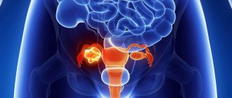
One of the main pathologies that are detected during X-ray examination are tumors. Cysts or neoplasms can develop covertly for a long time, without disrupting the functioning of body systems. The most informative method used to diagnose bladder tumors is computed tomography.
Doctors can make an accurate diagnosis of cancer only after cystoscopy with a biopsy, but there are a number of factors that make radiation examination necessary. Firstly, malignancy of papillomas most often occurs deep in the wall of the organ, and this cannot be determined by examining a biopsy specimen. Secondly, cystoscopy does not provide information about tumor growth into the wall and neighboring organs, nor does it show the presence of metastases in regional lymph nodes.
Therefore, if there is a suspicion of cancer, it is better to start the diagnostic search with radiation diagnostics. When an artifact is discovered, one cannot say whether it is benign or malignant. A reliable sign of cancer on radiographs is its germination deep into the wall of the bladder and invasion into the paravesical tissue.
Tomography is valuable because it can accurately visualize tumors in the fundus and apex of the bladder. Cystography is also capable of visualizing the process, however, for this, a double contrast procedure must be performed.
Lacunar cystogram of a patient with prostate adenoma, papillary tumor and bladder stones
Another use of radiography in bladder examination is to diagnose congenital anomalies.
The most common of these is diverticulum. It is a cavity formation that is connected to the bladder through a thin neck. It is best determined by cystography. A ureterocele may also be present. At first glance, it may be similar to a diverticulum, however, its main difference is that it does not lie away from the organs, but is, in fact, a hernia of the ureter.
Decoding the research results
A radiologist interprets the results. The conclusion will indicate the size of the kidneys, structural features, and the presence or absence of pathological formations. If there are stones, cysts or tumors, the doctor will definitely indicate their location and size in the test results.
After the conclusion is formed, it is transmitted to the attending physician. Here, a nephrologist or surgeon compares the results of the decoding with other patient tests - blood tests, urine tests, and ultrasound data.
Taking into account all the indicators, the doctor makes a diagnosis and outlines a further treatment plan, taking into account the indications for surgical intervention - planned or urgent.
Indications and contraindications
Diseases of the kidneys and urinary system are numerous. Their symptoms differ in many ways, so urography is prescribed only after initial diagnosis. Urine and blood tests together indicate the presence of pathology in the urinary mechanism. X-ray examination of the kidneys allows you to clarify the diagnosis and create a clear clinical picture. It is prescribed when:
- renal colic;
- urinary tract infections;
- periodic occurrence of lower back pain;
- the need to monitor the condition after surgery, etc.
Urography is an important diagnostic method for the treatment of pyelonephritis and other kidney diseases. Often such an examination is prescribed if blood is detected in the urine or the presence of stones in an organ is suspected. Diagnostics with contrast is contraindicated in:
- renal failure;
- pregnancy;
- lactation;
- allergies to contrast agent;
- hyperthyroidism.
Contraindications to urography are relative. After eliminating the conditions that impede the procedure, the doctor can prescribe it. In case of an allergic response of the body to the administration of Urografin, other medications are used, selected individually.
Possible complications of the procedure
Complications after the kidney x-ray procedure are minimal. Most complications are associated with intolerance to the contrast agent. Before conducting the study, doctors test for allergies to contrast, and most patients do not experience problems with routine x-rays.
Minor deterioration in health, weakness, nausea, allergic reactions may occur in patients who were diagnosed without preparation - when it was necessary to quickly obtain data on the condition of the kidneys. In this case, doctors try to minimize complications and alleviate the patient’s condition.
X-ray of the kidneys is an extremely important test for health. It is prescribed for diagnosing diseases of the excretory system, which is responsible for the evacuation of waste products from the body, maintaining metabolism, and water-salt metabolism.
These processes ensure the normal functioning of the body. Therefore, when ordering an x-ray, you should not delay the procedure - it helps to make a diagnosis and save the patient’s life.
Contraindications
Like any medicinal method, this procedure has a number of contraindications in which it is strictly forbidden to carry out this examination procedure.
Contraindications to intravenous renal urography are presented in the following list:
- hyperfunction of the thyroid gland (hyperthyroidism);
- excess iodine in the body or intolerance to substances containing iodine;
- feverish condition.
However, if the patient’s health and life are at risk, the attending physician may decide (in exceptional cases!) to refer the patient for examination.
For the fair sex, there is another conditional contraindication - the menstrual cycle.
It is strongly recommended to reschedule the scheduled examination until the end of the menstrual period if the attending physician decides that this examination is not urgent and that it may be delayed for several days.
Women during pregnancy and lactation (breastfeeding) require special, increased attention and careful treatment. In case of pathology of the renal and genitourinary systems, the attending physician must decide to refer the patient for intravenous urography with special precautions!
Possible side effects
The procedure itself rarely leads to any side effects. The main risks when performing intravenous or retrograde urography using a contrast agent are associated with the following phenomena:
- allergic reaction to the substance Urografin;
- swelling in the area where the drug was administered;
- redness of the skin;
- itching;
- increased body temperature (usually in young children);
- deterioration of health.
If unpleasant symptoms persist, mandatory treatment is carried out. To eliminate the risk of allergic reactions, blood tests, urine tests, and a sensitivity test to a contrast agent are taken.
Conducting research
After the patient enters the office, he needs to remove all jewelry, metal jewelry and parts, and change into the hospital clothes provided. The procedure lasts about an hour.
The contrast agent is injected slowly through a vein or Foley catheter, over about 1-2 minutes. All this time the patient remains in a sitting position, after which a series of photographs are taken while lying down. The definition of the projection of the images, the time interval and the patient’s posture depend on the nature of the disease.
It is very important to evaluate your own well-being. A slight burning sensation and nausea are allowed. The appearance of severe itching, coughing, and redness of the face is a reason to stop the procedure.