The study of the kidneys using X-rays - urography, is aimed primarily at diagnosing nephrolithiasis (calculi, otherwise stones) and determining changes in the structure of organs. Depending on the suspected disease, the patient is prescribed excretory or survey urography.
The first type of examination is more focused on assessing the excretory functions of the kidneys, is carried out according to individual indications, and takes a fairly long period of time. The procedure is performed using a special contrast agent, the intravenous injection of which provides a more detailed image on the X-ray.
Types of urography
There are several types of x-ray examination of the kidneys. The urologist decides which method to use in each specific case. The doctor must choose the optimal way to study kidney tissue and their functionality, depending on the nature of the disorders.
Survey urography
Content:
- Types of urography
- Possibilities of survey urography
- Indications and contraindications
- What can a survey urography show?
- How to take a photo correctly
- How is the procedure performed?
- Special training
- Features of the procedure for children
- Algorithm for describing the resulting image
- Alternative to survey urography
This procedure is prescribed to the patient if the doctor sees any signs of kidney disease. As a rule, it precedes all other, more complex studies. Survey urography is a regular x-ray of the area of the body where the kidneys are located.
This diagnosis cannot be called particularly informative, but it can be used to check the location of organs, as well as identify large stones and some other problems. The undeniable advantage of this method is the fact that this study does not require any additional equipment, and it is carried out in any clinic. This method requires certain preparation of the patient, which will be discussed below.
Survey urography does not use a contrast agent. This method gives an idea of the overall picture of pathologies: it allows you to detect tumors, foreign bodies and large stones, as well as identify changes in the anatomical location of the kidneys.
Survey urography allows a specialist to examine the structural condition of the kidneys, starting from their upper section to the beginning of the bladder outlet. In addition, the doctor sees the bones of the pelvic area, and can also “at a first approximation” assess the condition and performance of the ureters and bladder.
Intravenous diagnostics
Intravenous urography is an X-ray examination that involves the use of a contrast agent. As a rule, it contains iodine. This solution is injected into a vein to the patient, provided that his bladder is empty. After this, several photographs are taken.
Preparation for intravenous urography is that the patient must empty the bladder. There are three methods of intravenous urography: excretory, compression and infusion.
Excretory urography
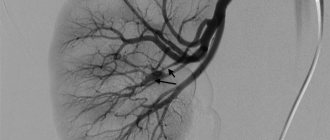
In many ways it resembles a review. The only difference between these diagnostic methods is that excretory examination uses a contrast agent. The first picture is taken one to two minutes after its introduction, the second after five minutes, and the third after seven minutes. The study allows you to assess the intensity with which the bladder, ureters and renal pelvis are filled with fluid, and also allows you to assess the shape and structure of the kidneys, the location of stones and cystic tumor neoplasms.
Compared to survey urography, the method is more informative, and the image itself is clearer. Among the disadvantages of this technique is the possibility of an allergic reaction to the contrast agent. The doctor must find out whether the patient suffers from individual intolerance to any drugs. Even if the answer is negative, an allergy test is performed before administering the substance: 0.1 ml of a contrast agent solution is injected intradermally into the forearm. Then the doctor looks and evaluates the reaction.
Compression urography
This type is a more complex procedure. During this procedure, the patient's ureters are artificially clamped. The image is very clear, but it is impossible to assess the condition of the ureters because they are compressed. Therefore, experts recommend combining the compression method with the excretory method so that the result is as informative as possible. A significant disadvantage of the procedure is the fact that this research method is very painful.
Infusion urography
Usually prescribed to people with disabilities, as well as those who cannot move. The contrast agent is administered through a catheter, and the examination is carried out with the patient lying down.
Depending on where exactly the contrast agent is injected, urography can be retrograde or antegrade. In a retrograde study, a contrast agent is injected directly into the ureters under general anesthesia. After this, a survey urography is performed. This method makes it possible to study the ducts as clearly as possible.
In antegrade percutaneous urography, a contrast agent is injected through the skin into the upper part of the ureters. Going down them, the drug allows the doctor to study them properly. This method allows you to identify ureteral ruptures, inflammation, and neoplasms.
Each of the above methods has its own advantages and disadvantages. Thus, survey urography is fast and does not require the introduction of contrast, but it cannot boast of high information content. During excretory examination, the image is not very accurate, but this method is simple and reliable. Compression urography also does not take much time, but the procedure is very painful. Infusion therapy is suitable for people with disabilities, but is time-consuming due to the need to be on a drip. Retrograde and antegrade methods provide a comprehensive amount of information, but require general anesthesia.
Contraindications and restrictions
Urography is contraindicated in patients:
- during pregnancy;
- with renal failure (decrease in specific gravity of urine below 1.005, urea content in blood serum above 11.6 mmol/l, azotemia above 70 mg%);
- if the patient is in a state of shock or vascular collapse;
- with intolerance to iodine-containing drugs (in the case of a study with contrast).
Consult with a SM-Clinic specialist if you have been prescribed excretory or intravenous urography. If you have an allergic reaction to a contrast agent containing iodine, the procedure cannot be performed. Thanks to the wide variety of diagnostic methods used at SM-Clinic, the doctor will select the most suitable procedure for you.
Possibilities of survey urography
The main purpose of a survey x-ray examination of the kidneys is to identify stones at different levels of the urinary system. These can be stones in the kidneys, bladder and ureters. However, it should be taken into account that this method does not allow visualizing all types of stones, but only stones of urate and oxalate origin. Phosphate neoplasms are most often invisible on photographs. In addition, the image shows tuberculosis lesions and echinococcal cysts.
Also, using this method, the doctor can identify other pathological processes based on the data obtained. Thus, visualization of the renal shadow will allow us to draw a conclusion about the size of the organ and its location. Based on the outlines of the psoas muscle, one can judge the state of the fiber surrounding the kidneys. The doctor may also look at the lumbosacral spine, hip joints, lower edges of the ribs, and pelvic bones.
Varieties
In medical practice, the following types of urography are distinguished:
- intravenous (excretory);
- overview;
- retrograde.
Intravenous (using contrast)
The essence of the procedure is the introduction of a contrast agent through a vein. Some time after the injection, an x-ray is taken, in which, thanks to this substance, the internal organs are clearly visible.
The following contrasts are used for the study:
- Visipack;
- urografin;
- triomblast;
- Cardiotrust.
Four x-rays are taken, which clearly show:
- renal pelvis;
- ureter;
- bladder;
- urethra (urethra).
The following indicators are important:
- contrast reagent release rate;
- the time of its action;
- preservation in the urinary tract.
Overview
Survey urography is carried out if necessary to examine the condition of the bone tissue of the skeleton, the shadow of the kidneys, their morphology and localization, as well as to evaluate the functions of other organs of the urinary tract. This examination method is often prescribed for pain in the pelvic area. Usually this is the first study that is performed on a patient with pathology of the urinary system.
Retrograde
Retrograde urography is very similar to intravenous examination. The difference is that in the first, the contrast agent is injected directly into the bladder using a catheter.
In this case, the unpleasant consequences of the intervention, for example, allergic reactions to contrast, are minimized, since it does not enter the patient’s blood.
Indications and contraindications
As noted above, survey urography is the first procedure to which a patient is referred if pathological processes in the kidneys are suspected.
Indications for the study are:
- Congenital or acquired hydronephrosis of one or both kidneys.
- Stones in the urinary tract: in the tissues of the kidneys, bladder and ureters.
- The presence of foreign bodies in the urethra or in the lumen of the bladder.
- Lower back injuries, including back muscle tears.
- Tumors and other pathological foci (cysts, abscesses, etc.).
- Abnormalities in the development or location of the kidneys
At the same time, like any x-ray examination, the procedure also has its contraindications. Thus, the study is not carried out for pregnant women, patients suffering from radiation sickness, as well as for those who have had one kidney removed and the functionality of the second is impaired; urography with the introduction of contrast is contraindicated.
In addition, they try not to refer patients for urography who have recently undergone a study of the gastrointestinal tract with a barium suspension. This is because barium remaining in the body significantly impairs visualization of the kidneys and urinary tract. Therefore, the interval between studies should be at least a week so that the barium suspension is completely removed from the body.
Contraindications
Urography provokes undesirable effects, so it is not suitable for all patients. Contraindications to x-ray examination are:
- pregnancy;
- renal failure;
- lactation period;
- myocardial infarction;
- thyroid diseases;
- pheochromocytoma;
- treatment with metformin;
- diseases of the hematopoietic system;
- low blood clotting;
- allergy to iodine preparations;
- hemorrhagic stroke;
- exacerbation of glomerulonephritis;
- tendency to internal bleeding.
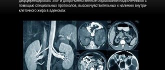
If urography is contraindicated, alternative examination methods are used - MRI or ultrasound.
The decision on the advisability of performing an x-ray examination is made by the doctor. If the risk of adverse reactions is high, less informative but safe methods of diagnosing urological diseases are used. The correctness of the diagnosis and further therapy depends on the quality of the examination and the completeness of the data.
What can a survey urography show?
This diagnostic method, as noted above, is the initial stage of studying the condition of the kidneys. The doctor prescribes this procedure if he has suspicions about organ dysfunction or the presence of certain pathological processes in it.
Thus, survey urography helps to identify:
- Benign or malignant neoplasms
- Changes in the structure of organ tissue.
- Stones.
- Anomalies in the structure of the kidneys.
- Pyelonephritis, glomerulonephritis, hydronephrosis, renal tuberculosis.
- Causes of blood in urine.
- Possible complications after surgery.
What does the procedure show?
Urography is one of the most informative methods for diagnosing kidney and urinary tract diseases. This is how pathological changes in tissues and the functional state of the kidneys are assessed. Urogram visualizes the organs of the excretory system, their size and shape. From the photographs it is determined:
- the extent of inflammation;
- localization of stones;
- condition of the renal collecting system (PSS);
- congenital anomalies of the ureters;
- scarring of the bladder;
- changes in the parenchyma;
- narrowing of the urinary ducts;
- neoplasms.
Based on the data received, a nephrologist or urologist assesses the performance of the excretory system. If there are suspicious tumors, additional hardware tests are prescribed - biopsy, histological analysis, scintigraphy.
Urographic examination is based on the ability of renal tissue to pass radiopaque solutions. The urogram determines:
- impaired renal blood supply;
- motor functions of the ducts;
- anatomical features of the kidneys;
- congenital and acquired pathologies.
Urography is one of the most useful methods for diagnosing urological diseases. The examination is performed routinely or urgently to quickly assess the performance of the urinary system.
How to take a photo correctly
There are several principles that must be followed in order for the X-ray image to be as informative as possible and for the specialist to be able to diagnose the pathological process.
First of all, you should not do an x-ray of just one kidney. Doctors are aware of situations where changes in the site of pain cannot be detected, but disorders are detected in the opposite organ.
A high-quality image is considered to be one that maximally covers all the structures that are components of the urinary system: the kidneys with the ureters and the bladder. The image should also show the contours of the eleventh and twelfth ribs and the upper edge of the pubic symphysis. You should use a film measuring thirty by forty centimeters.
Excretory urography: basics of the method
The urinary system not only cleanses the blood of waste and toxins, but also serves to remove them out. For this purpose, nature provided for the presence of several organs at once:
- The kidney is the main structure designed to purify the blood. This process occurs in two stages. Filtration is carried out in small vessels collected into dense balls. The filtrate is subsequently converted into urine by convoluted tubules;
- renal calyces and pelvis - the structures from which the outward journey of urine begins;
- From each kidney there is a narrow tube called the ureter. It constantly transports urine from the pelvis further along the urinary system;
- bladder - an organ designed to temporarily store urine;
- The urethra is a narrow tube designed for the final removal of urine to the outside.
The urinary system consists of several organs
Kidney function - video
Excretory urography is a way to study the anatomy and functioning of several parts of the urinary system: the renal pelvis, ureters and bladder. This method is based on the ability of the kidneys to cleanse the blood. By adding a certain type of substance to it, you can ensure that it goes through all stages of purification from filtration in the vascular glomeruli to being excreted through the urethra. The substance, in addition to being able to penetrate the blood and be filtered through the kidneys, must be visible to X-rays. With their help, you can see the contours of the organs of the urinary system.
The nephron cleanses the blood of waste and toxins.
The ability of a substance to be filtered determines another feature of excretory urography: this study allows you to evaluate the functioning of the kidneys over time. The method documents the passage of the drug through the pelvis, ureters and bladder, which allows one to calculate the rate of blood filtration and draw a conclusion about the functioning of the kidneys. Urography is used to examine adults and small patients. The technique in this case is the same, only the dose of the administered X-ray contrast agent differs.
How is the procedure performed?
Survey urography is a simple and not time-consuming procedure. Immediately after the patient enters the office, he is asked to remove jewelry and metal objects. After this, the patient is asked to lie on his back, with a pillow placed under his head. The X-ray beam is directed strictly perpendicular to the surface of the body, several centimeters below the xiphoid process. Immediately at the time of the photo, the patient must hold his breath. This will help avoid “double” images.
Special training
In order for the results to be as reliable as possible, the patient must prepare for it in advance.
Thus, it is often normal to visualize the processes that occur in the organs of the urinary system, but sometimes swollen intestinal loops interfere. Therefore, it is extremely necessary to clean it of feces in advance.
For this purpose, patients are asked to exclude foods that can cause flatulence from their diet at least three days before the test. This includes bread made from white flour, cereals and legumes, potato dishes, fruits and dairy products.
Dinner the night before should be light. You should sit down at the table no later than six o'clock in the evening. On the day of urography, it is better to refrain from breakfast.
It is recommended to start taking sorbents a few days before the procedure. An enema should be performed in the morning before the kidney x-ray.
You should refrain from drinking large amounts of liquid. On the day of the test, you can drink a glass of unsweetened tea.
What is required from the patient
In order to obtain reliable data after the diagnostic procedure, it is necessary to follow the rules of preparation for survey urography. Inflated loops of the gastrointestinal tract complicate the process of visualizing pathological syndromes developing in the area of the urinary system.
The emphasis is on cleansing the gastrointestinal tract of feces. 3 days before survey urography, the patient must adhere to the following recommendations:
- Foods that cause flatulence should be completely excluded from the diet. These include baked goods, as well as milk, kefir, and cottage cheese.
- Dinner before the procedure should be light. It is recommended to skip breakfast.
- Doctors also prescribe a sorbent drug and an enema to the patient 5-6 hours before a visit to a medical facility.
- You should limit your drinking regime 8 hours before undergoing a diagnostic examination. In the morning before the survey urography, you are allowed to drink no more than 200 milliliters of tea without sugar.
Features of the procedure for children
The method of conducting survey urography for children is no different from how the study is carried out on adult patients. It is not recommended to conduct the study on babies under one month of age.
Note that when performing urography with a contrast agent, the amount of the drug should be carefully calculated for small patients.
However, even when conducting survey urography, which is absolutely painless, parents must prepare the child in advance and explain to him that he will not experience any discomfort, and therefore should not be afraid.
Contraindications to urography
Despite the many advantages, urography using a contrast agent has a number of specific contraindications that cannot be ignored:
- allergic reaction of the body to iodine (part of contrast agents);
- severe renal, hepatic or heart failure;
- pregnancy;
- glomerulonephritis;
- serious endocrine pathologies (decompensated diabetes mellitus, hyperthyroidism);
- history of stroke or heart attack;
- poor blood clotting;
- absence of one kidney (an exception may be survey urography).
ATTENTION! Contrast urography , as a type of x-ray, involves radiation exposure of the body in small doses, therefore frequent examinations, as well as examinations against the background of other x-ray diagnostic or treatment methods, are undesirable.
Algorithm for describing the resulting image
The first stage of describing the image is to study the state of the skeletal system. The thing is that pathological processes in the organs of the urinary system also affect the bones. For example, in chronic kidney diseases, a so-called “compensatory scoliosis” of the spine towards the healthy organ is observed.
If the patient has carefully prepared for the procedure, the shadows of the kidneys are clearly visible in the image. The norm is the location of the left kidney at the level of the body of the twelfth thoracic and to the second lumbar vertebrae, and the right kidney - from the first to the third body of the lumbar vertebrae. The fact that the right kidney is located lower than the left is explained by the fact that the liver puts pressure on it.
When describing the shadows of the kidneys, the radiologist must indicate their shape, size and contour condition, and also comment on the density of the organ tissue.
The doctor is also interested in the condition of the lumbar muscles, their contours and symmetry. Any changes may indicate that an inflammatory process is occurring in the retroperitoneal space or that there are tumor foci.
As a rule, the ureters are normally not visualized because they are hollow. If there is inflammation in their lumen or there are stones, then shadows are noticeable in the images, which correspond to the location of the ureters.
Best materials of the month
- Why you can't go on a diet on your own
- 21 tips on how to avoid buying stale food
- How to keep vegetables and fruits fresh: simple tricks
- How to curb your sweet cravings: 7 unexpected products
- Scientists say youth can be extended
The bladder is visible in the image if concentrated urine is present in its lumen.
After the specialist has compiled a description of all the “natural” shadows and structures, he begins to study pathological and additional shadows that may indicate dysfunction in the area being examined.
X-ray results
The results are decoded by a doctor from the radiology department. The image is described sequentially according to a specific algorithm:
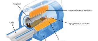
Magnetic resonance imaging of the kidneys
- condition of the spine and pelvic bones. In chronic renal pathology, there is a high probability of developing a frontal curvature of the spinal column;
- shadows of the buds and their location. According to the standard, the left shadow should be located from the 12th thoracic vertebra to the 2nd vertebra of the lumbar spine, the right one - slightly lower - from the 1st to the 3rd lumbar vertebrae. When assessing shadows, the diagnostician describes their outlines, sizes and shapes;
- kidneys directly. Healthy organs should have clear outlines and a homogeneous structure. In the presence of a single cyst, a tubercle is visible; in polycystic disease, the kidney is enlarged, and the outline of the organ is wavy. The tumor is determined by the growth of the kidney and curved contours;
- lower back muscles. Blurred outlines indicate the presence of tumor formations and internal hematomas;
- hollow tubes connecting the kidneys to the bladder or ureters. These organs are visualized on the image only if there are other concomitant diseases;
- bladder. Normally, the urinary reservoir is not visible; its visibility is due to the presence of urine with any impurities in the organ.
According to the doctor's description, the patient receives examination results, including information: about the condition of the kidneys (size, shape, shape, location, structure) and the presence (absence) of stones. And also, about the degree of deformation (if any) of the pelvic bones, the lower part of the spine, and possible injuries to the lower back muscles. For an experienced radiologist, the description process takes no more than a quarter of an hour.
Analysis of the urography results is carried out by a radiologist, but the final diagnosis is made by the attending physician