Mammography is a standard method of examining the mammary glands, used as a screening and diagnostic test. The advent of mammography gave a serious impetus to the development of oncology, since the examination helps in almost 100% of cases to timely diagnose and treat breast cancer in older and middle-aged women.
Mammography is performed using X-rays. The resulting image is called a mammogram. Using a mammogram, an experienced specialist can easily identify the existing pathology of the mammary glands and prescribe treatment.
What is mammography and why is it needed?
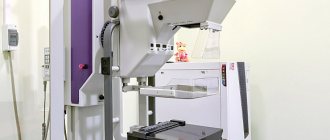
Mammography is a non-invasive screening technique for examining women (including nursing mothers), used in outpatient clinics and inpatient medical institutions. A mammograph is a special X-ray machine that is used to take mammograms (images).
The operating principle of this device is not fundamentally different from a simple x-ray. A special tube sends X-rays precisely to the area of the mammary glands on the chest, where they are absorbed with varying intensities. Denser tissues absorb X-rays well, which will be reflected on the mammogram as an area of differentiation of large ducts.
Research technique and methodology
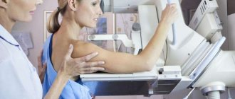
Before conducting the examination, the woman undresses to the waist and removes jewelry from her chest. The nurse or doctor places the woman's breast on a special support, after which another plate is compressed on top of the breast. This is necessary for several reasons:
- For a more even distribution of the breast, which creates the same breast thickness in different areas.
- For clearer visibility of pathological neoplasms on a mammogram.
- For lower radiation dose.
- To keep the chest in one position. When turning and moving, the mammogram may become blurred and unclear.
Two photographs are taken: in frontal and lateral projections. Body rotations can only be performed during breaks between shots. During the “photographing” itself, you should not move; it is advisable to hold your breath so that the picture turns out clear.
Results are ready within minutes. The mammogram is given to the patient, after which she can immediately go to a specialist.
Read also:
How to properly care for your breasts while feeding your baby
Features of mammography in nursing mothers
X-rays of the mammary glands in nursing mothers are performed in extremely rare cases. Here, preference is given to ultrasound techniques, since they have less impact on feeding and the condition of milk. However, there are situations when mammography is necessary for nursing mothers.
A feature of feeding is the constant accumulation of milk in the mammary glands. If a nursing mother, after the baby has eaten, does not express the remaining milk, then she is automatically included in the risk group for the occurrence of lactostasis and mastitis.
Important! Almost all cases of mastitis detected in nursing mothers require confirmation of the diagnosis using mammography. Breastfeeding is not an absolute contraindication to the study. Mammography for nursing mothers is done strictly according to indications and in a situation where it is impossible to confirm the diagnosis otherwise.
It is not necessary to stop breastfeeding during the examination. If mammogram results confirm mastitis, the doctor will usually prescribe antibiotics. In this case, the nursing mother temporarily stops breastfeeding.
Since the functioning and morphology of tissue changes during lactation, assessing mammogram results is often difficult. For a more accurate diagnosis, nursing mothers are recommended to express milk from the gland being examined before the examination, then the mammogram will be clearer and more informative.
Is a mammogram necessary while breastfeeding?
Mammography is a method that is considered one of the most informative among diagnostic and screening tests used for the early detection of breast tumors.
Mammography gave impetus to the development of oncology, making it possible to timely diagnose cancer in women of any age and begin therapy to combat the disease before complications incompatible with life occur. A mammogram is an image obtained as a result of X-rays of the breast during mammography.
Any radiation, if used uncontrolled, can cause pathological changes in the tissues of an adult. X-ray techniques are even more dangerous during pregnancy and breastfeeding.
Let's consider whether it is possible to do mammography while breastfeeding, why it is necessary.
When is a mammogram necessary for a nursing mother?
Screening tests are carried out in the age group of women over 40 years of age. The need for annual additional thoracic irradiation is dictated by the increasing incidence of breast cancer throughout the world.
It is recommended to start the examination at an earlier age:
- if a woman is at risk of developing breast cancer due to a family history of this disease;
- there is evidence in the anamnesis of existing precancerous pathological changes in the mammary gland.
Let's consider in what cases it is necessary to do a mammogram during breastfeeding.
Reasons for performing diagnostic mammography in breastfeeding mothers:
- Having diagnosed breast cancer.
- Inflammation of the gland tissue - mastitis.
- Lactostasis.
- Purulent diseases of the mammary gland.
- Benign breast neoplasms: fibroadenoma, lipoma, intraductal papilloma, soft tissue tumors.
- Benign dysplasia: cysts, nodular and diffuse mastopathy, adenosis, fibrosclerosis, ductal ectasia, etc.
Contraindications to the procedure
There is only one absolute contraindication to the procedure – pregnancy at any stage.
X-ray radiation, even in small doses, can disrupt the normal course of fetal development.
Mammography of a nursing mother is allowed if the information content of other methods is questioned.
Preparing for the examination
The procedure does not require careful preparation. Before the examination, it is necessary to consult a mammologist, because You will not be able to determine on your own whether you need this method of examination or whether you can get by with more gentle methods that do not involve radiation exposure to the body (for example, ultrasound scanning).
It is necessary to exclude the use of deodorizing agents, as well as perfumes and cosmetic gels and creams in the area under study. The use of these tools may affect the quality of the image.
Features of mammography in nursing mothers
When examining nursing mothers, ultrasound techniques are more often used due to the lesser influence of ultrasound on the quality of mammary gland secretions during lactation.
When breastfeeding, milk constantly accumulates in the glands, which leads to the risk of inflammation or lactostasis. The reason for these conditions is that the woman does not express the remaining milk from the breast after finishing feeding.
When signs of mastitis are detected, in almost all cases there is a need for diagnostics for nursing mothers using mammography.
An X-ray examination is prescribed according to strict indications if a situation arises in which it is not possible to confirm the diagnosis by other means.
During the study, weaning from breastfeeding is not necessary.
https://www.youtube.com/watch?v=628Tc87NjAA
If mastitis is still confirmed, it must be treated using antibacterial agents, which are the reason for temporarily canceling natural feeding and expressing milk.
To obtain more informative data on a mammogram, it is better to express milk right before the procedure: filling the gland with secretion changes the morphological picture of the organ, causing difficulties in assessing the image.
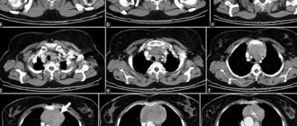
Results are normal and pathological
In the presence of a pathologically changed lesion on a mammogram, even in the first stages of the process, its size, localization, spread to nearby areas and tissue density are assessed
The x-ray picture of the mammary gland is normal: uniform density of tissue areas in all its parts.
For pathology:
- Abscess, neoplasm: clearly limited darkening of a round shape. The difference between an abscess and a breast tumor is the presence of a capsule filled with purulent contents.
- Diffuse darkening of areas of organ tissue is characteristic of mastitis, phlegmonous inflammation or lactostasis (changes in the intensity of darkening along the ducts filled with secretions).
With mammography, both false-positive and false-negative results can be observed, which arise due to improper preparation or conduct of the study, as well as due to errors in recording the result by the radiologist.
The presence of a mammogram does not allow a definitive diagnosis to be made with certainty. To confirm the disease, additional clinical, laboratory and instrumental examinations must be performed.
Pros and cons of mammography
The advantages of mammography include:
- In the images you can see minute changes in breast tissue that are inaccessible to other research methods.
- The exact localization of microcalcifications present in the gland is determined.
- The condition of the ducts is assessed.
- Allows a more accurate assessment of the size, nature and localization of cystic formations in the gland tissue.
- The technique does not harm women over 50 years of age.
- Mammography is 95% accurate.
Negative aspects of the examination:
- Mammography is not very informative in young women under 30 years of age, because the glandular tissue is still too dense, and the image does not provide complete information, unlike an ultrasound.
- Mammography is not recommended more often than once every 12 months due to the radiation exposure this technique carries.
Consequences after mammography
The main fear of women who have to have a mammogram is the risk associated with x-ray exposure.
The consequences of mammography performed as a screening test can be:
- Identification of false-positive results in examined women.
- The need for a painful biopsy due to the diagnosis of suspected breast cancer.
- 1% of women examined are at risk of receiving treatment for a cancer they do not have due to diagnostic errors. And especially tragic will be death that occurs as a result of depletion of the body by unnecessarily prescribed chemotherapy drugs, surgeries and radiation therapy.
The risk itself, which is associated with the use of X-ray radiation, does not pose the danger perceived by patients, because low intensity rays are used.
Is it possible to breastfeed after a mammogram?
You should not immediately start breastfeeding after a mammogram. It is recommended to express milk for several feedings, replacing natural feeding with artificial feeding.
Source: https://iDiagnost.ru/issledovaniya/rentgen/est-li-neobhodimost-v-mammografii-pri-grudnom-vskarmlivanii
Preparing for the examination
Mammography does not require any special preparation. A few days before the test, consult a specialist and get a referral from him. A preliminary consultation with a doctor is advisable, since not all cases require an x-ray examination.
The day before the procedure, do not use deodorants or other cosmetics in the chest area so as not to spoil the picture.
Remember! Your doctor will decide whether you need a mammogram or not. Since mammography is performed using X-rays, unnecessary examination is an unnecessary radiation exposure to the body.
Read also:
The more effective way to treat a sore throat during breastfeeding without harming the baby
Advantages and disadvantages of the method
Benefits include:
- The procedure takes little time, results can be obtained immediately and treatment can begin as soon as possible. This is especially important if a serious pathology is diagnosed during pregnancy.
- Does not require preliminary preparation.
- It is not prohibited to carry out as often as necessary.
- Has no contraindications.
- In certain cases, it is a more informative method than mammography.
The disadvantage is the impossibility of conducting high-quality diagnostics after menopause, since during this period the glandular tissue is replaced by adipose tissue. Ultrasonic waves cannot pass through the fat layer.
When is a mammogram performed?
The optimal timing is the first week after the last menstruation. During this period, the breasts are least susceptible to cyclic hormonal changes in the body.
Important advice! It is not recommended to have a mammogram one week before your expected period. At this time, the mammary glands of most girls swell slightly and become coarser due to increased production of prolactin. The mammogram may not be informative, and after the examination the mammary glands will ache very much.
As for nursing mothers, there are no time restrictions. Nursing mothers are not subject to cyclical hormonal changes, and they produce prolactin daily at a constant level. The study is carried out at any time convenient for them.
Indications and contraindications for nursing women
An examination is necessary if a woman’s medical history contains evidence of pathological changes in the mammary glands or there is a risk of hereditary predisposition to cancer. Other indications:
- breast cancer;
- lactostasis;
- benign dysplasia (mastopathy, cystic formations, ductal ectasia, fibrosclerosis, etc.);
- mastitis (inflammation of the gland);
- purulent lesions of the glands.
An examination is also necessary for breast hardening and abscesses. Contraindications include only pregnancy, since X-rays can disrupt the normal development of the fetus.
A nursing mother is examined if the results of other methods are in doubt.
When is a mammogram necessary?
Mammography is used as a screening and diagnostic method of examination.
Mass screening tests to determine the condition of the mammary glands must be completed annually by every woman after 40 years of age. This is necessary for early diagnosis of breast cancer, the incidence of which in our country is steadily growing every year.
Important advice! Annual screening examinations, in consultation with a doctor, can be carried out at an earlier age if there is a family history of breast cancer and a high risk of its occurrence.
Diagnostic mammography is indicated for all girls and women if they suspect:
- Mammary cancer.
- Mastitis.
- Abscess and other purulent diseases of the mammary gland.
- Lactostasis (especially important for nursing mothers).
- Benign breast tumor.
- Other rare pathologies of the mammary glands.
Results are normal and pathological
Using a mammogram, you can assess the size, density and prevalence of the pathological focus even in the initial stages of the disease. Normally, breast tissue is the same in consistency in all parts; darkening of the lobes and ducts is not observed.
With a tumor and abscess, a rounded darkening will be detected in one of the lobes with clear boundaries, while at the abscess, a pyogenic capsule is visible along the edge of the pathological focus, delimiting inflammation.
Read also:
Methods for increasing the calorie content of mother's milk
With phlegmon, mastitis, lactostasis, diffuse darkening of one or several areas or the entire gland as a whole is determined. Moreover, with lactostasis, the darkening is determined along the ducts.
Important! It is important to be aware of false-positive and false-negative mammography results. Incorrect examination, improper preparation for the study, carelessness of the doctor when writing a report, or other diseases hiding under the mask of mastitis can give incorrect results. The result of a mammogram is not a diagnosis, but only a sign that confirms or refutes a presumptive diagnosis made on the basis of clinical symptoms or other studies.
Ultrasound of the mammary glands during pregnancy
Discharge from the breast during pregnancy, with the exception of the third trimester, is an indication for examination
The onset and course of pregnancy is accompanied by continuous hormonal surges in the body, which can cause the formation and development of pathological conditions in the body. To identify them at the initial stage of development, it is necessary not to delay routine diagnostics of the body. Ultrasound examination does not have a negative impact on the course of pregnancy and fetal development. Another important advantage of instrumental diagnostics is the absence of the need for preliminary preparation.
Pregnant women require an ultrasound scan of the mammary glands in the following cases:
- the appearance of discharge from the breasts - only in the last trimester of pregnancy can spontaneous discharge from the nipples be the norm;
- limited mobility of the skin in the area of the mammary glands, the skin has become denser and rougher;
- retraction of the skin or nipples is detected, which indicates the presence of a neoplasm, the nature of which should be identified as soon as possible;
- pain or discomfort;
- when palpating, the woman clearly feels a tumor, nodes, cysts, etc.
During the examination, the doctor will be able to find the following pathological conditions:
- inflammatory processes;
- cysts, tumors and other types of neoplasms;
- fibroadenoma;
- mastopathy.
Diagnostics allows you to become familiar with the structure of the gland, as well as any, even the slightest, deviations from the norm.
Ultrasound examination of the mammary glands involves the use of special equipment and a sensor that produces high-frequency pulses with parameters from 1 to 18 MHz. The image on the monitor is formed due to the reflection of sound waves and different acoustic resistance at the border of soft tissues.
To perform an ultrasound, a woman will need to undress to the waist and lie on her back near the diagnostic equipment. The doctor applies a special gel to the chest to improve the conductivity of the rays produced by the device.