Radiography is one of the research methods, its basis is obtaining a fixed image using x-rays. The result is usually obtained on X-ray film or displayed (if digital devices were used) on a monitor screen or paper. The study is based on the passage of X-rays through body tissue. X-rays are usually used as a diagnostic method. To obtain more accurate results, use an x-ray image in two projections.
Chest X-ray
X-ray of the chest organs (chest organs) is the most common examination method, which allows us to identify pathologies of the respiratory, as well as cardiovascular systems, ribs, and thoracic spine that arise from various injuries and diseases.
How do X-rays work? As they pass through the body and organs, they are absorbed in different ways. The result is an x-ray image. Tissues with a denser structure look white on it, those that are softer look dark. After development and drying, the radiologist evaluates the resulting picture. An X-ray of the lungs will show all pathologies, if any, and indicate possible diseases.
Modern digital devices simplify the procedure, while the radiation dose is significantly reduced. There is also mobile equipment that allows you to examine bedridden patients.
Protocol for describing a chest x-ray by a doctor
A therapist, pulmonologist and other doctors will be able to see obvious pathological formations in the image. But in order to decipher the X-ray of the lungs in detail and identify the slightest deviations from the norm, a regulated algorithm is used - a protocol for describing the X-ray. This is done by a highly specialized doctor – a radiologist. According to this protocol, the analysis of the radiograph includes the following points.
- The name of the procedure is the name of the area under study, projection (direct, lateral).
- Assessment of the symmetry of the pulmonary fields.
- The presence of shadows indicating pathology, their type - focal, infiltrative, diffuse. If there are enlightenments, they also need to be indicated in the description.
- Analysis of the pulmonary pattern. A discrepancy with the normal picture indicates problems with the blood vessels of the lungs.
- Condition of the roots of the lungs. A violation of the structure of the lymph nodes is recorded, the condition of the large bronchi is assessed.
- Shadows of mediastinal organs. Particular attention is paid to the shadow drawing of parts of the heart and large vessels - ventricular arches, aorta, pulmonary artery.
- The condition of the diaphragm and pulmonary-diaphragmatic angles - symmetry of the position of the organ, sinus angle, fullness (the presence of effusion indicates pleurisy).
When deciphering an image of the lungs, it is very important to describe everything correctly. Radiologists must consider the quality of the image, the presence of shadow areas on the radiograph, their location, size and other parameters.
A standard x-ray of the lungs is done in two projections. They clearly show lung tissue, bones (ribs, collarbones, shoulder girdle), domes of the diaphragm, overlapping shadows of the heart, vascular bundle, mediastinal organs, spine, sternum.
Assessing the quality of the image
Various factors can affect the quality of an x-ray image. If you violate the technology for its implementation, you may end up making an incorrect diagnosis. Here's what affects image quality:
- asymmetrical body position. If you violate the requirement of symmetry, you can mistakenly diagnose “displacement of the thoracic vertebrae”;
- hardness/softness of the image;
- the presence of artifacts (additional shadow areas) and diseases that affect the condition of the patient’s chest;
- coverage. The image should show the costophrenic sinuses inferiorly and the superior pulmonary fields;
- The patient's shoulder blades should be positioned outside the chest. Otherwise, decoding becomes more difficult;
- images must be single-contour. Dynamic sharpness indicates that the patient was breathing during the examination. Such a photograph cannot be interpreted; it needs to be redone;
- The contrast level is determined by the fields that produce darkening and highlighting. Based on this, you can understand whether a dark or light area is a pathology, or whether it is a feature of the image developer.
The direction of the X-rays affects the image of the lungs.
Interpretation of lung x-rays - features of chest analysis
The examination procedure for radiography of different organs and systems varies. X-ray of the lungs has such features.
- The correct position of the patient's body and its positioning are of great importance. If this condition is violated, the location of the pulmonary fields, the opening of the shoulder blades, and the overall picture at the time of the respiratory act may be distorted.
- It is important to exclude artifacts from entering the photo - jewelry, piercings and similar details. They can obscure pathological foci and interfere with the integrity of image perception.
- It is necessary to check the completeness of capture - the apexes, fields, and diaphragmatic sinuses should be present in the final picture.
- To obtain an informative image, the strength of the X-rays should be adjusted based on the person’s physique and the presence of fat deposits. For obese patients, it is necessary to increase the dose of X-rays.
Analysis of anatomical structures on a normal chest radiograph
The lungs are an organ filled with air. X-rays pass through it almost unhindered. Therefore, the pulmonary field will look like a cleared area, colored black on the x-ray. Hard tissues (bones), on the contrary, are visualized as white. The roots of the lung, being denser than the lung fields, block some of the rays, so they are a little lighter.
Shadows on an x-ray can indicate both normal formations and pathological ones. The doctor's task is to determine whether the shadowing is something dangerous for the patient. Here's what you can see on healthy lungs:
- bone skeleton;
- pulmonary fields and pulmonary pattern;
- roots of the lung;
- mediastinum;
- heart shadow;
- diaphragm dome.
The fields are uniform, the pattern is clear. The division into shares or segments will be indistinguishable.
Features of lung field analysis
If there are no dark or light areas on the x-ray, this does not mean that there are no diseases. The doctor should carefully examine the lung fields. Here are the features that need to be taken into account during decryption:
- the right margin should be short and wide, and the left margin should be narrow and long;
- because of the heart, the median shadow should be slightly expanded to the left;
- when describing, three belts are evaluated - lower, upper and middle;
- the level of transparency can be determined by air and blood filling, the volume of parenchymal tissue of the organ;
- in women, the image may be slightly overlapped by the mammary glands;
- The pulmonary pleura is not visible in the normal state, but becomes noticeable during inflammatory processes or with a tumor (in the presence of pathologies, an additional lateral radiograph is required).
For convenience, the pulmonary lobes are divided into segments: in the right lung there are 10, in the left – 9. If deviations are identified when interpreting the radiograph, the conclusion indicates the number of the segment where they are located and the type of pathology in accordance with the codifier:
- 01 – cavity;
- 02 – darkening, in the projection of the lungs;
- 03 – focal shadows, ibid.;
- 04 – increase in mediastinal shadow;
- 05 – pleural effusion;
- 06, 07 – fibrosis of lung tissue;
- 08 – increased transparency of lung tissue;
- 09, 10 – damage to the pleura;
- 11-18 – petrification;
- 19 – pathology of the diaphragm;
- 20 – residual postoperative signs;
- 21 – skeletal pathologies;
- 22 – foreign body;
- 23 – pathology of the cardiovascular system;
- 24 – deviations that are not standard;
- 25 – patient is healthy;
- 26 – the photo is rejected.
The tumor on X-ray looks like “plus tissue” or a characteristic darkening. Its contours are usually unclear, as the cancer progresses, spreading to healthy cells. The darker the image, the more severe the tissue damage. The presence of rounded outlines indicates the presence of metastases in the organ.
At the second stage of cancer, local lymph nodes are affected, this is expressed in a violation of the structure of the root of the lungs. Lymphatic tissues increase in size and metastases form.
At later stages, the tumor is already quite large and spreads to a significant part of the organ. The pulmonary pattern intensifies, since the growing tumor requires increased blood supply, and therefore signs of hypertension are observed. Pleurisy is often observed - the contour of the pleura is visualized, pathological transudate is visible - fluid in the sinuses of the organ.
The diagnosis is confirmed in the presence of accompanying symptoms - sudden weight loss, cachexia, symptoms of intoxication (fever, weakness, lethargy, fatigue, drowsiness, apathy). The disease is often diagnosed in smokers.
Features, errors
On an x-ray, the inflammatory and tumor processes look almost identical. When interpreting the image, the following features of a typical tumor radiograph are taken into account:
- pulmonary pattern – does not change;
- in the adjacent sections the shadows are absent or single;
- the affected tissues have a bumpy surface and radiant contours;
- the roots of the lungs are changed only in the last stages;
- the shadows have a uniform shade.
X-ray for pneumonia
Pneumonia, or inflammation of the lungs, is clearly visible on x-rays. True, difficulties may arise at the initial stage. Here's what you can see in the picture during the inflammatory process:
- foci of inflammation in the form of shadows of various sizes;
- level of organ damage;
- inflamed pleura and bronchial tree;
- expansion of the root of the lung as a reaction of the lymphatic system to the disease;
- strengthening of the pulmonary pattern.
Foci of inflammation can be up to 1.5 cm in size. The infiltrate can have any form. The size of the affected area is assessed on frontal and lateral photographs.
Long-term smokers usually develop chronic obstructive pulmonary disease. In this case, the pulmonary pattern will be enhanced, especially in the lower sections. There are compensatory areas of clearing - emphysema.
In smokers, the size between the ribs increases, the diaphragm gradually lowers, and the domes smooth out. The angle between the fin and the diaphragm gradually turns from acute to straight. As a result, hypoxia occurs, or deficiency of pulmonary ventilation. In this case, the organ cannot perform its function normally.
X-ray capabilities and interpretation of the result
A chest x-ray helps detect the following pathologies in the body:
- Respiratory system: bronchitis, pneumosclerosis, pleurisy, tuberculosis, cancer, pulmonary atelectasis, pneumonia. The doctor interprets the X-ray images and immediately sees the probable disease.
- Cardiovascular system: myocarditis, pericarditis, changes in heart size.
- Mediastinum: displacement of structures, mediastinitis.
- Musculoskeletal frame of the chest: fractures of the sternum or ribs, vertebrae, hemothorax, pneumothorax, wounds of the mediastinum, heart.
X-rays are also used to track the dynamics of recovery in the treatment of pneumonia. However, x-rays cannot be called a universal diagnostic method. For example, x-rays cannot assess the nature of the tumor, and this study is also limited to immobile patients. For such exceptional cases, computed tomography is used.
When deciphering the result of an X-ray image of the OGK, the doctor evaluates the size and shape of the mediastinum, the structure of the chest and soft tissues, the transparency of the pulmonary field, the intensity of the pattern, the position and structure of the roots of the lungs, the shape of the pleural sinuses and diaphragmatic domes.
Preparation and carrying out the procedure
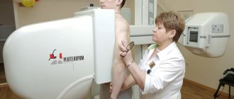
To perform the X-ray procedure of the OGK, no special preparation is required. The doctor only recommends removing clothing and jewelry from the area that will be irradiated. You also need to remove all objects that may interfere with the examination (glasses, dentures). If there is a need for the presence of a relative of the patient, a protective lead apron is put on him.
Having taken off his clothes, the patient is positioned in front of the photographic plate. The doctor leaves the room to the console; on his command, you need to raise your shoulders, press against the plate and hold your breath for a while. You can't move while doing this. If the patient is unable to assume a vertical position, he is placed on a table. Relatives or a nurse help him with this.
The examination is painless and does not cause any discomfort. The only discomfort is the cool temperature in the room. The x-ray will be ready within 15 minutes. It will be given to you immediately along with a description. Based on this, the doctor will make a diagnosis or refer you for further examination.
Indications and features of the procedure
An X-ray of the chest and lungs is prescribed when a person develops the following symptoms:
increased general weakness,- sudden loss of weight by a person,
- a hard-to-clear dry cough that literally exhausts a person,
- secretion of blood along with sputum,
- prolonged increase in body temperature for no apparent reason,
- if a person feels pain in the back towards the lungs.
In addition, a chest x-ray must be done in case of such pathologies.
- Pneumonia.
- Tuberculous organ damage.
- Tumors (benign and malignant).
- Fungal pathologies.
- It is also necessary to take an X-ray of the chest cavity if there are foreign bodies in the lungs.
Preparation
Before performing a chest x-ray on a patient, the doctor takes a detailed medical history. How an X-ray of the lungs is done depends on its results. Depending on the disease and the purpose of the diagnostic examination, the doctor selects the necessary film, selects the necessary X-ray modes, the required radiation time, etc.
Preparing for such a study is simple. A chest x-ray is performed with clothing removed. The patient must be positioned correctly in front of the x-ray equipment for the chest x-ray to be most accurate.
In addition, proper positioning of the patient in front of the equipment can significantly reduce the negative effects of X-ray radiation.
During this procedure, the patient must take a deep breath and no longer breathe. While the chest x-ray is being taken, the person must remain still. How important is such a requirement? If it is not performed, then in some cases the patient will have to undergo another examination. In the same way, children are given an X-ray of their lungs.
Frequency
How often can an X-ray of the lungs be taken? This question interests many patients. It must be said that this method of examining the chest organs is permitted, so it is not so easy to answer it unequivocally. The fact is that, despite the hundred-year practice of using X-rays, research into their effect on the body continues. Today it has been proven that X-rays cause:
mutation of the cell's genetic apparatus,- death of platelets, leukocytes and erythrocytes,
- increase capillary permeability.
At the same time, X-ray methods for studying the respiratory organs are characterized by a relatively low load. From this we can conclude: there is no need to refuse x-ray examination. And it is performed only when there is a need for it. At the same time, all radiation protection measures are observed.
According to many clinical studies, the acute stage of radiation sickness develops with a single 25 thousand fluorography of the chest organs per year, or 1000 radiographic and fluoroscopic examinations of the lungs in several projections. This suggests that a single x-ray of the lungs cannot cause significant harm to the human body.
And one more thing: doing fluorography once a year is also harmless. And this is the basis of an annual preventive examination.
X-rays of teeth
X-ray examination has become widespread in dentistry. The image not only makes it possible to track pathologies, but also reveals deviations in the structure of the jaws. X-ray diagnostics is important when choosing optimal treatment options.
There are several types of x-rays in dentistry:
- Panoramic. This image allows the doctor to evaluate the entire panorama of the location of the teeth, determine their number, and see unerupted teeth and rudiments. The anatomical structure of the jaw and nasal sinuses is also visible. A panoramic photograph is important for dental implantation, bite correction, and wisdom teeth removal.
- Biteful. Otherwise, such a picture is called interproximal radiography. A common type of photo. It is used to detect periodontitis and caries. Sometimes a bitewing photograph is taken after the crown is placed to verify the correctness of the procedure.
- Sighting. With the help of a targeted image, you can see exactly what the diseased tooth looks like and establish the correct treatment regimen. A targeted photo allows you to see no more than four teeth.
- Digital. Safe modern diagnostics. 3D X-ray makes it possible to get a clear picture of the entire dentition and individual teeth. The three-dimensional image is displayed on the screen, after studying it the doctor determines treatment methods.
Dental disease on x-ray
Radiography can reveal pathologies such as:
- carious cavities and abscesses;
- malocclusion
- jaw fracture;
- periodontitis;
- source of infection in the root canal;
- cyst, granuloma;
- tumor, abscess;
- unerupted teeth;
- deviations in the growth of wisdom teeth.
Caries on x-ray
Caries is one of the most common dental diseases, so many patients are concerned about what caries looks like on an x-ray. Caries is a lesion of the hard tissues of the tooth, which begins with the destruction of enamel. The main cause of the disease is the destructive effect of bacteria on the hard tissues of the tooth.
The rate of development of carious lesions varies: from acute caries in children, causing rapid destruction of baby teeth, to sluggish chronic caries in adults. The initial stages of the disease may be asymptomatic.
Caries in most cases is recognized clinically. Dentists resort to X-ray examination to diagnose a hidden carious cavity, which is inaccessible for examination and instrumental examination. Most often, there is a need to study caries on an x-ray if there is a suspicion of the presence of a cavity under a filling, crown, or on the root.
Mild degrees of caries, not accompanied by significant destruction of the enamel, do not appear on x-rays. As a rule, cases of fissure (in the natural recesses of the teeth) and cervical (root) caries do not require x-ray diagnostics.
Caries is visualized on an x-ray depending on the severity of the pathology. The image will show a carious spot, medium and deep degree of caries, complicated pathology. X-ray signs of caries appear depending on the listed cases as follows:
- a carious spot on an x-ray is visible as an area with limited darkening of reduced density;
- medium and deep degree of dental caries damage looks like a decrease in surface density at the site of enamel damage;
- Complicated caries on an X-ray image is visualized as a deformation of the anatomical structure of the tooth with multiple granulomas (focal growths of inflammatory connective tissue) and denticles (hard formations in the dental pulp).
Pulp diseases
The pulp is a living, soft tissue containing nerves and blood vessels that is located in the center of the tooth. The most common disease of the dental pulp is pulpitis. The disease most often develops as a result of the formation and progression of a carious enamel defect, which has spread to the dentin. Sometimes pulpitis occurs due to tooth trauma, exposure to thermal or chemical factors.
Pulpitis in an X-ray image is determined by darkening in the central and lower part of the tooth and is determined by indirect symptoms: a carious cavity, which is combined with gum and/or granuloma. In case of severe damage to the pulp, denticles may be visualized in the image, which look like multiple or single dense cavities localized against the background of the root canal.
Knowing how pulpitis looks on an x-ray, the doctor can use the probing method (instrumental examination) to clarify the diagnosis.
Cysts and granulomas on x-ray
A cyst is a pathological neoplasm ranging in size from 0.5 to 2 cm, which is a cavity with a fibrous wall and contents, in other words, a bag of pus. The neoplasm occurs as a response of the body to the development of an infectious process in the canals of the tooth.
A cyst on the root of a tooth, thanks to the fibrous wall of the cavity, retains dead bacteria, preventing them from spreading, but without treatment, as the disease progresses, rupture is inevitable. In the initial stage, the disease is asymptomatic; as the size of the cavity increases, acute pain, swelling of the gums, and swelling appear. Flux or fistula may form.
The most informative and accurate diagnostic method to determine the cystic formation of a tooth is radiography. On an x-ray, a dental cyst looks like a cleared area located near the tooth root in the form of a round or oblong cavity with a fairly clear outline.
Often, pathology can be diagnosed completely by accident, during an X-ray examination of adjacent teeth.
In complex cases, on the contrary, additional examination may be required, for example, using computed tomography (CT).
Knowing how a cyst looks on an X-ray of a tooth, the doctor, after studying the X-ray image, determines the type of tumor, its exact location, and also determines the cause of the cyst in order to prescribe adequate treatment.
Granuloma is a limited inflammation of the periodontium (tissues surrounding the tooth on all sides and preventing it from moving in the socket). Granuloma is a round formation, 3-5 mm in size, located in the area of the tooth root.
The long asymptomatic course of the disease often does not allow timely diagnosis and treatment of the disease, so it is often detected in an advanced stage.
Diagnosis is carried out mainly by radiographic methods. On an x-ray, a dental granuloma appears as a dark, rounded, limited area located in the area of the tooth root.
Snapshot Procedure
An X-ray of the teeth is performed on the recommendation of a dentist: in cases of caries, malocclusion, diseases of the periodontal tissues, pulpitis, cysts, jaw injuries, abscesses.
Before the examination, it is recommended that the patient remove all metal items and jewelry: they may distort the image data. The procedure depends on the type of image. The study takes a few minutes. Radiation has a minimal dose. The session takes place in a special room. The patient bites on the photosensitive film; it should be placed between the device and the tooth being examined.
When examining using a computer radiovisiograph, a special apron is put on the patient, the sensor is installed on the area under study and connected to the device. The result is displayed on the computer.
When using an orthopantomograph, an x-ray is performed as follows: the patient stands next to the device, the chin is fixed on a support. A block is clamped with the teeth, which prevents the jaws from closing. The patient must stand still. The device rotates around the head several times. Photos can be received on the same day.
Photo interpretation
Based on an X-ray of the teeth, the doctor writes a conclusion indicating the number of teeth, size and location. All detected pathologies will also be displayed in the report.
The picture shows the location of each tooth, the inclination, and the condition of the bones. Darkening in the image indicates the presence of pulpitis and denticles. Defects in tooth enamel mean caries. Where the density is reduced, brightening is noticeable. If the caries is complex, the tooth structure is deformed and granulomas are formed.
A cyst may be detected - a clear outline of a homogeneous structure of an oblong shape. The cyst is located at the root of the tooth, it can be small or large. Large cysts can affect two teeth at once. Chronic periodontitis is visible as a sharp darkening in the image at the apex of the root. With periodontal disease, a reduced bone marrow area is visible, atrophic processes and sclerotic changes are visible.
X-ray of the spine
In what cases does the doctor recommend taking an X-ray of the spine?
- For pain in the cervical, thoracic and lumbar region.
- For lumbar muscular pain of unknown origin.
- With limited mobility of the limbs.
- For injuries, falls and bruises.
- If you suspect degenerative changes in the bones.
- When diagnosing curvatures, osteochondrosis, scoliosis.
X-rays are recommended to be taken in two projections: lateral and direct. Descriptions of X-ray images are made by a radiologist; he evaluates the contours of the vertebrae, the spaces between them, the intensity of color, and the presence of growths. After this, an experienced specialist is able to immediately make a diagnosis, determine the likely prognosis and the need for surgical treatment.
Possibilities of using x-ray diagnostics
Radiography for pneumonia helps not only to establish a diagnosis and exclude other pathologies with similar symptoms, but also to monitor treatment, promptly identify possible complications, and establish the effectiveness of therapy. It is mandatory to conduct research in two projections.
Main objectives of the study:
- to confirm the diagnosis of pneumonia, type of disease, extent of damage,
- for diagnosing a protracted course, the effectiveness of therapy,
- to monitor recovery and complete restoration of the lung structure,
- to avoid the development of complications.
X-rays are recommended for children after 14-16 years of age. Contraindications are:
- pregnancy,
- extremely serious condition of the patient,
- the presence of concomitant ongoing bleeding.
These contraindications are relative. If the condition requires immediate diagnosis due to a direct threat to life, then the study is still carried out. At the same time, negative consequences are minimized. For pregnant women, additional protection of the abdomen and pelvis with shielding aprons is used.
The disease is characterized by the leakage of fluid into the alveoli, swelling of the tissues, and the presence of a large number of cells in them, primarily leukocytes and macrophages. This manifests itself clinically (in the form of an acute infectious inflammatory process) and radiologically.
Unambiguous signs of pneumonia on an x-ray are darkening of some part of the lung field.
Focal shadows or a widespread, confluent decrease in transparency are revealed. Characterized by fuzzy, blurry contours.
Pneumonia is classified as:
- Focal (a small area of lung tissue is affected),
- Segmental (one or more segments are involved in the process),
- Lobar (large, involving a lobe),
- Total (damage to the entire lung).
The degree of involvement of various lung structures and the prevalence of the pathological process affect the prognosis of the disease and determine treatment tactics.
How is the procedure performed?
No special preparation is required to take pictures of the upper spine. If the lumbosacral region is being examined, it is recommended to prepare in advance:
- It is necessary to completely cleanse the intestines, otherwise it will be difficult to make a correct diagnosis.
- Eliminate foods that promote fermentation from your diet two days before the procedure: bread, milk, legumes, coarse fiber.
- You should exclude dinner the day before and breakfast before the procedure.
- Quit alcohol and smoking.
- Before the procedure, cleanse the intestines with an enema.
- There should be no metal objects on the body at the time of shooting.
- Stay still.
The examination is absolutely painless for the patient. It is carried out for 10-15 minutes. Photos with descriptions are immediately available.
How is an X-ray of the lumbosacral spine performed?
This is a painless procedure lasting only 15-20 minutes. The main thing is that the patient must remain calm and simply follow all the nurse’s commands. The patient will receive the results of the examination later during a consultation with a neurologist:
In the X-ray room, a person will be asked to remove outer clothing and metal objects: a cross, a watch, a piercing, a belt.
X-rays are taken on a special table with the body in a horizontal position: on the back and on the side. You should lie still for some time while the radiation is being administered. Otherwise, the pictures will turn out blurry;
To avoid receiving an extra dose of X-ray radiation, areas of the body above and below the lumbar region are covered with a protective lead coating.