It happens that a person seriously injures his leg. But the most unpleasant thing is considered to be injury to the small toe or little finger. This is the smallest and most fragile toe, which has a very complex structure. By injuring him, a person experiences severe pain. He believes that he broke his finger. But injury to the small toe does not always lead to a fracture. Sometimes it's just trauma. And here it is important for the patient to figure out whether he received a bruise or a fracture. Whether or not he turns to a specialist depends on his decision. We will tell you further about what to do if your little finger is broken.
Causes
Elderly people are more susceptible to this phenomenon than others. As they age, their bone structure changes and their bones become more fragile.
But a young man can also break his little finger. The reasons for this phenomenon are different:
- diseases localized in the lower extremities. They are the ones who reduce the strength of bone tissue;
- outdoor games. During games, a person can easily twist his leg and break a finger;
- hitting a heavy object. For example, a blow to the little toe on furniture, after which the first signs of a fracture appear;
- falling a heavy object on your leg.
In addition, a fracture of the little finger can occur during a stampede. When another person steps on a person's foot.
Types of finger fractures and characteristics
The classification of fractures and their general characteristics are based on the causes, direction of the fracture and the number of fragments.
Types of fracture depending on the cause:
- pathological;
- traumatic.
Pathological ones can cause even minor impacts or awkward movements. This is possible with the following diseases: tuberculosis, osteoporosis, osteomyelitis, etc.
With age, the risk of fractures increases significantly because bone density decreases in proportion to age.
Towards some form of fracture - this classification is based on the projection of the fracture line:
- longitudinal;
- oblique;
- helical;
- transverse
Other types of fractures:
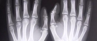
- Comminuted if there is no clear fracture line, and the x-ray shows fragments.
- Wedge fractures occur when one bone presses directly into another, deforming it.
- Hammered - when one fragment is directly hammered into another.
Comminuted fracture
Symptoms
It is very easy to identify a fracture of the little toe. After a blow, the following symptoms appear in a person: a fracture of the little toe. In the patient:
- the limb swells greatly;
- a hematoma appears on the leg. It can be red or blue;
- severe pain occurs when trying to move the affected limb;
- The shape of not only the little toe changes, but also the entire foot. The length and curvature of the little finger may change;
- the skin is broken by bone fragments.
Remember: if a patient, after a broken leg, has one of the above signs, then he should immediately contact a specialist who will tell you in detail what a broken little toe looks like.
Only a doctor can make an accurate diagnosis and carry out competent treatment.
A closed fracture of the little toe is much more difficult to determine. It is accompanied by the following symptoms. The patient has:
- when you try to step, the limb immediately swells and sharp pain appears;
- During the examination, extensive internal bleeding is revealed, a crunch is heard when palpating the sore limb;
- when pressing on the sore finger, its position changes sharply and it takes on an unusual shape;
- During the day, increasing pain appears, which leads to increased swelling. Moreover, the swelling can even spread to neighboring fingers.
Remember: large swelling of the toe occurs when the phalanx adjacent to the foot is fractured. Understanding what a broken toe or pinky finger looks like is very simple. The patient can try to straighten the sore finger. If he felt severe pain, then he was broken.
The most difficult thing to identify is a fracture of a finger or little toe without displacement, because There are no characteristic symptoms for this disease.
Typically, such a fracture can only be detected on an x-ray. In general, you can understand that your little toe is broken by the following symptoms. With it the patient:
- the structure of the bones is disrupted. And this is determined by feeling the diseased limb;
- the small finger looks much smaller than the others. Moreover, this symptom can be detected even with the naked eye;
- the limb acquires excessive mobility;
- the little finger acquires an unusual position.
Classification
Doctors conventionally classify little finger fractures into 2 types. They are:
- with offset;
- without displacement.
Let's look at each of them in more detail, and based on the symptoms described, we will decide how to understand that the little toe is broken.
No offset
When the little finger is injured without displacement, the patient only suffers a cracked bone. The axis remains in place, the little finger does not become shorter. If a patient wonders whether it is necessary to wear a cast if the little toe is fractured without displacement, then only a traumatologist can answer it. Also, if a person has a lot of unanswered questions, he can find answers on special forums that are available on the Internet. It is there that the victim can read about whether the little toe is broken, whether a cast is applied, and what patients who have broken their fingers on the lower extremities say when applying it.
With offset
When the little toe is fractured with displacement, it is the large bone tissue that is damaged. It cracks, bone fragments change their position. In this case, the bones become deformed, and the broken little toe becomes shorter. If the patient is interested in the question of whether plaster is applied to the little toe in this case, then the answer is yes.
Object of study
With the help of X-rays, a competent specialist will be able to carefully examine various anatomical parts of the lower extremities. These primarily include large structures of the musculoskeletal system, namely:
- phalanges - tubular bones that make up the fingers;
- metatarsus and tarsus, forming the foot;
- calcaneus and talus;
- fibula and tibia of the leg;
- patella - kneecap;
- hip.
In addition, radiography also allows us to examine mobile bone segments - joints that are often subject to injury, especially the ankle. It is equally important to study the condition of individual soft tissues - blood vessels, ligaments, muscle fibers, tendons and nerve endings. Only a comprehensive study can bring a complete, redundant result that determines the further actions of the treating doctor and his patient.
Diagnostics
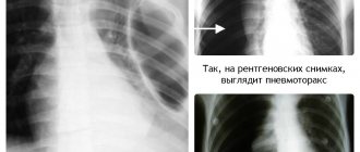
Initially, a patient with a broken little finger is referred for an X-ray examination. It is in the image that bone fragments will be visible, which indicate the presence of a fracture in a person. Sometimes the patient is sent for a computed tomography scan. It helps determine the nature and complexity of the damage. After the examination, the doctor can make a diagnosis, prescribe the correct treatment and tell you whether it is possible to break the little toe.
In children
Children can also break their little toe. In a small child or in children in general, a fracture of the little toe is characterized by the fact that one piece of bone tissue enters another. They develop a so-called “angular displacement”. But due to the rapid growth of bone tissue, when the little toe is broken, the bones heal faster and the bone fragments fall into place on their own. But a traumatologist will be able to answer whether children should be given a plaster cast if their little toe is broken.
In adults
Also, an adult who hit his foot on a heavy object can independently understand how to recognize a fracture of the little toe. He needs to do the following:
- Step on a limb. The broken finger does not participate in walking; it bears little weight. But it is precisely because of the weight that the little finger is deformed, and the patient immediately sees that it is broken. Remember: you shouldn’t feel the sore spot yourself. This can cause the bones to shift a second time.
- Place two limbs side by side and look at them. If it is externally noticeable that they are different, then the patient should immobilize the injured limb and seek help from a doctor.
- Look at the broken limb. If the phalanges are different, a crunching sound is heard when moving a finger, then the foot should also be immobilized. You cannot wear shoes on her, because... this can cause sharp pain.
If the patient discovers a fracture, he must be taken to a medical facility. Only medical professionals will be able to answer his question about how to quickly treat and what to do if his little toe is broken.
How is the examination carried out?
The prepared cassette is placed on the working table of the X-ray machine. The limb intended for examination is placed on it, the fingers are straightened and closed as much as possible.
For complex injuries, photographs are taken in two projections (direct and oblique, at an angle of 45 degrees). It is possible to take pictures in atypical projections to get a clear result. To study small structures (phalanxes of the fingers), images are taken at a short focal length (close-focus radiography, or plesiography).
X-ray of the thumb
Thumb injuries are quite common. To correctly assess the damage, it is recommended to take more than just a general photograph of the hand and fingers
, but a targeted shot of the thumb. To do this, the palm is placed on a special table or stand, the thumb is moved to the side as much as possible. In addition to direct projection, lateral or axial projection can also be used.
If digital X-ray equipment is used for filming, a targeted photo of the thumb may not be taken, since there is a possibility of enlarging the previously taken general one.
X-ray of a toe
The limb required for examination - the leg - is exposed and placed on a special stand. In some cases, the picture is taken with a load. To do this, the patient lifts the healthy leg up, bending it at the knee so that the body weight falls on the leg being tested.
X-rays of the toes or feet are often performed in several projections; this will allow the doctor to obtain the maximum amount of information about the nature of the damage or the course of the pathological process.
First aid
Before doctors arrive, relatives of a person with a fractured little toe should provide him with first aid. You need to do the following:
- Examine the injured limb. If there is an open wound, treat it with an antiseptic composition.
- If the patient has a severely displaced fracture, immobilize the injured finger. It requires a splint. You can make it from available materials. Also, relatives can simply attach the damaged finger to healthy fingers. This is done with a bandage.
- Give the victim a pain reliever.
- Apply a cold compress to the injured part of the leg. It could be ice from the refrigerator compartment. Remember: it is better not to apply ice to an open area of skin. This can cause the patient to get frostbite. It is advisable to wrap the ice in a cloth and only then apply it to the injured limb. Such a compress will help narrow blood vessels, stop bleeding, and reduce swelling. Apply ice for 15-20 minutes.
Help at home
Also, at home, a person who has a broken little toe should observe a gentle mode of movement and read in advance about what he can do.
For movement you can use:
- crutches;
- shoes that properly support the foot and ankle.
While lying down, the patient should raise his leg. This will prevent the appearance of new swelling. Additionally, it must be transported to a medical facility.
How to fix bandage fixators
For rapid healing, the affected limb should be immobilized. The pharmacy now sells many different fixations for the little toe in case of a fracture. Many patients wonder how to fix a cast in case of a broken little toe.
This is done like this: if the patient does not have displacement, then a medical bandage can be secured with a plaster. The last two fingers are secured together with a bandage. It is recommended to place a cotton pad between them first. This will prevent irritation and cracks on the little toe.
A medical splint made of plaster is used to fix a finger in case of a fracture. It is fixed on the lower part of the leg. Keep it like this for 1 month.
In especially severe cases, the patient is recommended to wear a special bandage that secures the leg.
First aid for a broken finger
Before the victim is examined by a traumatologist, provide all possible assistance on the spot.
Call an ambulance, and before arriving:
- Assess by eye the severity of the injured person’s condition and the location of the damage.
- Stop bleeding, if any, using clean bandages or cloth.
- If you need to splint a broken finger , do so. The broken phalanx must be fixed to avoid possible displacement. Any hard object or even a nearby finger will do.
- If the fracture is open, you need to disinfect the wound with any antiseptic at hand: hydrogen peroxide, chlorhexedine solution. And be sure to apply a sterile bandage to prevent possible infection of the wound. This must be done, especially if the medical institution is not close.
- An important step is pain relief . You can use any painkillers you have on hand.
Treatment
If a person has a broken little toe, the symptoms of which are difficult to identify, then he should not move the injured limb.
If the patient is diagnosed with a displaced fracture, then he must be given a plaster cast. In case of a fracture of the little toe, plaster fixes not only the damaged toe, but the entire leg. Conventional fixation is sufficient for the bones to heal on their own.
If the nail phalanx of the little toe is fractured, it is necessary to perforate the nail plate. It is also performed if there is blood under the nail. There is no need to plaster your finger after the procedure. It is simply fixed with a plaster or bandage to the adjacent finger. This bandage is worn for 2 weeks.
Remember: the foot should always be slightly elevated during healing. It is strictly forbidden to step on it. A fracture of the little toe takes 1.5-2 months to heal. In order for the bones to heal faster, you need to find out in advance from a doctor how to treat a fracture of the little toe.
Surgical methods
If the patient has severe destruction of bone tissue, then he undergoes open reduction. It is also carried out when conservative treatment methods have not given the desired result. This operation helps return the finger to its previous shape.
This method is very accurate. With its help, doctors compose bone remains and eliminate all damage. Doctors fix the remains using osteosynthesis. Knitting needles help keep the bones in the same position. They are placed directly on the bone and other parts are already attached to them. After a couple of weeks, the bones and plates are removed.
Remember: 1 week after surgery, the patient must undergo an X-ray examination. It helps prevent re-displacement of bone fragments.
Medications
The following medications will help relieve the patient from severe pain. This:
- "Nurofen".
- "Ibuprofen."
- "Ketanov."
- "Dexalgin".
The drugs are produced in tablets and are intended for oral administration.
Additionally, the patient will need to take vitamins and vitamin complexes with a high calcium content. In food, preference should be given to products that contain a lot of calcium, phosphorus, vitamin D, fermented milk products, seafood, fish oil, broths, jellied meat.
Ointments
Special ointments help relieve pain and hematoma. Commonly used:
- "Nurofen".
- "Sinyakoff."
- "Nise."
- "Bodyaga."
- "Voltaren."
- "Traumel".
What is discovered using radiation research techniques?
As a result of the diagnosis, the radiologist gives a conclusion in which he describes in detail the pathological changes, their location and the nature of the damage. This helps the attending physician gain a complete understanding of the patient's condition and create a treatment plan.
X-ray images can reveal:
- fractures (describes the exact location, extent, levels of displacement, the presence of fragments and splinters, the degree of damage to soft tissues);
- cracks (look like areas of clearing);
- “green stick” type fractures (detected in childhood, when the periosteum is preserved);
- osteoarthritis of the joints (detect narrowing of the joint space, degeneration of articular surfaces, inflammatory changes in soft tissues in the form of thickening and compaction);
- ankylosis (fusion of the joint space with the formation of a fixed joint);
- replacement of the center of tubular bones with an inflammatory infiltrate during osteomyelitis, the appearance of osteolysis (melting of the bone structure in the form of blurred contours, clearing of the bone);
- identification of a heel spur (pathological bone spike on the talus bone);
- oncological disease of bone tissue (Ewing's sarcoma, osteosarcoma, chondrosarcoma and others);
- metastases to bone structures;
- bone deformation;
- formation of pathological joints (formation of articular surfaces in atypical places, for example, with improperly healed fractures);
- degree of flat feet.
X-ray diagnostics of the leg helps not only to confirm and establish the diagnosis, but also to outline treatment tactics, including surgical and orthopedic, and also to predict the outcome of the disease.
Thanks to x-rays, the severity of the process is determined: chronic or acute (with a chronic course, the changes are more pronounced, not only the soft tissue component is affected, bone degeneration occurs in the form of osteochondrosis changes - the bone density in the image decreases).
Features of the clinical course of injury
As mentioned above, when a patient is injured, the typical symptoms of a fracture of the little toe appear: swelling, pain, bruises, bruises. In this case, the damaged finger is deformed in several places, its configuration, angle and length are disrupted, it becomes shorter, bone fragments rub against each other, and produce an unpleasant crunch. With an open fracture, a wound forms in a person at the site of displacement of bone fragments.
With a closed fracture of the little toe, there is no wound on the foot, so it is very difficult to identify it. A fracture is called traumatic when the force injuring the leg is equal to the severity of the injury.
A fracture that is caused by a minor traumatic force is called pathological. This could be osteoporosis, tumor, myeloma, tuberculosis.
If the bone fragments in the wound are displaced, then the fracture is called “displaced”.
If the fracture was diagnosed 2 weeks after the injury, then such a fracture is called “old”.
Is it possible to walk
If the little finger is fractured, the patient is advised to lie in bed more. Stepping on or moving on a broken limb is strictly prohibited. Do not forget that the patient's physical activity will be minimal. Therefore, he may become constipated. Carrots, beets, cabbage, and coarse fiber products help to avoid it. They improve the functioning of the gastrointestinal tract.
Types of orthopedic devices used for a broken finger
A patient with a broken toe needs to wear a plaster cast. It is worn for 2-3 weeks. There are also special orthopedic products that help fix the injured limb. This:
- A semi-rigid device that secures the foot on both sides. It is made from very dense material. It is attached to the leg with special tight fasteners.
- Hard bandage. It is worn in case of a displaced leg fracture. This device has special fasteners. They securely attach the toe when the little toe is fractured and provide proper tension to the foot.
Complications
A fracture of the little toe is not as safe as it might seem at first glance. It, like other illnesses, can lead to dangerous consequences in a person. These complications:
- Callus formation. It is formed when bone fragments fuse incorrectly and the body loses its former strength. It interferes with normal development of the damaged finger.
- Formation of a false joint. Such joints block bone canals, bones, and lead to friction of supporting surfaces. Typically, such a joint is formed instead of cartilage. In this case, the finger acquires an unnatural position and an inflammatory process develops in it.
- Development of ankylosis. The spread of the inflammatory process inside the finger leads to ossification of the internal cartilage, connection of the phalanges of the fingers and the formation of solid bone. Joint prosthetics helps to partially get rid of ankylosis, but usually it remains and patients suffer from this disease all their lives.
- Incorrect fusion of bones. This usually occurs when the bone remains are incorrectly repositioned. This also leads to the formation of callus, bone deformation, and impaired support function. Doctors can remove the callus and re-operate to connect the bone fragments.
- Development of osteomyelitis. Its development is caused by the spread of the inflammatory process in the bone marrow. Inflammation is caused by pathogenic bacteria that enter the blood. Therefore, with an open fracture, pathogenic bacteria should not be allowed to enter the wound. If this happens, the wound must be treated with an antiseptic.
- The development of gangrene, in which tissue dies. This occurs due to strong squeezing of the fingers. Oxygen begins to flow into them worse, tissue cells begin to die. Moreover, if pathogenic bacteria enter dead cells, an inflammatory process develops in the body. Remember that gangrene constantly spreads to neighboring areas of the skin, so a person cannot do without surgery.
Is it possible to have an X-ray of the leg during pregnancy?
Pregnant women are like porcelain figurines that must be treated with special reverence and caution. Since the developing baby’s body is closely connected with the mother’s body, it is worth understanding that exposing one of them to radiation will cause consequences in the other. When radiation is applied to, for example, a woman's pelvis, abdomen, or lower back, the fetus is at greatest risk of harm.
At the moment of contact with ionizing rays, the process of internal destruction of eukaryotic cells occurs, first of all, nucleic acids are eliminated: RNA, which is involved in the formation of protein structures and DNA, which is responsible for the storage and further transmission of hereditary genetic data. The risk of the formation of mutating cellular structures, giving rise to the formation of various pathologies and anomalies, increases.
X-rays are especially dangerous in the early stages of gestation: this is due to insufficient development of the placenta - the natural membrane that protects the child from external negative influences. The fetus is in an extremely vulnerable position, its tissues and organs are just beginning to form. Unless there are compelling reasons, pregnant women should not be tested.
If the expectant mother needs to take a photo of the lower leg, foot or toes, there is no need to worry about a threat to the baby’s life. X-rays will have a localized effect only on the area being examined, without causing damage to other parts of the body. Sometimes doctors prescribe a radiation procedure to their patient, who is in a special situation, without justifying their decision.
In this case, blind faith in a man in a white coat is very inappropriate. If the mother’s heart feels mistrust of the doctor, it is advisable to make an appointment with another, more qualified specialist who will comment on the situation from a professional point of view and suggest further actions.
Recovery
During rehabilitation, the patient will also have to follow the following simple rules:
- Perform therapeutic exercises developed by your doctor.
- Go for a massage to a specialist and perform it at home. Massage with warming oils. They accelerate the flow of blood and nutrients to the injured limb and speed up rehabilitation.
- Get rid of highly salted foods, bread, flour products, sweets. These products slow down metabolism, settle in the kidneys, and increase the patient’s weight.
Physiotherapy
Physiotherapeutic procedures also help speed up the healing process. Used:
- UHF. They have an analgesic effect on the body, help remove swelling, and improve blood circulation.
- Magnetotherapy. It is carried out with devices that emit a useful magnetic field. Moreover, the procedures are carried out directly through a plaster cast. Such procedures speed up the process of bone fusion after surgery.
- Electrophoresis. It is used when the patient has severe pain. With the help of the device, the cells of the drug better penetrate into the tissues and improve all processes.
- Interference currents. They also speed up the healing process of a broken finger. The procedure is carried out with 4 electrodes, which are connected directly to the patient. A current of 100 Hz is supplied through them. This device has an analgesic effect and helps remove swelling.
Remember: if a patient, after a broken finger, has a dense tumor at the site of injury, then he should visit a massage therapist’s office. He can also give himself a massage at home on his own. But you shouldn't do it too much.
This will allow the bone fragments to move further away from each other.
Physiotherapy
After keeping the finger in a bandage for a long time, the patient’s motor activity sharply decreases. The following simple set of exercises helps restore it. This is how it is done. Patient:
- Sits on a chair and makes rotational movements with his feet.
- Collects pencils directly from the floor into a box. This exercise improves body coordination and restores the previous performance of the muscular system.
- Spreads the fingers of the injured limb. Remember: there should be no pain. If it appears, then the exercise should not be done.
To summarize: injury to a small toe is not as harmless as it seems at first glance. It is important that the patient does not diagnose himself or try to cure himself. Such actions can be very dangerous and will lead to serious illnesses. It is better to entrust your body to an experienced doctor in this matter, who will help get the patient back on his feet quickly.
Rehabilitation after a fracture
As soon as complete healing of the broken bone occurs, the patient turns to rehabilitation, which allows one to restore impaired functions after such a long period of immobility. The main methods of rehabilitation are physiotherapy and therapeutic exercises, which can be used both comprehensively and individually. Let's look at each of the methods in more detail.
Physiotherapy
Physiotherapeutic methods are used exclusively under the guidance of a specialist who will think through the entire treatment program in advance down to the smallest nuances. There are many physical therapy techniques that are effective in recovering from a broken toe. Among the most popular we note:
- Electrophoresis - is prescribed for severe pain that occurs when trying to move a finger. Medicines are injected deep under the skin, producing an analgesic effect.
- Ultra-high-frequency inductothermy procedure. In addition to the analgesic effect, the procedure relieves inflammation and also prevents the appearance of swelling.
- Interference currents - 4 electrodes are connected to the patient’s body, and a current with a frequency of 100 Hz is supplied through them. As a result, swelling occurs and an analgesic effect is achieved.
- Magnetic therapy allows bones to grow together faster and restore their integrity.
Each type of physiotherapy has a beneficial effect on the affected finger, improves necessary processes, and prevents pain when trying to develop the limb. Whatever option the doctor prefers, the course of physiotherapy lasts at least 10 procedures, and really has a positive effect on the recovery process.
Physiotherapy
When the little finger is injured, a person loses the ability to move calmly for almost a month. As a result, some muscle groups responsible for joint mobility atrophy. To recover from an injury, you need to perform the following set of exercises.
- You need to sit on a chair, straighten the leg with the injured toe and try to squeeze/unclench the toes of the entire foot together;
- Then, in the same position, you need to try to lift small objects from the floor with your fingers, for example, pencils. This will improve coordination, restore the functionality of the little finger, and speed up recovery processes;
- The next exercise will be to spread your fingers to the maximum permissible amplitude. If severe pain occurs in the little finger, you can skip this exercise or reduce the amplitude of spreading;
To restore the functions of the little finger, it is enough to perform these 3 exercises 2 times a day for 15 minutes. They will allow you to restore function, reduce pain, and bring you closer to the full recovery phase. When all the exercises can be easily performed and do not cause discomfort, you can turn on the pressure on the foot and then step on it. Recovery will come only when pain completely disappears, the foot can perform any actions in a free position, and the little toe can be flexed and extended.
After the return of all functions, it is necessary to remember that in order to maintain the result and avoid a relapse, you need to wear the correct size shoes that will not squeeze the little toe, creating pain in it. Also, at first you should avoid contact sports, which can re-deform the little finger.
