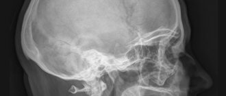
X-ray of the hand and wrist is a way to study their internal structure without invasive intervention. The hand and fingers are complex anatomical parts of the body, consisting of many small bones and performing important tasks in human functioning. This is the ideal and precise tool we use. Every day they are exposed to mechanical stress, their condition is affected by diet, quality of metabolism and other life processes. With the help of radiography, diseases in this area, cracks, dislocations and fractures are detected. Timely diagnosis prevents the development of severe diseases of the hands and wrists.
What does a hand x-ray show?
In everyday life, the human skeleton is subject to constant loads, including the hands. Excessive physical activity, infectious diseases, and mechanical damage change the structure of this part of the body and can sometimes lead to partial or complete loss of functionality.
To diagnose the disease and its cause in a timely manner, radiography is used. This method has been known for a long time and is still popular among traumatologists, surgeons, and therapists.
Content:
- What does a hand x-ray show?
- Indications for radiography
- Features of the study
- Research results
- Contraindications for hand radiography
- Alternative to radiography
- Where to take the study
- The myth about the dangers of research
The technique works on the principle of x-ray radiation. Beams of rays pass through the object and leave a contour mark on the film. Depending on the density of the tissue, the beam passes through it differently. Thus, bone tissue is reflected best, while soft tissue is reflected poorly.
However, based on the shadow in the image, a radiologist or other doctor can see changes in the lymph nodes, tendons, and muscles. The x-ray image shows the entire internal structure of the wrist and hand.
In the picture you can see all parts of the hand: wrist, phalanges of fingers, metacarpus. In turn, each of these sections contains several subgroups of bones, ligaments, joints, and muscles. Damage to even one unit of this system leads to pain, swelling, and deformation of the entire arm or its parts.
An x-ray clearly shows any damage to the bone tissue: thinning, cracks and microcracks, dislocations, inflammatory processes. To accurately diagnose problems with soft tissue, the doctor may prescribe a more informative computed tomography scan, or angiography to examine blood vessels.
Today medicine uses several options for examining the hands and wrist; they are more modern, more accurate, and faster. These include MRI, CT, MSCT and others. However, conventional X-rays continue to be used because they are simpler and more accessible.
Every clinic has such equipment; it costs much less or is completely free. In addition, the radiation dose for all progressive options and radiography is almost the same.
First aid after injury
After injury, the wrist must be immobilized. Any movement of the hand brings pain to the victim. To immobilize the wrist, a splint is placed on the wrist, which is made from improvised means: a board, a stick, a metal rod. Do not apply the splint directly to the skin. First, a cotton cloth or gauze is placed on the hand; in an emergency, a scarf or handkerchief will do.
Of particular concern is an open fracture, when bleeding begins from the wound. If possible, damaged tissues are treated with an antiseptic solution, and a pressure bandage is applied above the wrist. It is important to stop the bleeding; blood loss is life-threatening.
You should not try to replace a bone fragment protruding from the damaged area. The fragment is covered with gauze or any clean rag.
To relieve swelling, apply ice or a cold compress to the wrist. The patient is given a painkiller tablet: analgin, nise, ketones. If the patient is shivering, then he must be wrapped in a warm blanket or any available clothing. Warm drinks in the form of tea or water are allowed.
Indications for radiography
Like every part of the body, the hands and wrists are subject to illness and injury, and the latter happens to them most often.
To accurately establish the diagnosis and future treatment plan, the doctor prescribes an x-ray of the hand and wrist. The images also help to clearly establish the location of the injury, determine its severity and recovery methods.
X-rays of the wrist and hand are prescribed for:
- mechanical damage: bruises, fractures, dislocations;
- arthritis and arthrosis;
- rheumatism;
- swelling and hyperemia of the joints;
- osteomyelitis;
- congenital anomalies in the structure of the hands;
- neoplasms: tumors, cysts, papillomas;
- systemic lupus erythematosus;
- scleroderma.
It is impossible to accurately determine the cause of the disease and its stage by visual examination, so the doctor first prescribes a diagnosis. Any changes in the structure of the hand and wrist skeleton will be visible in the photographs. In addition, radiography is prescribed for children if a lag in physical development and too rapid development is noticed. The patient can undergo this procedure if he experiences pain in any part of the hand, notices curvature of the fingers, numbness, and swelling.
Possible complications
Complications can arise as a result of improper fusion, which causes deformation. If you experience persistent aching pain in the damaged area, as well as stiffness of movement, then immediately consult a specialist.
One type of complication is post-traumatic arthrosis, which is characterized by pain when bending the wrist, as well as a crunching sound in the injured area. Most often, this complication occurs a month after injury.
Another equally dangerous complication is arthritis. It is not difficult to diagnose, since the appearance of the injured area of the arm changes: a very noticeable swelling or depression appears, which is associated with muscle atrophy. Unlike post-traumatic arthrosis, with arthritis pain does not depend on the position of the hand. Pain is felt even if the hand is in its natural position.
Another complication may be contracture of the wrist joint after a fracture. Its result will be a violation of the flexion-extension function of the hand. The following symptoms will help identify contracture:
- increased limitation of joint mobility,
- the presence of dense nodules on the palm.
Features of the study
Pictures of the hands are taken quickly and painlessly. No preparation is required for the test; you need to take with you to the session directions from the doctor who prescribed it. X-rays are carried out in a separate room equipped with equipment. Before diagnosis, the patient removes jewelry so that it does not shade the image.
It is worth warning the doctor if there are metal inserts in the palms and wrists. To protect the rest of the body, the technician provides a lead membrane that blocks X-rays.
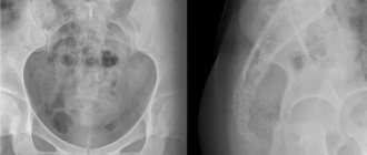
The installation consists of an X-ray table and a chair. The patient sits at the table and bends his limbs at the elbows, placing his hands on the table. Depending on the desired area of study, the projection of the images is changed. The patient's hands can be placed in different positions to obtain the most informative data.
For wrist x-rays, use:
- Direct projection. A cassette is placed under the palms, on which the photograph will then be reflected. Pictures can be taken from the back or from the palm, sometimes from two projections at once. The patient takes a stationary position, the laboratory assistant takes a photo using a remote button. This way the doctor will be able to examine all segments of the wrist: scaphoid, triquetrum, capitate and others. The phalanges and metacarpal regions are also visible. However, there are several segments that are not visible with this installation.
- Lateral projection. The hand is placed sideways on the cassette, with the thumb protruding slightly forward. This arrangement allows you to view the phalanges of the fingers, carpal and metacarpal bones from a different angle.
- I slant my palm. For this projection, the patient's hand is positioned so that the palm with the cassette creates an angle of 45 degrees. It is important that during the examination the hand does not move or shift; the laboratory assistant will monitor the correct positioning. Images from this position show the trapezium, scaphoid, and trapezius bones.
- I slant the back. In this case, the back of the hand creates a 45-degree angle with the cassette. In this way, the pisiform, hamate and triquetrum bones are best visible.
In addition to these methods, other types of styling can be used to examine the wrists. The radiologist can take targeted pictures of each individual bone. I also have my own radiography methods for reflecting fingers.
Each phalanx is placed in a direct and lateral perspective; the doctor can assess the condition of each of them separately. The accuracy of the results depends on the experience and competence of the doctor; he must set the minimum level of radiation and ensure the correct position of the patient.
For the patient, this procedure takes place without discomfort and lasts 5-15 minutes. If necessary, the doctor can take pictures in all projections for a detailed and complete study of the hands; for example, in case of infectious diseases, it is necessary to establish the entire affected area.
Also, in case of injuries or impaired development, a set of images is needed. No matter how many images need to be taken, the process will be quick and painless.
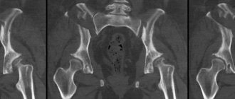
Diagnosis of fractures
Naturally, the basis for an accurate diagnosis is a qualified X-ray examination. In any case, irrefutable evidence of a fracture will be the detection of both the fracture line and individual displaced fragments. In children, a fracture with the beautiful name “green twig” often occurs when the young and flexible periosteum remains intact.
In this case, as well as in impacted fractures, identifying the fracture line presents certain difficulties. But, “fortunately”, for a beam injury in a typical place, an impacted mechanism is not typical, and yet variants are very rare.
The radiologist then determines the position of the fragments. Sometimes the distal fragment is not whole, but fragmented. In some cases, a fracture of the styloid process of the ulna is detected. This “surprise” is noted in 70% of all cases.
It is very important to determine the type of fracture - what mechanism caused the injury, and specifically on a lateral X-ray. When repositioning, you need to put the fragment in place so that it does not stand anteriorly or posteriorly. If this is not done, then after the bone heals, either flexion or extension of the hand may be limited.
Research results
After the images are taken, they must be interpreted and described by a radiologist. This will take 15-40 minutes, depending on the severity of the condition. Based on these data, the attending physician will determine the treatment regimen.
For hand and wrist injuries, therapy is carried out and monitored by a traumatologist; for other diseases, this can be a surgeon or therapist. The patient must take the results of the x-ray to the doctor who referred him for research. The images may reveal mechanical damage or rheumatic diseases.
Rheumatic diseases include scleroderma, joint inflammation, rheumatoid arthritis and other diseases. X-rays can also reveal dangerous joint psoriasis, bone calcification, and cysts. Advanced types of such inflammation lead to erosion of bone tissue and loss of functionality. In severe cases, X-rays indicate surgery, but systematic treatment is usually sufficient.
Symptoms
Each bone fracture has its own clinical symptoms, despite the fact that they are located in sufficient proximity. It is not always possible to make a correct diagnosis based on symptoms, but they can lead to an understanding of the essence of the issue. So, the scaphoid bone is characterized by:
- pain in the area of the “anatomical snuffbox”, it appears at the base of the first finger, between the tendons, if you lift it up;
- pain when tapping the first and second fingers;
- pain during dorsal extension of the hand;
- swelling and subcutaneous hemorrhages at the site of injury;
- signs of a broken hand in the wrist are complemented by pain when trying to clench into a fist;
- in a displaced fracture, the site of injury is deformed;
- fragments can crepitate, pathological mobility is observed;
- active and passive movements of the hand are limited.
Symptoms of damage to the lunate bone:
- pain at the site of injury;
- painful dorsiflexion of the hand;
- swelling and bruising in the projection of the bone;
- pain when putting pressure on the third and fourth fingers.
Symptoms of a fracture of the trapezium, triquetrum, trapezoid, pisiform, capitate and hamate bones:
- pain and swelling when palpating the damaged area;
- pain when putting pressure on the finger, the axis of which passes through the damaged bone.
Contraindications for hand radiography
Radiation examination carries a dose of radiation to the patient, so patients are often afraid of this procedure. However, when examining the hands and wrists, the dose is very small, so there are no categorical contraindications. This procedure is carried out even for small children; for maximum protection, they are completely covered with a lead apron, freeing only the hand.
Pregnant women are still not taken pictures unless absolutely necessary, but in case of fractures, the patient is also covered with a membrane. Breastfeeding mothers also undergo x-rays if necessary.
The session may be complicated if the patient has serious mental problems. To obtain clear images, the patient requires complete temporary immobility, which is not always possible in case of mental disorders.
Also, in a critically ill patient, such a study cannot be performed. If x-rays cannot be taken for some reason, alternative methods of examination are prescribed.
Causes
Each bone has its own factors that lead to damage. Thus, a fracture of the hand in the wrist , namely the scaphoid bone, occurs when falling on an outstretched arm. Road traffic accidents often lead to damage. Another reason is hitting a hard object with your fist or during a fight. Damage occurs as a result of a blow to the palm. Bilateral injury occurs less frequently and is caused by a fall on both upper limbs at once. In parallel, with a fracture of the scaphoid, dislocation of the lunate may also occur.
Injuries to the lunate are rare, occurring mostly from a fall on an outstretched arm or from a direct blow. Injuries to the trapezium, triquetrum, trapezoid, pisiform, hamate and capitate bones are rare in the practice of a traumatologist and are the result of a direct blow.
Alternative to radiography
Radiological diagnostic results can be obtained in other ways. The main competitor to radiography is computed tomography. It is based on the same radiation, but at a lower dose.
The CT scan process is faster and the results are more comprehensive. Thus, with a regular x-ray, soft tissues are poorly distinguished, although they are also often susceptible to disease. CT scan reflects vessels, muscles, and ligaments much better. However, the conventional technique is better suited for diagnosing bones.
Also, sometimes the X-ray method is replaced by MRI. This option is considered, like computed tomography, to be very informative. It provides comprehensive information about the condition of bone tissue and its surrounding elements. In this case, the patient does not receive radiation exposure at all, since the process is based on electromagnetic resonance. For a detailed examination of individual systems, such as blood or lymphatic vessels, lymphangiography or angiography is prescribed.
Rehabilitation activities
After the cast is removed for a wrist fracture, the rehabilitation process begins, however, some methods are used even before this. The patient is shown:
- physiotherapy;
- massage;
- physical therapy;
- nutrition.
It begins on the second day after the injury and the application of a plaster cast. All exercises should begin with gymnastics for the fingers; the elbow and wrist joints are additionally worked out. The second stage is performed after the plaster is removed under the guidance of an experienced rehabilitation therapist.
Where to take the study
If a simple x-ray is prescribed, it can be done at any emergency room, clinic, or hospital. The procedure requires conventional equipment, which is available in private and public medical institutions. More advanced techniques such as MRI or CT will have to be sought in private clinics.
The price for a regular photo of one brush in one projection will be 5-10 dollars. The cost may vary depending on the reputation and financial policies of the institution.
Two projections will cost twice as much. The price includes the examination and photographs; interpretation of the results in a public clinic should be free, in a private clinic you will need to pay an additional 5-8 dollars.
Rehabilitation
After the cast is installed, the person will have to live with an almost immobile arm for several months. Of course, this negatively affects the condition of the entire limb, and often the entire body. Blood circulation is reduced to a minimum, and it is the blood flow that is the main transport vector for nutritional components.
Muscle contractions, that is, physical exercise, help best. How to develop a hand without harming it? It is better to start the first training through passive movements, that is, it is necessary to contract different parts of the arm through willpower, and not through direct movement. After just a couple of days, you may notice slight fatigue in your hands, which indicates muscle function.
On average, a set of exercises can be like this:
- Clench and unclench your fist with force. Repeat this several times.
- Place your thumb and index finger together and relax the rest of your fingers.
- Connect all fingers, with the exception of the little finger, which must be set aside.
- Interlace the fingers of both hands, do this with some effort.
The simplest actions will also be useful: folding figures from matches, tying shoelaces. Once your bones have fully recovered, you can begin weight-bearing exercises to develop sufficient muscle strength.
Massaging the hand will help prevent contractures and difficulty moving the hand. You can contact a professional, but many patients perform simple movements at home. Stroking, rubbing and kneading are exactly what can improve capillary blood flow and tissue metabolism.