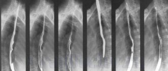
X-ray of the stomach is one of the methods for diagnosing various diseases and functional disorders of the gastrointestinal tract. The stomach is a hollow organ, so it is difficult to obtain sufficient information to make a diagnosis with conventional x-rays. To determine the size, shape, position of the stomach and its pathological changes, contrast fluoroscopy is used. This means that a contrast agent – a suspension of barium sulfate or air – is injected into the stomach before the examination. X-ray of the stomach differs from fluoroscopy in that it is a simultaneous image without dynamics. During fluoroscopy, the doctor observes a radiopaque image of the stomach in real time on a special screen. In this case, a series of images are taken reflecting the dynamics of the passage of barium suspension from the esophagus to the stomach and beyond.
Indications and contraindications for fluoroscopy of the stomach
Fluoroscopy is indicated in the presence of symptoms of gastrointestinal disease:
- pain behind the sternum and in the epigastric region associated with eating (or night “hungry”);
- periodic nausea and vomiting;
- dysphagia;
- stool disorders (diarrhea, constipation);
- belching;
- heartburn;
- the presence of blood in the stool;
- rapid weight loss;
- anemia of unknown origin;
- signs of obstruction of the esophagus or stomach.
Also, contrast fluoroscopy of the stomach with barium can be prescribed in the presence of diagnosed gastrointestinal diseases to determine the dynamics of their course (stomach ulcer, benign and malignant tumors, achalasia of the esophagus, pyloric stenosis, etc.). Using this method, you can obtain data on the effectiveness of therapy or the results of surgical treatment. Another indication for research is the presence of precancerous diseases of the gastrointestinal tract or a hereditary predisposition to neoplasms (polyposis, cancer).
Contraindications
- serious condition of the patient,
- pregnancy,
- persistent esophageal or gastrointestinal bleeding.
These contraindications are relative, since after the condition improves and bleeding stops, an X-ray contrast examination of the stomach is possible. If necessary, fluoroscopy is performed in the second and third semesters of pregnancy after agreement with the obstetrician-gynecologist. Fluoroscopy is performed only in cases where it is impossible to perform esophagogastroscopy or its data is insufficient to make a diagnosis.
How are barium x-rays of the stomach and esophagus done?
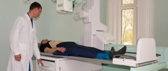
Many people are interested in what and how they drink during an X-ray of the stomach with barium. For diagnostics, barium sulfate is used, the drug is taken orally, dissolved in water. It is completely safe for the body, looks like a white suspension, tastes like chalk or calcium. The first photo is taken before barium is consumed. Then the patient drinks a few sips of the drug and the examination of the esophagus begins. After a few minutes, the patient finishes the solution, after which the radiologist continues to examine the stomach, taking pictures as the barium moves. During the diagnosis, the patient is asked to lie on a table and periodically change position, following the doctor's instructions. For a clearer image, the radiologist may ask the patient to hold his breath while taking the picture. The study lasts from 20 minutes to an hour. Next, the doctor examines a series of images and writes a conclusion.
When diseases are detected, many patients are interested in how often X-rays can be taken, waiting for a repeat examination to be scheduled. There is no clear answer to this question, since the doctor choosing a diagnostic method is guided by the individual circumstances of a particular patient. Age, condition, radiation dose and the appropriateness of the procedure are taken into account.
Preparation for the procedure
Useful article? Share the link on VKontakte
Preparation for fluoroscopy of the stomach does not require any special measures. The study is performed on an empty stomach; before this, you should not eat or drink for 8-10 hours. To obtain objective data, it is recommended to follow a diet for 3 days before the procedure. Foods that cause flatulence (legumes, brown bread, fatty, fried, smoked foods, fruits, vegetables) should be excluded. Preference is given to lean boiled meat (chicken, beef), lean fish, white stale bread, water-based porridge, and eggs. If you are constipated on the eve of the examination, you should do a cleansing enema. If the patient has pyloric obstruction, then before taking an x-ray with barium, the stomach is washed with a probe. Immediately before the procedure, you must remove any jewelry and remove removable dentures.
X-ray of the esophagus of the stomach and duodenum
For three days you should not eat:
- legumes;
- black bread;
- fruits;
- vegetables;
- dairy products;
- flour products;
- fried and smoked foods;
- sparkling water.
You can include in your diet:
- boiled lean meat;
- fish;
- white stale bread;
- porridge cooked in water;
- boiled eggs.
X-ray of the stomach is performed on an empty stomach, that is, 6-8 hours before the examination the patient should not be allowed to drink or eat.
In case of constipation, an enema is performed. If the patient has pyloric obstruction, barium probing is performed before fluoroscopy. Before the procedure itself, you need to get rid of jewelry, removable dentures, and other things.
Preparation
The day before the test, it is recommended to avoid eating dense, poorly digestible or gas-forming foods. From the evening before, eating is excluded altogether. Only drinking water is allowed.
On the recommendation of the attending physician, if an x-ray of the stomach is to be performed, a cleansing enema may be prescribed.
If you plan to study the rectum or large intestine, then the diet changes another 2-3 days in advance. In addition, it is recommended to cleanse the intestines with the help of special drugs: Fortrans, Lavacol. Cleansing enemas are also prescribed.
note
After all the preparation and enemas, you need to get clean rinsing water. The quality of the research directly depends on the quality of preparation for it. If feces remain in the intestines, the informativeness of the study may suffer.
If the patient is prone to chronic constipation, then more serious preparation is recommended. This may be an additional use of laxatives or enemas.
In the morning, at the beginning of the study, a survey radiograph of the abdominal cavity is taken. Then the patient drinks the barium mixture. The radiologist monitors its progress through the gastrointestinal tract on the fluoroscope screen. Barium should fill the stomach and thus define its internal relief.
If necessary, for better visualization, the radiologist can ask the patient to turn over, lie on the other side, and can press on the stomach so that the barium fills and marks all the bends of the stomach. An X-ray of the stomach takes about a quarter of an hour.
If only an X-ray examination of the large intestine is necessary, then barium can be injected directly into the intestine through the anus, through a tube inserted into it. The intestinal examination will take about an hour.
The barium mixture has a slight chalky taste. Mostly patients do not experience difficulties with its use, but in rare cases nausea and vomiting occur. There are no painful sensations when the barium suspension moves through the intestines.
While an x-ray is being taken of the stomach and other parts of the digestive tract, the radiologist takes a series of pictures.
Advantages of the method
Fluoroscopy of the gastrointestinal tract has a number of advantages, which explains its widespread use today:
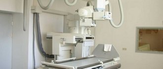
- accessibility (X-ray equipment is available in almost any hospital and is widely used);
- ease of implementation;
- reasonable price;
- safety (the risk of allergic reactions is low).
During the examination, the radiologist first evaluates the quality of the images and the degree of preparation of the gastrointestinal tract for the examination. Then the size of the gastrointestinal tract organs, internal relief (intestinal folding, smoothness of folds), time of barium movement, and the presence of obstacles are determined.
When a defect is detected, its size, clarity of contours, and shape are assessed. Peristalsis is also assessed, that is, the time during which barium must leave each organ sequentially. After a certain time, pictures are taken.
So, after 3 hours there is no barium in the stomach, after 6 hours in the intestines.
There are a number of specific radiological signs of gastrointestinal diseases, which are described by a radiologist:
- Change in lumen. Its narrowing may be caused by a formation growing inward, for example, an oncological tumor or polyps. Compression from outside the intestine or stomach may reduce the lumen. Increasing the lumen is also possible. Uneven expansion is observed with diverticula.
- Displacement of the organ itself. An X-ray of the stomach may show prolapse, or gastroptosis, and in severe cases this can cause it to be located in the pelvis.
- Violation of the integrity of the organ.
- "Niche" symptom. A defect in the stomach wall, filled with contrast, gives an intense degree of darkening in the image, possibly extending somewhat beyond the perimeter of the organ. This manifestation may indicate a peptic ulcer. More often it is detected when an x-ray of the stomach and duodenum is performed.
X-ray sign of a stomach ulcer
- Filling defect. It shows a place that the contrast has bypassed and has not penetrated. It could be a polyp that grows inside the lumen, a tumor, or a foreign body.
- Violation of the direction of folds. If folds are visualized that come together and are directed towards the “niche”, then an ulcerative defect can be assumed.
- Smoothness of folds. This symptom is determined by inflammatory phenomena. With inflammation and, as a consequence, swelling of the intestinal wall, its folding becomes less pronounced.
- Liquid levels (Kloiber bowls). This symptom is characteristic of intestinal obstruction, when fluid is retained in the intestine at the bends of the intestine, forming “cups” with liquid intestinal contents.
- The following symptom also indicates intestinal obstruction: the intestinal loops up to the site of obstruction in the intestine are stretched and inflated with gas, and after the site of stenosis they collapse.
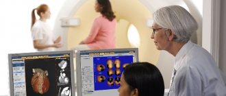
The radiologist describes all the radiological symptoms he has identified. All images and their descriptions are transmitted to the attending physician and subsequently stored in the patient’s medical history.
Contraindications
X-ray examination with barium is contraindicated in the following cases:
- acute stage of the inflammatory process;
- pregnancy;
- perforation of a hollow organ of the gastrointestinal tract.
Thus, X-ray examination remains today one of the leading methods for diagnosing diseases of the digestive tract. It will allow you to reliably determine the presence of ulcers, polyps, diverticula, intestinal obstruction, and suggest tumors of the digestive tract.
Indications for fluoroscopy:
- signs of a stomach ulcer;
- dysphagia;
- control after gastric surgery;
- bowel dysfunction;
- blood in stool;
- profuse vomiting;
- sudden weight loss;
- pain symptoms after eating;
- malformations of the stomach;
- diverticula;
- pain in the gastrointestinal tract on an empty stomach.
- Fluoroscopy should be performed if a blood test shows a sudden decrease in hemoglobin levels.
- Diagnosis of gastric diseases using radiology (video author - Medical company ilaya).
- Contraindications to the procedure are:
- pregnancy;
- continuous bleeding in the stomach and intestinal tract.
The purpose of radiography for diagnosing diseases of the gastrointestinal tract is an individual choice of the doctor, because there is probe gastrography, which is not accompanied by radiation exposure.
Due to the high background radiation, x-rays of the duodenum and stomach are prescribed according to strict indications. However, the technique can detect bowel cancer in 75% of cases in older people, and there is no more effective alternative.
Photo of a targeted radiograph: a large adenomatous polyp (indicated by an arrow)
X-rays of the stomach and duodenum are prescribed if the following indications exist:
- gastrointestinal discomfort;
- dysphagia;
- vomiting and nausea;
- unexplained weight loss;
- stomach ache;
- ascites;
- liver enlargement;
- occult blood in stool;
- anemia of unexplained etiology.
Some European doctors conduct X-ray examinations of the intestines in patients who are at high risk of developing stomach and duodenal cancer.
In practice, our radiologists are convinced that the value of the study is different for each patient. Sometimes contrast X-rays do not provide valuable information, and examination can only reveal disturbances in intestinal motor function.
Photo. Fibroplastic gastric cancer (SFGC): concentric narrowing with an uneven contour (indicated by arrows)
We also note that when choosing radiography tactics, it is not so much the experience of the radiologist that plays a great role, but rather his professional qualifications. A rational plan for the sequence of x-ray procedures allows for maximum efficiency with minimal patient exposure.
There are several types of x-ray examination of the small intestine:
- Verification.
- Urgent.
- Classic.
- Double contrast.
- Two-phase.
Test X-rays are carried out in medical institutions on gastrographic units under the control of an X-ray television monitor. In the absence of special equipment, the examination is carried out using a RUM-20 device, which was produced in Soviet times and has a television path sufficient for a full diagnosis.
Using the double contrast technique, radiologists are able to study the structure of the relief of the mucous membrane. In this case, it is possible to detect not only ulcers and duodenal cancer, but also superficial gastritis.
What is double contrast gastric contrast:
- 2 days the gastrointestinal tract is cleansed (fortrans, enema, activated charcoal);
- 30 minutes before the procedure, the patient takes 2-3 Aeron tablets under the tongue;
- for contrast, a solution of barium sulfate with an antifoam is used (for example, 1 gram of dimethylpolysiloxane);
- to swell the stomach, a person takes a gas-forming substance (urodan);
- after transillumination of the duodenum, a series of targeted radiographs are performed in the supine and standing positions;
- On average, the duration of the study takes 5-7 minutes.
A two-phase examination of the stomach involves a combination of a classic examination of the stomach and double contrast. In the first phase, the radiologist performs double contrast according to the scheme described above.
In the second stage, a tight barium filling is carried out.
To properly conduct a two-phase examination, it is necessary to prepare a barium suspension of high (for the 1st phase) and low (for the 2nd phase) density.
If a perforated ulcer or perforation of the wall of the small intestine is suspected, contrast is performed with water-soluble substances - gastrografin or verografin.
- Detection of stomach pathologies;
- Included in the comprehensive diagnosis of ulcers, tumors, enteritis;
- The structure of the mucous membrane is studied;
- Detection of gastrointestinal motility disorders.
Technique for X-ray contrast examination of the stomach
First, a survey X-ray of the chest and abdominal cavity is performed, which allows one to identify gross pathological changes. The patient is then asked to drink a contrast agent. Barium for X-rays is used in the form of sulfate. The barium sulfate suspension is white in color and tastes like chalk. The first photo is taken after the first two sips of the suspension. At this stage, the relief of the walls of the esophagus is determined. Then the subject drinks the remaining barium (about a glass). The images are taken on an x-ray table. The patient is asked to change position during the procedure.
The duration of fluoroscopy is about 40 minutes.
If it is intended to examine the lower intestines, then the images are repeated at certain intervals throughout the day.
Important: after fluoroscopy of the stomach, constipation may occur for 2-4 days. The stool is white or gray for some time - this is normal and goes away on its own. After the study, it is recommended to drink 1.5-2 liters of clean water to speed up the removal of barium.
X-ray as one of the first prescriptions for gastrointestinal pathologies
Because of its excellent diagnostic ability, x-rays have long been recognized as the number one study for suspected pathological processes in the gastrointestinal tract (gastrointestinal tract). Therefore, at the first manifestations, such as:
- pain in the chest or abdomen;
- difficulty swallowing;
- occasional belching or heartburn;
- vomiting for unknown reasons:
- disruption of the digestive process;
- blood particles in the stool (a sign of bleeding), the doctor recommends an X-ray of the esophagus and stomach.
X-ray examination of the stomach and other parts of the gastrointestinal tract is the process of visualizing the organs being studied using X-rays.
This diagnostic method is almost always prescribed in a number of the first tests, because x-rays of the stomach and esophagus show quite a lot of pathological changes, including:
- erosive and ulcerative lesions of the mucous membrane and underlying muscle layers;
- neoplasms of both benign and malignant nature;
- inflammatory processes localized in the duodenum;
- hiatal hernia;
- violation of motor-evacuation function;
- formation of pathological scars on the walls of the stomach and esophagus;
- obstruction of the entire digestive tract.
Thanks to an X-ray examination of the gastrointestinal tract, the doctor is able to detect until a certain time unknown causes leading to a particular pathology, and, if possible, choose the most appropriate therapy. And also evaluate the quality of functioning of the digestive system.
This method of examining the gastrointestinal tract allows, using X-rays, to obtain a complete picture of the examined organs with all pathological changes in their shape, structure, and even evaluate them over time. But it is possible to take an x-ray of the stomach and other organs only under certain conditions that the patient will definitely need to fulfill in preparation for the study.
Due to the structural features of the digestive organs - the esophagus, stomach, small and large intestines, which have a hollow shape that does not block X-ray radiation, contrast is used to successfully carry out the procedure. As a contrast agent, a drug made from barium is used, diluted with water to the consistency of a kind of suspension.
Barium coats the walls of the esophagus, which makes it possible to detect their defects
In some cases, in order to straighten the folds of the mucous surface of the gastrointestinal tract, immediately before the start of the study, the patient is asked to drink a solution of crystalline soda, which increases visualization. Sometimes, for the same purposes, air is introduced under pressure or using a perforated tube, through which the patient simultaneously receives a barium suspension.
This examination allows you to assess the condition and functional capacity of the stomach and esophagus based on studying the image on x-rays. Ingestion of a barium mixture, which tends to accumulate in the mucous membrane of organs, provides their detailed visualization. When a barium solution passes through the gastrointestinal tract, it will be easy for a diagnostician to recognize pathological changes in their shape and structure.
By noting the time of movement of the barium suspension through the digestive tract, the doctor will be able to draw a conclusion about stenosis (narrowing) or obstruction of any part of the system.
Determining the presence of esophageal stenosis by barium passage rate
There are several techniques for performing fluoroscopy of the stomach and esophagus. Depending on the indications and existing symptoms, the diagnostician chooses the most informative one. Research methods are divided into traditional, Trendelenburg examination and double contrast.
As a rule, X-rays of the gastrointestinal tract are done in the morning to relieve the patient from the discomfort associated with prolonged fasting. After carrying out all the necessary measures to clean the organs being examined, the patient drinks a barium solution in the X-ray room and is placed on a functional table.
This technique involves the patient lying down on a lowering table. During the diagnostic process, if there is a need for tight filling of the examined organs, additional intake of barium mixture may be required. During the procedure, the patient is asked to change body position, which allows the doctor to obtain information about all parts of the organs being studied - pharynx, esophagus, diaphragm, stomach, etc.
The doctor evaluates the overall picture and takes targeted photographs from different angles. The contours of the shadows are the inner surface of the organ. To ensure that the barium is evenly distributed, the patient is first placed in a horizontal position. The mixture does not stay in the esophagus for long - when you exhale, the esophageal sphincter relaxes and the solution enters the stomach.
It envelops the walls of the stomach, fills the cavities between the folds and makes it possible to visualize them. Better distribution of the solution inside the stomach is facilitated by stroking movements with your hands with slight pressure. They can be performed either by the patient himself or by the medical staff present. This technique lasts approximately 40 minutes. During the examination, the patient may experience attacks of mild nausea, which quickly pass without leading to vomiting.
After the procedure, barium sulfate is released naturally, thereby discoloring the stool. Given the potent astringent effect of barium, the patient may be constipated. Therefore, after diagnosis, to speed up the elimination of barium sulfate, you should drink at least 1.5–2 liters of water, which will ensure that the body quickly gets rid of both the drug and disturbances in peristaltic activity.
If stools have not returned to normal over the course of several days, you should definitely notify your doctor about such disturbances and receive recommendations for solving problems with restoring intestinal function. Do not delay your visit to the doctor under any circumstances to minimize the risk of intestinal obstruction!
The main purpose of this technique is to visualize a diaphragmatic hernia. An important point in this examination is to place the patient on his back with the pelvis elevated at an angle of 45° in the so-called Trendelenburg position, in which images are taken. In this position, the intestinal loops and other gastrointestinal organs are shifted towards the diaphragm and are visualized using a contrast agent, providing the diagnostician with the opportunity to determine the presence of a diaphragmatic hernia. When this defect is present, the organs prolapse into the chest cavity.
A hiatal hernia protruding into the diaphragm is diagnosed using the X-ray contrast method
Diaphragmatic hernia when using the Trendelenburg technique is determined by direct signs:
- change in the X-ray shadow of the heart;
- airiness under the diaphragm;
- darkening of the pulmonary field near the cavity;
- the lumen in the lateral photograph is round in shape;
- change in shadow shape when breathing;
- filling a separate cavity with contrast;
- the presence of gastric folds in the esophagus.
And also by indirect manifestations, characterized by: the absence of gas bubbles in the stomach contents, tortuosity of the esophagus above the diaphragm, displacement of the stomach to the lower third of the esophagus, rotation of the heart along the longitudinal axis, flattening of the vault and a decrease in the volume of the ventricles. This examination method is contraindicated for elderly and senile people with sclerosis and functional disorders of the heart and lungs.
It is also prohibited to carry out it in the presence of blood, pus, exudate and oncological processes in the peritoneum.
The technique involves conducting an examination using two contrasts - barium and gas. To do this, the patient is asked to drink a barium solution through a perforated tube, which allows him to simultaneously swallow air. Massage of the anterior abdominal wall promotes the distribution of the barium mixture, and the air ensures the straightening of tissue folds.
To relax the smooth muscles of the gastrointestinal tract and reduce peristalsis, antispasmodic drugs are administered. This technique allows you to obtain a complete picture of the esophagus and abdominal organs, which helps to identify various pathological processes, including oncology in the early stages.
Oncological diseases detected using radiography
Of course, it is not easy to carry out such a procedure for a child, but there are no special problems with older people. If a small child refuses to drink barium, then contrast is administered through a special tube. To make it easier to examine the baby in all projections, he is placed on a rotating platform that provides an inclined position.
X-ray examination remains a relevant method not only for the study of bone structures, which absorb X-rays well and provide clear images. The gastrointestinal tract is no exception. Moreover, despite the fact that the digestive tract is not radiopaque. X-rays of the stomach and intestines are performed today in almost any hospital.
Duodenum: ulcer (A) and tumor of the descending section (B)
To make the study informative, the lumen of the gastrointestinal tract only needs to be filled with a substance that is radiopaque. For this, a barium solution is used.
This substance, entering the cavity of the stomach and then the intestines, envelops its walls, fills the lumen, making all its internal bends and defects visible. Barium testing is safe.
The drug is tasteless, is not absorbed at all in the intestines, and is excreted without loss. Allergies to it are rare.
Sometimes double contrast is used. For this, not only barium contrast is used, but gas is also injected. This allows you to somewhat “stretch” the collapsed walls of the hollow organ and “straighten” its folds.
Indications
X-ray examination of the stomach and other parts of the gastrointestinal tract is prescribed to diagnose various pathological changes:
- ulcerative defects of the walls of the stomach and intestines;
- hiatal or diaphragmatic hernia;
- diverticula of the esophagus, intestines;
- oncological formations;
- intestinal obstruction;
- gastroesophageal reflux (disease);
- irritable bowel syndrome;
- disturbances of peristalsis;
- Crohn's disease and other inflammatory bowel pathologies;
- foreign bodies of the gastrointestinal tract.
Fluoroscopy analysis of the stomach
The results of fluoroscopy of the stomach are interpreted by a radiologist and gastroenterologist, and sometimes by a surgeon. With fluoroscopy, you can obtain data on the speed of movement of barium suspension through the esophagus and stomach, and the time of its entry into the duodenum and colon. Based on this sign, one can judge the presence of motor disorders and difficulties in evacuating a food bolus from the esophagus or stomach. Based on the relief of the mucous membrane and the uniformity of barium distribution, the presence or absence of its changes is determined.
Symptoms determined by fluoroscopy of the stomach
- Changes in the position of the esophagus and stomach (with congenital anomalies of the structure of the digestive organs, prolapse of the stomach, previous operations, tumors of the mediastinal organs, myocardial hypertrophy).
- Narrowing of the lumen of the esophagus or stomach (tumors, strictures, cicatricial narrowings due to ulcers, chemical burns, increased tone of the muscle wall).
- Expansion of the lumen (with achalasia cardia - expansion of the esophagus, decreased wall tone with Hirschsprung's disease and functional disorders).
- Violation of the integrity of the wall (perforation of the walls of the esophagus or stomach due to ulcers).
- Symptom of “niche on the contour” (ulcers).
- Filling defect (polyps, papillomas, foreign bodies, malignant tumors).
- Reducing the folding of the mucous membrane (with atrophic gastritis).
- Radiation-like convergence of mucosal folds (together with the niche symptom indicates a peptic ulcer).
The listed symptoms, when detected, are not always the basis for making a diagnosis. Fluoroscopy data are assessed taking into account complaints, objective examination data, results of laboratory and instrumental diagnostic methods. There are many private clinics and diagnostic centers where you can do fluoroscopy of the stomach. But you should not prescribe this study yourself, because excess radiation exposure can negatively affect your health.
Important: if you discover symptoms of gastrointestinal diseases, first consult a doctor who, if necessary, will prescribe an x-ray examination of the stomach.
One of the most common ways to diagnose stomach pathologies is x-ray. It can be used to identify neoplasms and peptic ulcers. An X-ray of the stomach will show what is happening with the sphincters, what is the integrity of the walls, what are the dimensions of the organ. The procedure is prescribed only by doctors.
What does a two-phase study of the stomach show?
A two-phase examination of the stomach involves a combination of a classic examination of the stomach and double contrast. In the first phase, the radiologist performs double contrast according to the scheme described above.
In the second stage, a tight barium filling is carried out.
To properly conduct a two-phase examination, it is necessary to prepare a barium suspension of high (for the 1st phase) and low (for the 2nd phase) density. If a perforated ulcer or perforation of the wall of the small intestine is suspected, contrast is performed with water-soluble substances - gastrografin or verografin. When using them, the cocktail is prepared as follows: 200 grams of contrast with 350 grams of Borjomi and half a bag of vanillin.
Biphasic contrast shows:
- stomach and duodenal ulcers;
- violations of the motor function of the organ;
- esophagogastric reflux;
- cancerous filling defects.
Indications for the procedure
An X-ray of the stomach will show what exactly is happening in the organ. This type of diagnosis is prescribed to patients in the following cases:
- suspicion of developing an ulcer;
- inflammatory diseases;
- neoplasms;
- organ deformation;
- developmental defects.
Indications include symptoms of rapid weight loss, anemia, and blood in the stool. There are other indications for the examination. However, not all patients are allowed to undergo the procedure.
Features of the procedure for children
Unfortunately, stomach diseases and organ pathologies occur not only in adults, but also in children. Any health problems in a child also require informative diagnostics in the form of an X-ray of the stomach. Children over 10-12 years old are examined with a barium suspension. If we are talking about a small child or even an infant, then a contrast agent is used that dissolves in water. Often, fluoroscopy of the esophagus and stomach is performed in young children without any contrast agent at all. Children are prescribed procedures for the following indications:
- the child has swallowed any foreign body;
- symptoms of gastric intussusception are observed;
- the doctor suspects the presence of an inflammatory process in the gastrointestinal tract;
- The child is suffering from acute pain in the stomach.
Preparation rules
All patients are conditionally divided into two groups: those whose intestinal functioning is impaired and those whose intestinal function is normal. Each group has its own preparation rules.
An X-ray of the stomach will show what exactly caused the pain and other clinical manifestations of the pathology, but before the procedure it is necessary to prepare the body. To do this, patients with no bowel dysfunction should abstain from eating for at least twelve hours.
If there are any disorders of the digestive tract, then preparation begins three days before the date of the x-ray. The patient should completely exclude foods that cause gas formation from the diet. The menu includes as much protein food and porridge as possible. If necessary, patients are prescribed a cleansing enema.
An X-ray of the stomach and intestines shows what exactly caused the digestive disorder and what changes have already occurred in the organs.
Why do they do it?
An X-ray method for examining the gastrointestinal tract is prescribed if the patient has symptoms:
- decreased appetite;
- feeling of heaviness in the upper abdomen after eating;
- nausea and/or vomiting several times;
- aching pain in the abdomen, which intensifies on an empty stomach or after eating;
- tendency to constipation;
- burning sensation behind the sternum;
- increased gas formation (flatulence);
- belching sour;
- tendency to constipation or diarrhea;
- weight loss.
The use of X-ray methods helps diagnose the following pathologies:
- congenital malformations of the stomach and lower esophagus;
- peptic ulcer;
- perforation of the stomach wall (violation of the integrity of the organ);
- deformation of individual parts of the organ;
- inflammation of the gastric mucosa (gastritis);
- benign and malignant neoplasms, metastases of tumors from other organs;
- narrowing of the pyloric region (pyloric stenosis) and gastric obstruction.
Examination methods
There are two ways to conduct an X-ray examination of an organ: with contrast and the double-contrast method.
The stomach is a hollow organ. To see it in the picture, it needs to be filled with a contrast agent. X-rays of the stomach show exactly what is happening and how it is developed. For clarity, the organ is filled with barium sulfate. This substance does not dissolve in gastric juice and is not absorbed in the intestines. Barium does not absorb X-rays. Because of these features, an X-ray of the stomach with barium shows what happened to the organ and what pathological foci are present in it.
An x-ray examination with double contrast is performed to more clearly examine the walls of the stomach and intestines. Together with the contrast, air is pumped into the digestive tract. If you need to examine the upper part of the organs, then air is introduced through the mouth. When examining the lower parts - through the anus.
To ensure that the muscles are relaxed during diagnosis, patients are prescribed antispasmodic medications. They suppress the peristalsis of the intestines and stomach.
How to perform an x-ray of an organ with barium
How are x-rays done? Many people who are about to undergo this procedure want to know the answer to this question. A stomach x-ray with barium lasts from 20 to 40 minutes on average. Before starting the procedure, the radiologist asks the patient to remove all metal jewelry from the body. First, 1 series of images of the organ is taken without the use of a contrast agent. The person must stand still.
For further examination of the stomach, you need to drink 250 ml of liquid, which contains a contrast agent - barium suspension. In terms of taste, this drink does not cause disgust. It has a delicate, pleasant taste, reminiscent of a milkshake. Immediately after ingesting the liquid, the doctor begins to examine the walls of the stomach, as well as its mucous membrane.
X-rays are taken in various positions. To begin, the patient is asked to lie down on the couch. For an informative examination, you need to take a series of photographs on the stomach, back, and side. The basic posture for fluoroscopy is supine with legs elevated and slightly bent at the knees.
Possible consequences
Scientists have proven that the effect of X-rays on the human body is absolutely harmless. As for contrast, barium must be ingested, the dosage is about 250 ml.
To avoid any consequences after this type of diagnosis, doctors recommend following a certain diet for 2 days after the procedure. Meals should be based on plant foods; solid foods, including meat, should be avoided. Preference should be given to liquid porridges and fermented milk products.
The main condition of this diet is to drink fluid in maximum quantities. If a person drinks up to 3 liters per day. pure water, in which case the barium suspension will leave the body and will not cause any trouble.
Carrying out diagnostics
The doctor explains how an X-ray of the stomach is performed and what it shows, whether this method will help in making a diagnosis.
Patients should come to the procedure with an empty stomach, as the study is performed on an empty stomach. Before the patient is given a contrast agent, the specialist must conduct a general examination of the abdominal cavity. This is done to identify possible problems with the functioning of the stomach. For examination, the patient is placed in an X-ray machine equipped with a screen. Then he is given a contrast. It is taken orally under the supervision of a specialist.
On the monitor screen, the doctor sees how the barium passes through the digestive system, entering the stomach and then the intestines. The specialist takes a series of photographs in different projections. First, the patient lies on his back, then turns over on his stomach, on both sides. After this, the patient gets up.
The duration of the procedure depends on the number of projections in which images need to be taken.
Order of conduct
At the first stage, preliminary photographs are taken without the use of a contrast agent. At this time the patient is standing. Such a preliminary x-ray is needed to identify gross pathologies in the gastrointestinal tract, if any.
The patient then sits down and is given an aqueous barium solution to drink. This liquid mixture has a fairly thick consistency. It is similar in color and taste to a solution of ordinary chalk. Barium sulfate is absolutely harmless to the body. It is easily eliminated from the body, since the stomach and intestines do not absorb it.
The contrast agent is drunk gradually as the procedure progresses. First, the patient takes a couple of sips, after which the doctor examines the condition of the esophagus. This happens while standing.
Then the remaining contrast liquid is drunk, then the patient is placed on the table and a fluoroscopy of the stomach is performed. In order for the barium to be better distributed, the doctor, in the intervals between images, presses the patient’s stomach.
The total duration of the procedure is 40 minutes. At this time, the patient is asked to take different positions: lying on his side or on his back. His body is fixed on the table. A photograph taken in the Trendelenburg position is required (the pelvis is raised at an angle of 45 degrees and breathing is held).
If there is a need to examine the lower intestines, the procedure is interrupted for some time and then resumed again. In this case, a series of photographs is taken during the day with breaks.
At the end of the diagnostic session, the patient is asked to drink about 2 liters of water so that the barium leaves the body faster.
If the patient followed all the recommendations exactly and the procedure was successful, then after its completion no complications arise. The patient may experience mild indigestion and gray or white stools. There is no need to be afraid of this, such phenomena occur due to barium sulfate, they will soon pass.
Side effects of the examination
During the examination, patients are exposed to x-rays. Every patient is concerned about the safety of the contrast agent. Due to the chemical structure and the inability to be dissolved by gastric juice, barium is not absorbed, but is released naturally some time after the procedure. In isolated cases, an allergic reaction to contrast may occur.
What does the diagnosis show?
X-rays of the stomach and esophagus show what is happening in the digestive system and what changes are present. The doctor can see the changed lumen in the pictures. When it narrows, endophytic or exophytic cancer is noted. Sometimes there may be a tumor that presses on the stomach, for example, when cancer cells grow in the intestines. You can see various deformations in the pictures. They can be congenital or acquired. Usually the stomach has the shape of a hook, but there are other types: horn, snail, shapes with constrictions, reminiscent of an hourglass.
X-rays clearly show all existing displacements, including gastroptosis. This is prolapse of the stomach. This variant of the disease develops with hernias and various injuries.
The doctor can see a niche symptom in the image. It is characterized by filling the damaged area of the stomach with a contrast agent. Most often, this clinic is characteristic of peptic ulcer disease. The x-ray shows intense darkening that occurs at the site of the defect in the organ wall.
If a dark area is visible in the image, this indicates a defect in the filling of the digestive organ. This clinical picture can be observed with a tumor, as well as with foreign bodies.
The thinning of the folds of the stomach caused by atrophic gastritis is practically invisible. But the doctor can clearly see the existing radial convergence of the folds of the mucous membrane towards the niche.
What does the barium procedure show?
As mentioned above, using x-rays you can identify many diseases, as well as the causes of their occurrence. Barium, in turn, is necessary in order to accurately determine the size of the mucous membrane of the organ and see some of the features of its structure. Let's look at what an x-ray of the stomach shows:
- change in the location of the stomach;
- increase or decrease in size;
- the presence of an inflammatory process;
- cancerous tumors;
- The duodenum or stomach is in the stage of intussusception;
- foreign bodies in the stomach;
- presence of polyps.
Reviews
According to doctors, when examining the images, a hook-shaped stomach with normal, not enlarged dimensions is normally visible. The contrast agent is evenly distributed throughout the entire cavity of the organ, filling all the bends. During X-ray examination, no filling defects or bends are observed.
Doctors say that with a peptic ulcer, there is a niche from which the folds of the mucous membrane diverge. The clear and round contours of the stripes that appear when the stomach fills occur when there are a large number of polyps.
X-ray examination of the stomach with contrast allows you to see a variety of pathological foci. Also, this diagnostic method allows you to determine the size of the organ, its bends, and assess the condition of the digestive system as a whole.
X-ray of the stomach and duodenum
X-ray of the duodenum refers to a radiation examination of the upper part of the gastrointestinal tract. Fluoroscopy is performed using a contrast agent.
This method allows you to determine organ disease if the pathological focus is captured by the device. If abnormalities are found during the X-ray, it is necessary to carry out endoscopic diagnosis with the collection of material for a biopsy.
X-ray of the duodenum is carried out as follows:
- First of all, you need to conduct a survey x-ray of all organs located in the abdominal cavity. This procedure is carried out in a direct projection. This method helps to identify acute surgical diseases: bleeding, organ rupture or intestinal obstruction. Next, the patient needs to take a barium solution orally. The suspension has the consistency of sour cream and the taste of limestone. As a result, a series of photographs are taken in various positions of the body, which changes during the course of the study. After the procedure is completed, the patient needs to drink a lot of fluids, in particular water, this helps to quickly remove barium from the stomach and intestines.
- The barium solution drunk should enter the duodenum without any obstacles. In the absence of disease, the duodenum should be covered with folds of mucous membrane. if pathologies occur during filling, deformation, or tumors of unknown etiology grow, then all this is a disease of the duodenum and is a serious reason for undergoing additional diagnostics.