The female breast is not only a source of pride for the fair sex, but also a subject of concern, and one of the main indicators of failures and improper functioning of the reproductive system, as well as a kind of litmus test for the state of a woman’s hormonal background.
This article highlights the main points and nuances of a common disease - fibrocystic mastopathy. This disease occurs in almost every second middle-aged woman, and is a borderline pathology, which, if neglected, has a risk of degeneration into cancer.
What is mastopathy?
Mastopathy is the most common pathological condition of the mammary glands. More than 50 percent of women are faced with this diagnosis sooner or later, and this statistics is inexorably growing, and the disease is getting younger.
Mastopathy is a pathological condition directly related to the hormonal state of the female body. Despite the seemingly remoteness from the reproductive organs, the female breast and its painful conditions are directly related to the condition of the uterus and ovaries.
Mastopathy affects the proliferation of glandular tissue in the female breast. The danger of this disease is that with initially sluggish symptoms, breast cancer may subsequently occur.
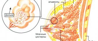
The structure of the mammary glands
The breast of an adult woman is a collection of connective, glandular and fatty tissues, conventionally divided into so-called “lobes”.
The mammary gland consists of about 16-20 lobes, and is an organ responsible for many processes in the female body, of course, the main one of which is breastfeeding.
As for the structure of the female breast, they consist of:
- chest;
- pectoral muscle;
- lobes of the mammary gland;
- areolas;
- mammary ducts;
- fatty tissue;
- skin.
Types of mastopathy
Experts have identified more than fifty subtypes of mastopathy at the present stage, however, from a clinical point of view, mastopathy is usually divided into three types:
- Diffuse , physiologically manifested in the formation of nodules and cords, and in the formation of compactions in the area of the mammary glands. This type of mastopathy is characterized by painful sensations in the chest area, as well as coarsening and enlargement of the glands during menstruation;
- Nodular , which is characterized by the appearance of neoplasms in the chest, which are palpable during tactile examination. These neoplasms can grow to quite noticeable sizes, reaching the diameter of a walnut. In addition to the discomfort in the area where these formations occur and their pain, women often have enlarged lymph nodes in the armpits, and the pain and enlargement of the breasts does not depend on the cycle and is almost constant;
- Fibrocystic , which is a mixed type, and is characterized by both the appearance of neoplasms, usually not very large in size, and the appearance of nodes. These pathologies together form cysts with fluid-filled cavities.
Causes
As mentioned earlier in this article, one of the main causes of mastopathy is hormonal imbalances in a woman’s body.
The work of the ovaries plays a huge role in this case. They produce most of the so-called female hormones.
They are mainly involved in the production of estrogen, androgen and progesterone. The slightest disruption in the production of these hormones affects the entire female body, and directly causes various diseases of the mammary glands, and mastopathy, as the most common breast disease in women.
In addition to this reason, it should be especially noted that mastopathy can occur as a result of failures in the normal functioning of the kidneys and adrenal glands, improper functioning of the liver, and improper functioning of the pituitary gland, which is responsible for the production of prolactin in women.
Many factors are to blame for the fact that over the past hundred years the number of women suffering from mastopathy has increased significantly:
- Increasingly older age of first-time mothers;
- Difficult childbirth;
- Increased number of abortions;
- Malfunctions of the ovaries;
- Refusal of breastfeeding or its sharp reduction, relative to the norms of feeding children in the past;
- The occurrence of lactostasis congestion;
- Excessive exposure to the sun's UV rays;
- Infectious diseases of the chest;
- Drinking alcohol and smoking;
- Use of hormonal contraception.
Until the 20th century, a healthy woman of fertile age was either pregnant or breastfeeding, which, according to many experts, had a beneficial effect on the natural hormonal background of the female body. The invention of various chemical and pharmaceutical agents, abortion at various stages, as well as other interventions in the reproductive system, had a sharply negative impact on hormonal levels, and continue to affect each individual female organ.
Diffuse fibrocystic mastopathy
The most common type of fibrocystic mastopathy is its diffuse subtype. This form is characterized by an increase in the amount of glandular tissue and the formation of edema.
According to the symptoms and characteristic course of the disease, it is considered the simplest form of diffuse mastopathy:
- component predominates in mastopathy, a woman experiences swelling of the mammary glands, and the interlobular connective tissue septa increase and put pressure on all surrounding tissues of the chest, the lumens of the ducts narrow over time, and sometimes fuse completely.
- There is also a variant with a predominance of the cystic component. With this variant of the disease, cavities are formed in the mammary gland, clearly separated from other tissues, filled with liquid, varying in composition, and depending on the degree of the disease.
- The mixed form of mastopathy is characterized by both an increase in lobes and the proliferation of glandular tissue in the mammary gland. This form is the most difficult to treat and requires an individual approach when prescribing a treatment regimen for the disease.
Nodular fibrocystic mastopathy
The nodular form of fibrocystic disease is much less favorable.
In this case, in addition to the disorders described above that are characteristic of the diffuse form of mastopathy, the presence in the mammary gland of one or several nodes, which most often represent a fibroadenoma or adenoma, is added.
Fibroadenoma is a fairly common benign tumor, which affects mainly women of reproductive age, less often adolescents.
This neoplasm can have different sizes, from a pea in diameter to a tumor reaching up to 15 cm.
Fibroadenoma rarely develops into a malignant tumor; according to medical analysts, this occurs in only two percent of cases.
The situation is much worse with the nodular form of nodular fibrocystic mastopathy, represented by nodes with the proliferation of glandular tissue. Atypical hyperplasia is the medical name for nodes of this type. In cases of manifestation of nodes of this nature, mastopathy turns into oncology in every fifth woman
Mixed fibrocystic mastopathy
Mixed fibrocystic mastopathy is a combination of nodes, cysts and compactions. Most often occurs in women under 35 years of age. The early stage is very difficult to determine due to the absence of any discomfort in the area of the mammary glands, and can most often be determined only during an annual examination by a mammologist.
Over time, all formations begin to grow, which leads to compression of the nerve endings, pain, a feeling of heaviness and fullness in the chest area.
Features of bilateral fibrocystic mastopathy
As the name of this form of mastopathy suggests, its main feature is that the disease occurs in both mammary glands.
A completely natural point is that with a bilateral course there can be the same different forms of the disease as with a unilateral course, but still more often women encounter bilateral mastopathy with a diffuse nature of mastopathy in the initial stage.
This is due to the fact that the form with the formation of nodes is characterized by the formation of single or multiple cysts or nodes in one breast.
Decoding the results
When interpreting a mammogram, the specialist not only looks at the image, but also takes into account medical history and previously identified symptoms.
When deciphering, the structure of the gland tissue, vessels, ducts, and lymph nodes are studied.
Normally, the gland looks like a uniform structure in the picture. There are no visible shadows or densities visible. The vessels and milk ducts of the gland are clearly visible, intertwining with each other, creating a network. Nearby lymph nodes are not enlarged.
With the development of any pathology, the normal structure of the gland is disrupted, and the regional lymph nodes are enlarged.
If pathological foci are detected during the deciphering of the mammogram, the doctor determines their number, uniformity, size, location and shape.
It must be taken into account that in young women the density of mammary gland tissue is always high, but after undergoing surgery to remove the ovaries or after menopause, the tissue density decreases.
Cysts and fibroadenomas appear on the image as round or oval formations with clearly defined boundaries. In cancer, on the contrary, the contours of the tumors are unclear and have uneven boundaries.
Mammography also reveals calcifications, which can accompany the formation of tumors (both benign and malignant).
A convenient standard for describing research results was developed in the USA. According to this standard, all results are divided into seven categories:
Zero category. Incomplete assessment. This means that for some reason the results were inconclusive and additional research must be carried out to make a final diagnosis.
First category. Negative. This result means that the woman is healthy. No anomalies or suspicious structures were found.
Second category. Benign tumor. It is also considered a negative result because no malignant tumor was detected and there are no signs of cancer.
Third category. A benign tumor that requires additional research. This means that the tumors found are almost certainly benign, but you need to be on the safe side and do an additional mammogram after six months. In addition, over the next two years the patient will be under the supervision of a mammologist.
Fourth category. The detected tumor was considered suspicious. With this result of mammography, a biopsy is necessary to determine the nature of the tumor. The prognosis is favorable, the likelihood of cancer is low.
Fifth category. A detected tumor is considered suspicious and the likelihood of cancer is high. In this case, a biopsy is also performed to make a final diagnosis.
Sixth category. Biopsy confirmed breast cancer. In this case, mammography is needed to monitor the results of cancer treatment and understand the dynamics of the disease.
It should be remembered that the doctor cannot make a final diagnosis based on mammography results alone. Rather, this study is only the first stage in diagnosing breast cancer.
Symptoms
Initially, the symptoms of mastopathy are very mild: the pain is mild, neoplasms may not be noticeable on palpation, and detection of mastopathy at the very beginning of the disease is possible only with regular mammological examinations.
The main symptoms may be aching pain in the mammary glands, with possible milk discharge from the breast.
These disruptions in milk secretion are called galactorrhea.
Galactorrhea with mastopathy can be of varying intensity:
- from meager and spontaneous,
- to discharge at the slightest touch to the breast,
- up to the presence of blood impurities in these secretions.
The presence of lumps in the chest upon palpation is the main symptom of mastopathy, in which it is necessary to contact a medical institution as soon as possible to establish a diagnosis and begin treatment at the very beginning of the disease.
In the initial stages, the symptoms of the disease are very similar to premenstrual syndrome.
Thus, it is observed:
- Headache
- Irritability
- Nausea
- Gastrointestinal tract disorders
- Flatulence
- Swelling of the arms, legs and face.
The most serious symptom, and the reason for urgent contact with doctors for mastopathy, is discharge from the nipples mixed with blood. Most often, these discharges indicate the presence of intraductal formations, papillomas, ulcerating and bleeding.
Pain
With fibrocystic disease, the pain syndrome may not be based in the mammary gland itself. So, they can radiate to the shoulder blade, shoulder and armpit area, and have varying degrees of strength.
The degree of pain is determined by various factors, such as:
- individuality of a woman’s pain syndrome,
- stage of the disease
- the size of neoplasms and their location in the mammary gland.
Discharge
Discharge from mastopathy can have different composition and intensity.
They can be:
- fickle;
- spontaneous;
- scanty;
- spontaneous.
Their nature depends on many factors, and each individual case of the disease.
Folk remedies
Treatment with folk remedies will not get rid of the pathology, but it can eliminate the symptoms of the disease. Herbs are used in combination with pharmacological drugs.
Alternative medicine doctors offer:
- Grind raw potatoes in a blender until you get porridge. Apply the resulting mixture to a cabbage leaf and apply to the chest for 2-3 hours, securing the compress with a bandage.
- Boil water and pour in boron uterus (15 grams), let it brew for 1–3 hours. The resulting mixture can be drunk to reduce the symptoms of FCM once a day for a week.
Treatment with folk remedies should take place intermittently. One to two weeks - take the medicine, 1-2 weeks - a break.
When the disease is at an early stage, or during the recovery period, traditional medicine can be used. It is allowed to use decoctions, infusions and compresses while taking medications.
Recipes:
- Infusions of immortelle, wormwood, and aloe have an immunomodulatory effect and are often used in the treatment of any type of mastopathy. You can buy them ready-made at the pharmacy.
- For 30-31 days, apply burdock or cabbage leaves to your chest at night.
- Infusion of burdock root for oral administration, 70 ml before meals, 3 times a day. Preparation: pour 50-60 g of roots with 350-400 ml of cold water. Let stand for at least 1 hour. Then boil for 15 minutes. Strain before use.
- With the permission of the mammologist, you can use rubbing with camphor oil. The procedure is also performed in the evening.
- Dill seeds (100 g) pour 300 ml of milk. Boil. Drink 100 ml 3 times a day.
- Wrap the steamed wormwood in a soft cloth and apply to the chest. The compress should not burn. Remove when cool. You can use up to 5 procedures per day.
- Applying coltsfoot leaves to the breast at night.
The described recipes help reduce pain and reduce tumor formation. When completing the full course (to be confirmed with a mammologist), a significant improvement in mammary gland tissue is noted.
Echography
Echography is nothing more than a study of the mammary glands using ultrasound machines. Echography to detect mastopathy is carried out using modern sensors with increased resolution up to 10 MHz.
Conducting echography contributes to:
- Detecting and measuring the presence of cysts of even the smallest size in the breast. Also, a highly qualified specialist, using ultrasound machines, can identify the nature of the cystic formation: its benignity, or atypicality, which may indicate the malignancy of the neoplasm;
- Assessing the condition of the lymph nodes in the chest , armpits and tissues closest to the chest, to identify the spread of the disease in the body;
- Detection of adenomas, fibroadenomas and cancerous nodes in the mammary glands
- Detection of dilations in the milk ducts.
Carrying out echography helps not only to record and analyze examinations, but also allows you to monitor dynamic changes in the course of the disease. In addition, it is worth noting that echography is a harmless and informative examination.
Hormone therapy
Hormonal drugs can be taken only after a blood test. The drugs are initially prescribed in a minimal dose, as side effects such as sleep disorders are possible. Under no circumstances should you self-medicate.
Treatment is prescribed by a highly qualified specialist, taking into account all the results of the preliminary examination:
- blood tests;
- Ultrasound;
- mammography;
- palpation.
For complete cure, hormonal drugs must be used for at least 4 months. It is preferable to use preparations that are herbal or for external use. Only in severe cases is it possible to use more serious hormonal medications.
Diagnostics
- Typically, mastopathy is diagnosed through ultrasound , mammological examination and examination by an oncologist.
- We also collect punctures and a set of standard tests to identify hormonal levels in each specific case.
- Carrying out an ultrasound is especially important in cases of formation of nodes in the mammary glands , that is, with nodular mastopathy
- An almost mandatory type of examination for women in the risk age group is mammography , which is an X-ray examination of the breast . This examination is also carried out at the slightest suspicion of breast cancer, regardless of age.
- If there are special indications after mammography, a woman may be prescribed ductography . This examination is carried out by introducing contrast agents into the ducts of the mammary glands, which make it possible to distinguish and identify foci of growth of tumors during repeated mammography.
The most favorable period for diagnosing mastopathy is days 5-12 of the menstrual cycle.
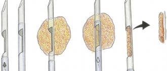
Puncture for fibrocystic mastopathy
Breast cysts are a kind of blisters filled with fluid.
And the liquid that fills them, depending on the stage and cause of their occurrence, may be different. To identify the nature of the fluid in mammary gland cysts, a puncture is used, or, as it is also called, a mammary gland biopsy.
This examination can be carried out in various ways. The most common method at the present stage is to perform a puncture with the additional help of ultrasound.
The largest of the cysts is located on the ultrasound machine, a sensor is pointed at it, and the needle punctures into the center of the tumor.
This allows you to obtain the most informative sampling of the material being studied. The results of performing a puncture in this way are much better, and the time for manipulations associated with the examination is noticeably reduced.
Puncture for fibrocystic mastopathy can be performed both in public medical institutions and in certified private medical centers. After the biopsy, the resulting material is sent for cytological and histological analysis.
A biopsy can also be performed to remove fluid from formations, or to administer medications directly to the site of the inflammatory process.
The puncture does not require any preliminary preparation for the examination, however, to obtain a better result, experts recommend not using drugs that have a blood clotting effect, such as aspirin, before the procedure.
Best time to explore
When undergoing an ultrasound procedure, it is worth considering one important nuance, namely when it should be done. Research for mastopathy cannot be carried out on any day of the female cycle. During its different phases, the concentration of certain hormones in the body is not the same. Such fluctuations affect the structure of the mammary glands.
The most reliable results will be from an ultrasound scan performed in the first half of the menstrual cycle. It is better if the study is carried out from 5 to 10 days. Doctors call this period hormonal rest. This is the period when estrogens have almost no effect on the body. It is at this time that pathological changes can be clearly identified, and the risk of obtaining a false positive or false negative result is minimized.
It is worth noting that these time limits are optimal for women whose cycle lasts 28 days, and the duration of menstruation is from 4 to 5 days. Not every woman can boast of a stable cycle, so in most cases only general recommendations are given. In order to determine as accurately as possible which period will be favorable for diagnosis, you can donate blood on the third day of menstrual flow to determine the level of estrogen.
Normally, the concentration of these hormones should be minimal at the beginning of the cycle, gradually increasing towards the day of ovulation. In the second phase, under the influence of estrogens, structural changes occur in the mammary glands. In particular, their ducts expand. A doctor, even using ultra-sensitive modern equipment, is unable to examine pathological neoplasms due to an increase in the diameter of the ducts. It is better not to do the procedure at this time.
All of these recommendations for choosing a day for performing a breast ultrasound procedure are given for women of reproductive age. With the onset of menopause, estrogen levels drop and these restrictions are lifted.
Therefore, it does not matter on what day of the menstrual cycle mastopathy is diagnosed after menopause.
Which doctor should I contact?
Visits to a mammologist for consultations and for preventive purposes, in principle, should be carried out annually to identify any pathologies, and in general to identify the disease at the earliest stages.
However, not all women follow this recommendation.
If you experience any discomfort or suspicion of mastopathy during self-examination, an urgent visit to a mammologist is necessary.
If suspicions are confirmed, the scheme for contacting further doctors and the general selection of treatment is determined strictly individually.
The attending physician independently determines the consultations and examination of which doctors are necessary in this case of the course of the disease.
Most often, in addition to mammological examinations, the patient will need to see a gynecologist, endocrinologist, neurologist and therapist. This will help establish all the information about the woman’s health, the causes of the disease, and determine further actions for the patient’s speedy recovery.
In advanced cases, or if characteristic signs are detected, the patient will need to be examined by an oncologist and undergo all the necessary tests
Benefits of Ultrasound
The most important advantage when performing ultrasound in women suffering from mastopathy is the health safety of this examination method, as well as painlessness and a high degree of reliability of the information obtained.
- Also, quick results.
- An ultrasound can determine whether there are lumps, cysts or other formations in the glands.
- Does not require special training to carry out.
- Ultrasound can be performed on different days of the cycle.
This type of examination does not always give an accurate result.
Therefore, if any suspicions arise during examination for the presence of tumors, the doctor prescribes additional diagnostic measures to check the woman for the absence or presence of cancer.
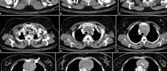
For this purpose, mammography, MRI, CT or biopsy may be prescribed.
Also, if you have mastopathy, you need to take tests. Which? Read more about this here.
Treatment
The methods and intensity of treatment largely depend on the time of detection of mastopathy and the degree to which the disease is located in a particular case. It is worth noting that surgical treatment of this disease is the most extreme method, which also does not eradicate the cause and source of its occurrence.
The first step in the treatment of mastopathy is considered to be the normalization of the functions of organs that affect the course of the disease, as well as the normalization of the patient’s hormonal levels.
The most common method of treating mastopathy is conservative observation with periodic examinations to monitor the development of the disease, and the prescription of a list of drugs that promote a speedy recovery.
Main purposes in the treatment of mastopathy:
- Administration of non-steroidal anti-inflammatory drugs;
- Prescription of drugs that improve blood circulation;
- Diet correction
- Prescribing sedatives;
- Prescription of strengthening vitamin complexes;
- Homeopathic remedies (Mastodinon, Mastofit);
- dietary supplements;
- To carry out competent treatment, doctors recommend excluding visits to saunas and baths, limiting yourself from physiotherapy, and categorically excluding even moderate visits to artificial tanning salons and direct sunlight on the mammary glands, as this can be dangerous.
- You can use folk recipes and make compresses to relieve severe pain symptoms.
Drugs
Frequently prescribed drugs for the treatment of different types of mastopathy include::
- anti-inflammatory drugs;
- drugs that stimulate the outflow of venous blood;
- antioxidant drugs;
- preparations with natural extracts that regulate prolactin balance and improve the hormonal balance of the female body.
The treatment regimen is always strictly individual and can be described in detail and calculated exclusively by the attending mammologist or mammologist-oncologist.
In cases of a very advanced stage of mastopathy, the mammary glands undergo surgical intervention.
Vitamins
With any disease, the body does not receive enough of the minerals and vitamins it needs, which is why complex treatment of fibrocystic mastopathy, in addition to taking medications, involves taking vitamin-mineral complexes. When choosing vitamins, you should avoid self-medication and consult your doctor.
The selected vitamin complex must meet the following criteria:
- minimize the entry of toxic substances into the tissues and cells of the mammary glands;
- help enhance the effects of medications prescribed by a doctor;
- strengthen the immune system in general;
- promote the normal functioning of the liver, which receives the greatest impact from taking medications;
- if possible, have a sedative effect and have a beneficial effect on the nervous system.
Vitamin complexes must necessarily contain vitamins of groups A, E, C, D and B.
Also, in addition to taking vitamin-mineral complexes, the body of a woman suffering from mastopathy should receive foods, fruits and vegetables enriched with natural, non-synthetic vitamins of these groups. Such as apricots, carrots, cheese, Brussels sprouts, sweet peppers, currants, rose hips, various types of fish, nuts and meat.
Massage
For quite a long time, scientists considered mastopathy a complete contraindication for massage not only of the chest area, but also of the back, but after conducting a series of studies it turned out that massage not only does not harm the health of patients, but can also improve the clinical picture of the disease.
It can also slow down the transformation of benign tumors into cancer. However, the decision on preventive massage for mastopathy should be made by a mammologist.
The goals of massage for diseases of the mammary glands are:
- Ensuring the outflow of fluid from inflamed breast tissue, and reducing and subsequently eliminating swelling. After achieving these goals, acute pain in the chest area significantly decreases or disappears altogether;
- Study of the complete clinical picture of the course of mastopathy , including the exact localization of pathological changes, their size and nature;
- Prevention of the formation of new foci of inflammation
- Normalization of the functioning of the mammary glands and getting rid of all the initial symptoms of the disease
- Prevention of neoplasms developing into cancer.
However, there are a number of clear contraindications for massage for mastopathy:
- Suspicion of malignancy of neoplasms;
- The presence of damage to the skin of the mammary glands, both traumatic and damage associated with various inflammatory processes;
- The presence of allergic rashes on the chest;
- The patient's temperature is elevated.
It is important to know that massage of the mammary glands should exclude the use of any force, and should be carried out exclusively with soft kneading movements, by a specialist who knows the technique and features of massage for mastopathy.
Massage is most effective for the cystic form of the disease.
There are also various self-massage techniques that allow you to identify the disease yourself even in the earliest stages. However, when performing a massage at home, a woman must strictly follow all the rules so as not to harm the mammary glands, and also have a clear understanding of palpation.
When performing a breast massage, it is recommended to use various soothing creams, tonics and moisturizing gels to enhance the effect and improve the condition of the breast skin.
Diet
Mastopathy as a disease requires certain changes in a woman’s diet and the introduction of a certain diet.
So, cocoa, chocolate and coffee should be excluded from the patient’s diet.
The principles of therapeutic nutrition must be introduced, and it is also necessary to exclude all products that in one way or another contain methylxanthines.
The diet of a woman with mastopathy should be rich in vegetables and fruits, which are the main sources of fiber and vitamins.
It is also worth adding fermented milk products, grains, products including bran and seafood to the menu as a source of vitamin E.
Removal of fibrocystic mastopathy
Medical removal of fibrocystic mastopathy is an extreme, radical way to eliminate the disease.
Mastopathy is a diverse disease that has many manifestations and forms of development, so there is no clear answer to the question of whether surgical intervention is necessary in the treatment of mastopathy.
Surgical removal of mastopathy is necessary only in cases where the presence of nodes and glandular tissues, as well as the size of neoplasms and the degree of neglect are a direct indication for removal of lesions.
Traditional medicine recipes for mastopathy
Herbal preparations and teas help to safely adjust a woman’s hormonal balance. Folk remedies that complement the prescribed treatment help maintain breast health.
Wood lice compress
Chickweed (chickweed) will help cope with the initial degree of mastopathy. Fresh herbs are washed, dried and applied to the chest. Wrap a cloth on top and hold the compress until the herb dries. The course continues for five days.
It is convenient to wear the grass during the day in a bra.
After a month, you can resume compresses. A greater effect is achieved when woodlice is added to food. Soups, borscht, and salads are prepared with woodlice.
Herb tea
There are ready-made mastopathy preparations, balanced according to the dosage of herbs. If you wish, you can make a collection for mastopathy at home.
● Yarrow
● Nettle
●Echinacea
●Coltsfoot
●Oregano
●Penny
All are mixed one part at a time. Brew a large spoonful in 300 ml. water. Drink half a mug twice a day.
Burdock root
Take one hundred grams of dry crushed burdock in a coffee grinder or blender. Pour in one and a half glasses of refined sunflower oil. Keep in a container in a dark place for ten days.
Apply the product to the nipples and entire breasts.
Dill with milk
One hundred grams of dill seeds are boiled in 500 ml of milk. After removing from the heat, cover the container for two hours. The mixture is filtered, take half a glass thirty minutes before meals.
The course lasts three weeks.
Beetroot with honey
Grate the beets, mix with honey three parts beets to one part honey. Place the pulp on a whole leaf of cabbage. Apply to your breast at night.
Black radish
Mix 3/4 cup of honey with half of grated black radish. Pour the mixture with 30 ml of vodka, mixing with table salt - a teaspoon.
Apply to the breasts every day for at least ten days.
Compresses on the chest
Grated carrots placed on cabbage leaves are used in the treatment of mastopathy. The chest with such a compress is wrapped in soft cloth.
Keep the product for several hours.
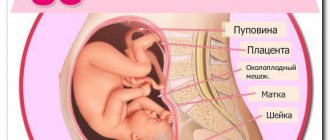
Mastopathy and pregnancy
Many doctors consider pregnancy the best way to cure fibrocystic mastopathy, and even recommend that women become pregnant to recover from breast diseases.
The thing is that during pregnancy, a woman’s body releases a large amount of progesterone, which helps treat the disease and restore hormonal balance in the female body.
An important factor is also that while carrying a child, the female body receives a kind of impetus to renew cells and improve the functioning of all vital systems.
Statistical studies show that more than 80 percent of women after pregnancy are completely cured of mastopathy. Long-term lactation also promotes healing.
This is explained by the fact that during breastfeeding, the process of renewal of mammary gland tissue accelerates, and fibrosis and compactions resolve on their own.
After the birth of a child, a woman with a history of mastopathy must have a preventive consultation with the attending mammologist, since the risk of lactostasis and congestion in the diseased breast is very high, and can cause further development of the disease.
Doctors' advice and treatment reviews
Timely treatment helps to completely get rid of the problem. Many who discover breast lumps do not take any further action. All this leads to emergency surgery, without which advanced cases of FCM of the mammary glands cannot be cured.
After the intervention, the breast becomes deformed and requires even more expensive surgery. Doctors recommend being examined by a mammologist twice a year to avoid complications.
Article design: Oleg Lozinsky
Mastopathy and menopause
There is no specific age interval at which a woman may experience menopause, just as there is no specific moment at which a woman may experience mastopathy.
However, doctors note that the signs of mastopathy in women during menopause appear much more clearly.
During menopause, they can no longer be confused with signs of pregnancy or premenstrual syndrome.
During menopause, women experience enormous hormonal changes, which can cause the formation of mastopathy, and the risk of this disease increases several times.
Treatment of mastopathy during menopause is developed exclusively on an individual basis, and most often represents a combination of various drugs, hormonal and non-hormonal in nature. To treat the mammary glands, courses of antioxidants, vitamin A or beta-carotene, and homeopathic remedies with proven effectiveness can also be prescribed.
Mastopathy and IVF
The opinions of specialists regarding in vitro fertilization for fibrocystic mastopathy are sharply different.
From the opinion that this step can serve to degenerate benign neoplasms into cancer, to the opinion that pregnancy achieved through IVF will serve to normalize the hormonal state of the female body and contribute to a complete cure for the disease.
The danger of performing in vitro fertilization for mastopathy is that during preparatory procedures for artificial insemination, a woman is prescribed strong stimulating hormonal therapy, which can cause both the emergence of new pathologies in the mammary glands and an increase in existing tumors.
Why is it dangerous?
Mastopathy of any type and at any stage can serve as a favorable environment for the development of cancer, and as you know, breast cancer is the most common and leading disease among women in terms of deaths.
Therefore, you should be attentive to your health and conduct annual examinations for diseases of the mammary glands, as well as conduct an independent breast examination.
When conducting a self-examination, it is recommended:
- Conduct examinations in the second week of the menstrual cycle;
- It is necessary to examine the mammary glands using circular movements of the opposite hand (the left breast with the right hand, and the right breast with the left);
- Conduct a visual examination to identify deformations of the glands, disturbances in their pigmentation, and the occurrence of areas of redness;
- Pay special attention to the nipples and areolas around them as indicators of hormonal changes. Identification of changes in the shape of the nipples, discharge of various types and frequencies, changes in pigmentation are a direct factor for an urgent visit to a medical institution for examination.
Carrying out the procedure
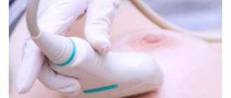
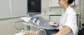
The entire procedure usually takes from 10 to 20 minutes. To carry it out, the doctor will ask the woman to undress to the waist and lie down on the couch. It is most comfortable to perform an ultrasound of the mammary glands while lying on your back. Hands should be placed behind your head. A special gel is first applied to the skin in the area to be examined. It will ensure the tightest fit of the sensor to the skin, eliminating the possibility of the formation of an air gap.
The computer converts the ultrasonic waves synthesized by the device into a picture. As a result, an image appears on the monitor screen, which a specialist will subsequently analyze. The result of the study can be obtained the next day.
This diagnostic method itself should not cause any discomfort. An exception would be the situation when mastopathy reaches a stage of development that is accompanied by pain in the chest. In this case, the woman will experience discomfort when the sensor touches the affected areas.
If mastopathy is advanced, touching the sensor will cause discomfort
Prevention
Prevention measures:
- The main preventive measures for detecting and preventing the development of any type of mastopathy are regular visits to a mammologist.
- Women who have had or have relatives with various diseases of the mammary glands , or with oncology, need to visit a mammologist annually.
- Women, both those included and those not included in the risk group, in addition to consulting with a doctor, need to conduct regular self-examinations to check for lumps or changes in the symmetry of the mammary glands.
- It is worth remembering that the risk of mastopathy increases after forty years , due to hormonal age-related changes in the body and changes in general hormonal levels. Therefore, after a woman reaches this age, regardless of whether signs of the disease are detected or not, annual examinations to detect the presence of tumors in the breast are mandatory.
- To prevent the occurrence of mastopathy, a woman must monitor her hormonal levels and lead a healthy lifestyle. Thus, the risk of developing breast diseases is reduced if a woman takes various vitamin and mineral complexes containing iodine and all properly balanced microelements.
- It is impossible to imagine strong immunity of the body as a whole, without healthy, at least eight hours of sleep. To prevent the occurrence of mastopathy, you need to avoid stressful situations, and if stress cannot be avoided, then give the body the time necessary to restore the nervous system.
- It is important to know that, according to many experts, products containing cocoa butter provoke the occurrence of tumors in the breast. Therefore, limiting the consumption of chocolate and other products containing cocoa will reduce the risk of disease.
- , large amounts of direct sunlight should be avoided
- The correct choice of underwear plays an important role both in case of mastopathy and in preventing its occurrence. An incorrectly selected bra can cause deformation of the mammary glands, and also with various squeezing and selection of underwear that is not the right size, various kinds of deformations of tissues and ligaments can develop, which can cause the development of mastopathy or other breast diseases.