Ultrasound examination - ultrasound of the salivary gland - is one of the leading and highly effective methods of additional diagnostics, widely used among most modern specialists who treat this organ. With its help, the doctor can determine whether the suspected disease is acquired, under the influence of any external factors, or whether it is congenital.
What does ultrasound show, why is this examination important? It allows you to determine the severity and extent of the pathological process, determine the size and location of the glands, and also assess the condition of their excretory ducts and blood supply.
Who is the study for?
The main indication for ultrasound examination of the salivary glands is the presence of any complaints in the patient. Most often, patients come to the doctor with the following problems:
- sharp pain in the submandibular or parotid area;
- local or general hyperthermia (increased temperature directly in the area of the lesion or in the entire body as a whole);
- increase in the size of the salivary glands;
- dry mouth (xerostomia);
- the presence of compactions (tumors, stones) near the salivary glands.
If the above symptoms are present, as well as after a visual examination, palpation examination and history taking, the doctor, in addition to laboratory tests, refers the patient to an ultrasound scan. The method is used if it is necessary to carry out differential diagnosis for such pathologies as:
- inflammatory diseases of the salivary glands (acute and chronic sialoadenitis, viral and bacterial);
- adenoma;
- salivary stone disease;
- tonsil damage;
- tumor neoplasms (malignant and benign);
- individual anatomical anomalies;
- enlargement of nearby lymph nodes;
- dystrophic lesions of organ tissue.
During the study, specialists can assess the condition of the group of large salivary glands: parotid, sublingual, submandibular.
Symptoms and possible diseases
An examination of the organs that synthesize saliva is indicated if the patient has the following pronounced symptoms:
- deformation of the glands, changes in their size, palpation of seals during palpation;
- sore throat without visible inflammation of the laryngopharynx;
- systematic low-grade fever of unknown origin (origin);
- a feeling of dryness in the mouth due to insufficient saliva production;
- impaired swallowing function (dysphagia);
- the formation of painful erosive changes in the area where the glands are located.
Together with general weakness, periodic headaches concentrated in the parotid region, these signs are a reason for ultrasound diagnostics. During the examination, the doctor evaluates the anatomical and functional changes of the glands, which are shown by ultrasound, which makes it possible to diagnose the following pathologies:
Ultrasound examination of lymph nodes
- inflammatory lesion (neuritis) of the facial nerve;
- expansion or stenosis (narrowing) of the canals;
- hyperplasia of secretory cells, not related to tumor pathologies (sialadenosis, otherwise sialosis);
- the presence of calculi (stones) or sand in the salivary ducts and glands (sialolithiasis or salivary stone disease);
- damage to the connective tissue of the lacrimal and salivary glands (Sjogren's syndrome);
- formations of a benign nature (adenoma, adenolymphoma);
- cancer (carcinoma, sarcoma).
The study also includes an analysis of the condition of adjacent lymph nodes (submandibular, mandibular, parotid). Since the symptoms of the diseases are similar, it is necessary to correctly determine the source of the patient’s complaints.
How to prepare for the procedure
Ultrasound of the salivary glands is an absolutely safe and painless manipulation, for which the patient does not require special preparation. Patients of almost any age tolerate the procedure well, without showing any anxiety before and during its implementation, and therefore it does not require premedication (pre-administration of sedative medications) even in children.
On the eve of the visit to the specialist (about 3-4 hours in advance), the patient is advised to refrain from eating, and immediately before the examination he needs to thoroughly clean the oral cavity.
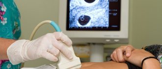
How to do an ultrasound of the salivary gland
The examination can be carried out in 2 ways: from the oral cavity and from the external surface. The procedure does not take much time (it usually takes no more than 20-30 minutes to complete). During an ultrasound scan of the salivary glands, the patient does not feel any pain or other discomfort.
During the examination, the patient takes a supine position, places his head on a specially prepared pillow and tilts it slightly back (or turns it to the left or right, depending on the location of the gland being examined).
If the parotid gland is being examined, the device's sensors are placed on the parotid area. When studying the sublingual and submandibular glands - into the oral cavity (sometimes the extraoral method can be used).
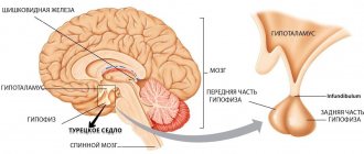
Anatomical location and functions of the glands
Glands that secrete saliva are classified into small and large. The first are localized on the lips, inner surface of the cheeks, tongue, and palate. The latter are divided into three types:
- Paired symmetrical parotid. Located between the lateral part of the lower jaw and cheekbones.
- Paired sublinguals. Based on the name, they are located under the tongue, on both sides.
- Submandibular. The location is the submandibular space, in close proximity to the lymph nodes.
The most voluminous is the parotid gland; it produces 26% of saliva. The work of the salivary glands is to maintain a normal level of humidity in the oral cavity and cleanse it, liquefy and grind food, activate taste buds, protect tooth enamel, and ensure proper functioning of the speech apparatus.
Layout of large paired organs that synthesize saliva
The ultrasound diagnostic method allows you to diagnose pathologies in a timely manner and prevent the development of complications.
Dimensions of the salivary glands according to ultrasound: normal
During the study, the doctor records all the necessary parameters of the organs, including their width, thickness and length. This table shows the sizes of the salivary glands, normally visualized by ultrasound:
Gland | Length, mm | Width, mm |
Sublingual gland | 18-22 | 10-16 |
Submandibular gland | 30-40 | 11-18 |
Parotid SG | 40-50 | 30-40 |
results
If tumor formations are detected during an ultrasound, doctors refer the patient to an MRI of the soft tissues of the neck with contrast as an additional examination.
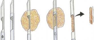
Sometimes, to establish the nature of the tumor, puncture, biopsy and histology are also required. All these procedures will help determine the nature of the tumor - whether it is malignant or benign. Material can also be collected during a biopsy or puncture under ultrasound guidance. Author: Telegina Natalya Dmitrievna
Therapist with 25 years of experience
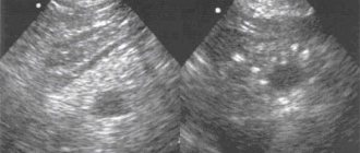
Normal parotid salivary gland on ultrasound
Normal ultrasound characteristics of the parotid gland are:
- The glands are located symmetrically and are visible in the form of capsules.
- The anterior surface of the organ is clearly visible, the deep parts are not identified.
- The posterior sections have unclear contours.
- Their structure is homogeneous, fine-grained.
The duct of the parotid salivary gland is not normally detected. It can be seen only in the presence of a pathological process (for example, in the case of obstruction - blockage - by a stone or compression by a cyst).
Current video:
Ultrasound of the submandibular salivary gland
The norm for an ultrasound examination of the salivary submandibular gland corresponds to the following parameters:
- Fine-grained homogeneous structure.
- The edges of the glands are smooth.
- The excretory duct is well visualized, small in size, without signs of blockage.
Determining the normal size of the submandibular glands is not particularly difficult precisely because of its clear contours. During inflammation (sialoadenitis), the organ increases in size and its edges become blurred.
Ultrasound of the sublingual salivary gland
Normally, the sublingual salivary gland has smooth, not very clear contours. They have a homogeneous structure, but echogenicity (the ability to reflect ultrasound) may be slightly increased. In the absence of any diseases, the device will not show the ducts of this organ.
The shape of the sublingual salivary glands displayed on the device screen changes depending on where the sensor is placed. When located in the chin area, the glands have oval contours. If the device is applied parallel to the body of the lower jaw, the glands will be slightly elongated.
Methodology for ultrasound of the salivary glands: protocol
Each gland is scanned separately according to the protocol. A protocol is a special document in which data about the examined organs and tissues is recorded.
Thus, the parotid salivary gland is examined in accordance with this document according to the following scheme:
- First, in cross section from the angle of the jaw to the tragus.
- Then in a longitudinal projection, including the cervical region.
Next, the organ is examined bilaterally, since in some cases there are bilateral lesions of the glands or it is necessary to compare the affected organ with a healthy one located on the opposite side.
The submandibular gland has the following examination plan:
- Transversely along the midline of the neck from the hyoid bone to the horizontal ramus of the mandible (when viewed bilaterally).
- For a detailed examination of an individual gland, the sensor is moved slightly to the side, parallel to the branch of the lower jaw.
The protocol for examining the sublingual salivary gland does not differ significantly from that applied to the submandibular gland. The sensor is placed in the midline of the neck just below the lower jaw, making both paired organs clearly visible.
Ultrasound examination of the salivary glands is a highly informative way to diagnose various organ diseases. With its help, you can timely identify a serious pathological process, which is much easier to stop in the early stages of the disease. However, not every specialist prefers to limit himself only to it. Increasingly, doctors have to turn to such additional methods as:
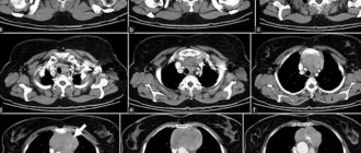
- computed tomography (CT);
- magnetic resonance imaging (MRI);
- X-ray and so on.
In some situations, a diagnostic puncture is necessary for further cytological examination of the obtained material if neoplastic processes are suspected. Ultrasound of the salivary glands cannot replace biopsy and histology, which determine the degree of malignancy of tumors. Only the combination of a whole range of measures can give the most complete picture of the pathology, based on which the doctor prescribes the most effective and targeted treatment.
Pathologies
During an ultrasound examination, the device can detect the following pathologies:
- dystrophic changes in the functioning of these organs. They are often represented by pathologies such as sialosis and sialodenosis.
- inflammatory changes in the functioning of the studied parts of the body. First of all, this is stone disease and sialadenitis. However, this can also be a consequence of an inflammatory infection transmitted from another organ (lymph nodes, throat, etc.)
- cancerous tumors. These can be both carcinomas and other types of cancerous tumors. The formation of benign tumors in the area of the salivary glands is extremely rare.
- developmental anomalies. Such anomalies are often either congenital or a consequence of the development of acromegaly (an abundance of growth hormone during puberty). Such anomalies can be corrected surgically. Also, such anomalies may not manifest themselves in any way during the life of the human body, which means that getting rid of them will be impractical. In this case, the presence of anomalies can lead to greater vulnerability of the glands.
- traumatic changes. Any maxillofacial injury can affect the organ being examined. Therefore, when the lower jaw is knocked out, or any other injury, during a preventive examination of internal organs, ultrasound diagnostics of the salivary glands is also used.
Do not forget that the organ being studied is quite small. Moreover, most diseases develop similar symptoms. Therefore, when making a final diagnosis, the doctor must evaluate not only the results of the ultrasound examination, but also a comprehensive diagnosis of the body. In particular, the level of white blood cells in the blood is used to confirm the diagnosis of cancer. At the same time, to confirm damage to these parts of the body due to trauma, ultrasound results are compared with x-rays.
Read also…. Irritable bowel syndrome
To obtain a final diagnosis, the following is also prescribed:
- Comprehensive blood test (general and biochemical)
- Urinalysis (in cases where the suspected swelling depends not on themselves, but on the kidneys)
- Sputum analysis
- Saliva analysis
- Biopsy of the relevant organ.