Manifestation of echogenic reflections
The ultrasound method of studying internal organs is based on the quality of absorption and reflection of ultrasound in tissues. In this case, depending on the density of these structures, different images appear on the device screen. Varieties of structures differing in echo density are divided into three main types. This:
- Hyperechoic formations. Such foci have an extremely high density. Against the background of the main tissues of the organ, they stand out in a lighter shade.
- Hypoechoic. Their acoustic density is low. On ultrasound they appear as dark lesions.
- Isoechoic. Detection of such areas is difficult because they have the same echogenic density as the tissue of the organ being examined.
When describing all types of reflections, the terms formation and inclusion are used. They denote the same phenomenon, that is, a focus characterized by the ability to reflect ultrasonic signals.
How is an isoechoic inclusion detected?
The similarity of the display of such formations with the surrounding structures of the uterus largely makes it difficult to identify foci of developing pathology. Such inclusions are detected if there is a capsule (shell) or there is a feeling of squeezing of the forming formation.
For a long time, the development of such processes does not remind of itself by any symptoms, that is, the patient is not bothered by anything. Most often, isoechoic inclusions are detected by chance during a routine examination or during examination for completely other diseases.
Echogenicity of individual organs on ultrasound
When performing the procedure, the ultrasound diagnostician evaluates the size of the organ, its contours, homogeneity and, necessarily, the degree of echogenicity, which can indicate the presence of various pathological processes in the object under study.
Changes in the structure of the pancreas
Normally, the pancreas is located in the projection of the epigastric region and has the following echo signs.
- The echogenicity of the pancreatic parenchyma is comparable to that of the liver and is designated as average. With age, the gland undergoes changes, and the parenchyma becomes more dense.
- Usually the organ is presented in a “dumbbell-shaped” or “sausage-shaped” shape (due to the fact that the gland consists of a head, body and tail).
- The contours are clear and even, well demarcated from surrounding tissues and structures.
- The echostructure is homogeneous and fine-grained (other options are possible: homogeneous or coarse-grained).
- The Wirsung duct looks like an elongated anechoic cord, the diameter of which normally ranges from 1.6 to 2.6 mm.
We can say that the echogenicity of the pancreas is increased when its color on the device screen has a whiter tint and is in brighter ranges than the color of the liver tissue.
Common causes of hyperechogenicity are listed below.
- Interstitial edema of glandular tissue as a result of acute reactive pancreatitis. In addition to changes in density, an increase in the size of the organ is also observed.
- Increased echogenicity of the pancreas will occur with pancreatic necrosis. In this case, against the background of heterogeneous hyperechoic changes, hypo- and anechoic areas are visualized, indicating necrosis.
- Diffuse fibrosis as a result of chronic (autoimmune, alcoholic, infectious, drug) pancreatitis. The changes are based on the replacement of normal organ tissue with connective tissue.
- The echogenicity of the pancreas will be significantly increased with lipomatosis (fatty infiltration of the organ). The gland has blurred contours and a fairly light or even white tint compared to other formations.
- Diabetes mellitus, in which more than 90% of the organ tissue is destroyed.
Read more The skin on your hands itches and cracks
A gastroenterologist makes a diagnosis not only based on ultrasound data, but also during a subjective examination; an ultrasound scan of the stomach is also indicated.
Echostructure of the uterus and its changes
Normally, the uterus undergoes monthly cyclic changes under the influence of pituitary and ovarian hormones. As a result of this, she has different indicators on ultrasound, correlating with the phase of the menstrual cycle.
The organ is pear-shaped, and in women who have given birth it tends to be round. Normal myometrium is characterized by average echogenicity, which is comparable to that of a healthy liver and pancreas.
The endometrium undergoes pronounced functional changes.
- On the 5-7th day of the cycle it has a lower echogenicity and a homogeneous structure. In the center of the uterus, a thin line with a hyperechoic signal is visualized, which represents the junction of the posterior and anterior layers of the inner membrane.
- By the 8-10th day, the echostructure of the endometrium practically does not change, only some thickening is noted.
- On days 11-14, its density increases, which corresponds to average echogenicity.
- Until the 15th-18th day, the density of the shell increases slowly.
- On days 19-23, the endometrium can be characterized as hyperechoic, making the central line almost invisible.
- By the end of the period, the internal lining of the uterus has a hyperechoic and heterogeneous structure.
The most common causes of increased echogenicity of the uterus are: inflammation, fibroids, polyps, endometriosis and malignant neoplastic process. The endometrium becomes hyperechoic on certain days of the cycle, as well as as a result of inflammation, the appearance of a malignant neoplasm or adenomyosis in it, or during pregnancy (hypertrophy of the functional layer and glands occurs).
Changes in the ovaries
This paired organ is located in the pelvic cavity and communicates with the uterus through the fallopian tubes. Similar to the endometrium, the ovaries also undergo a large number of changes associated with the menstrual cycle.
Normally, they have an ovoid shape, a tuberous outline due to growing follicles, and a hypoechoic structure with anechoic round inclusions along the periphery.
The echogenicity of the ovaries often increases with diffuse sclerosis (as in Stein-Leventhal syndrome), prolonged and sluggish inflammation, as well as with their malignant degeneration.
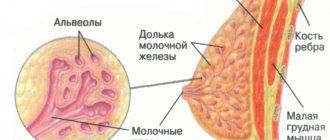
Changes in the structure of the mammary glands
A woman’s mammary glands are an important organ of the reproductive system that requires special attention. Due to the increase in malignant neoplasms, mammologists recommend annual breast screening examinations using mammography or ultrasound.
Such glands are also prone to cyclical changes, and their normal echostructure depends on the woman’s age.
- In the reproductive period (from 18 to 35 years), glandular tissue is represented by a homogeneous fine-grained formation of increased or medium echogenicity, in the thickness of which tubular anechoic structures (milky ducts) are visible.
- In late reproductive age, a fairly thick hypoechoic layer is visualized, represented by subcutaneous fatty tissue. Around it there is connective tissue, visible on ultrasound in the form of a hyperechoic rim.
- In women over 55 years of age, the breast tissue is replaced predominantly by adipose tissue, which is also reflected on the screen of the ultrasound machine. The gland corresponds to a hypoechoic area with rare hyperechoic round inclusions.
The causes of pathological increased echogenicity of the mammary glands are listed below.
- Mastopathy resulting from hormonal imbalance. In this case, the increase in echogenicity is associated with the proliferation of fibrous tissue (both diffusely and in the form of nodules).
- Fibroadenoma is the most common benign tumor of the mammary glands, occurring mainly in women of reproductive age. Most often, this is a solitary formation with a high content of connective tissue fibers, which makes it hyperechoic on echography. Although the literature indicates that this neoplasm may have different echogenicity.
- Advanced forms of mastitis - nonspecific inflammation of the tissue of the glandular organ. In the later stages of the disease, the mammary gland has a large number of hyperechoic inclusions with a similar dense capsule.
Increased echogenicity of the kidneys
The echostructure of healthy kidneys is heterogeneous due to the presence of the medulla and cortex. The contours are smooth and clearly demarcated from the surrounding formations. Normally, the pelvis and calyces are practically not visualized. The “contents” of the ureters have a reduced echogenicity, and their walls are represented by a light echo signal.
The reasons for the increased reflectivity of the kidneys are presented below.
- Neoplasms. Moreover, uneven contours indicate the malignant nature of the tumor.
- Moderately increased echogenicity of the kidneys indicates dysmetabolic nephropathy (that is, sand in the kidneys).
- Stones are defined as hyperechoic areas of varying sizes and shapes.
- Triangular hyperechoic zones in the renal parenchyma are a sign of hemorrhage.
- An increase in organ density (due to edema) is observed in acute pyelonephritis.
Increased echogenicity of the liver
On normal echograms, the liver parenchyma appears as a homogeneous structure of average echogenicity and is considered the standard for comparing the echogenicity of the pancreas and kidneys. Its contour is smooth and shows a clear linear hyperechoic signal on all sections.
The echogenicity of the liver is increased with:
- chronic hepatitis of various origins;
- hereditary Gaucher disease (based on lysosomal enzyme deficiency);
- Wilson-Konovalov disease (copper accumulation occurs in the liver);
- congenital and acquired liver fibrosis;
- cirrhosis;
- The echogenicity of the liver is also increased with antitrypsin deficiency;
Gallbladder structure
The shape of the gallbladder is quite variable: from pear-shaped to cylindrical or ellipsoidal. It has a homogeneous anechoic structure. The wall of a healthy bladder is within 1-3 mm.
Causes of suspended echogenicity:
- acute and chronic cholecystitis;
- bile stagnation (especially with the hypomotor type of biliary dyskinesia);
- calculous cholecystitis (the density of the echo image is due to the accumulation of hyperechoic stones);
Changes in the structure of the spleen
Located in the upper left quadrant of the abdomen, the spleen on the echogram is represented by a crescent-shaped formation with clear, even contours. Its parenchyma has a homogeneous structure and echogenicity, which is slightly higher than that of the liver and the renal cortex. Despite the fact that pathology of the spleen is quite rare, the following reasons for the increase in its echo signal are identified:
- “old” heart attack (hemorrhage);
- calcifications (most often they appear with long-term use of medications such as anticonvulsants, etc.).
Here you can also do an ultrasound at home, if you have such an opportunity, and additionally do an ultrasound of the spleen.
Features of formations
A characteristic feature of echogenic inclusions is their identity with the echo density of the uterine structure. That is, these neoplasms are practically no different from other tissues of the organ. This is why isoechoicity is considered a normal manifestation of acoustic density. Healthy reproductive organs have this degree of reflectivity.
Most often, such inclusions do not pose a threat to health.
But in some cases, the detection of foci with similar density in the uterus indicates the development of both benign formations and malignant inclusions. It is the same ability to reflect the sound signal that complicates the differential diagnosis of safe isoechoic formations from such serious diseases as:
- adenomyotic node;
- myomatous node;
- uterine cancer;
- Rokitansky syndrome, which reflects the isoechoic density of the uterus, which is not completely healthy.
In the presence of a pathology of uterine development such as Rokitansky syndrome, echographic signs of other inclusions that are hyperechogenic or hypoechogenic may be found in it.
The detection of an isoechoic inclusion indicates that a tumor has been detected in the uterus. Detection of its benign or oncological nature requires a mandatory thorough examination using laboratory and instrumental methods.
conclusions
Isoechogenic formations are detected as a result of ultrasound examination. They mean the development of tumor tumors in various parts of the body. Ultrasound is only the initial stage of diagnosis, and only further thorough research allows us to determine the nature of the disease and how to treat it. Do not forget to undergo regular medical examinations to identify changes in organ structures in the early stages.
Ultrasound examination has recently become an increasingly popular option for examining internal organs. Its use is especially effective in diagnosing pathological manifestations in the female reproductive system. Isoechogenic formation in the uterus is no exception. It has the same echographic density as the rest of the organ tissue.
Causes and symptoms of inclusions
Ultrasound makes it possible to determine the presence of a neoplasm in the uterus. Their echo density directly depends on the degree of development of the pathological process. At the initial stage, all focal pathologies in the uterus are visualized on the monitor screen as isoechoic.
If the nature of the inclusion is malignant, then as the process progresses (tumor formation increases), symptoms such as the presence of anechoic, hypoechoic or hyperechoic formations in the lesion appear.
This is how it looks on the monitor screen.
Whereas progression is not typical for benign inclusions.
Causes of isoechoic transformations in the uterus
Among the most common formations in the uterus, which initially have an isoechoic picture, the following inclusions pose a particular danger:
- Adenomyosis. It represents the detection of pathological foci in the muscular walls of the uterus, similar to the mucous membrane of the organ. Echographic signs of such changes are an increase in the size of the uterus to 13-14 cm (in length). There is an asymmetrical thickness of the uterine walls and a violation of clarity in the boundaries.
- Myomas. These inclusions in the uterine wall appear on ultrasound as thickenings with clear boundaries. In most cases, these are benign neoplasms that form from the tissues of the uterus itself. Varieties of such tumors are fibromyomas, leiomyomas, which also have an isoechoic density. An ultrasound diagnosis of fibroids is considered only an assumption and requires mandatory additional examination to exclude uterine cancer. Differentiation of such a formation is possible only after its removal. A histological examination of the changed tissues is mandatory. This makes it possible to identify the presence or absence of oncology.
- Uterine cancer. It appears as an adenomatous node on the mucous membrane of the organ. For such nodes, the form of manifestation can be both clear contours and shapeless outlines. Nodes with isoechoic signs account for more than 25% of all cases of oncological formations.
The detection of isoechoic inclusions on ultrasound becomes an indication for an in-depth study that reveals the benignity or malignancy of the neoplasms.
Timely detection of oncology gives a chance for a favorable prognosis.
Indications for treatment of isoechoic formations
If an inclusion detected by chance does not provoke pain or discomfort, then you can limit yourself to observing it.
Constant monitoring will make it possible not to miss the moment for timely surgical intervention if negative changes occur.
Myometrial changes during pregnancy
Heterogeneous myometrium during pregnancy is quite rarely the primary diagnostic finding. In most cases, the disease is detected at the planning stage or in the previous life period. And ultrasound during pregnancy is used not only for dynamic assessment of fetal development, but also to monitor the condition of the uterine wall.
This is necessary not only to determine the tactics of current treatment, but also to draw up a prognostic assessment. After all, such tactics make it possible to timely identify signs of incompetence of the uterine wall in areas of damage to the myometrium and the threat of its rupture during childbirth, and resolve the issue of the feasibility and admissibility of natural delivery.
It should be remembered that only a doctor can interpret ultrasound data. Diagnosed changes in the myometrium in themselves are not grounds for immediate initiation of therapy. Therapeutic tactics are determined individually, taking into account the dynamics of the clinical and echographic picture of the disease, etiology and severity of the identified pathology.
With previous adenomyosis, a pregnant woman is considered to be at risk for miscarriage and the development of chronic placental insufficiency. Myoma increases the likelihood of an abnormal course of the labor period and is a risk factor for insufficient uterine contraction in the postpartum period with the development of pathological bleeding. And fibrosis and scar changes in the myometrium can cause ruptures of the birth canal during the pushing period of labor or insufficient productivity of contractions.
The ultrasound examination method is widely used in the diagnosis of most somatic diseases. The constant progress of this field of medicine allows us to expand diagnostic capabilities and increase their significance and reliability. In an ultrasound report you can often find such a phrase as increased echogenicity of a particular organ. The reasons for this conclusion can be both functional, that is, reversible, in nature, and indicate a serious pathology.
Treatment
To avoid unpleasant consequences when diffuse changes in the myometrium are detected, early diagnosis and active treatment of adenomyosis are necessary.
Methods for getting rid of this pathology include medication, surgery and combination treatment.
Drug therapy
Treatment of diffuse changes in the myometrium with medications is aimed at:
- to prevent complications;
- to stop the progression of pathology by suppressing estrogen production;
- to preserve reproductive function;
- to eliminate pain syndromes and prevent cancer.
All groups of drugs have many side effects and serious contraindications, therefore they are taken only as prescribed by a gynecologist and after a detailed study of the complete (and not introductory) instructions for the drug by the patient herself.
Main groups of medications:
- Estrogen-progestogen contraceptives (Jess, Janine, Diane 35, Yarina, Demoulin, Non-ovlon, Marvelon).
Able to suppress ovulation and estrogen secretion. They help in the initial stages of structural changes in the endometrium and myometrium. Prohibited if you are prone to thrombosis or have high blood viscosity.
- Progestin drugs - Visanne, Duphaston, Norkolut, Utrozhestan, Getstrinone. Prescribed for varying degrees of myometrial damage.
- Antigonadotropic (agonists of gonadotropic releasing hormones), including Nemestran, Danol, Buserelin-Depot, Danogen, Lucrin-Depot, Zoladex, Diferelin.
When used as injections once every 28 days, significant atrophy of diffuse, nodular and focal changes in the body of the uterus is achieved.
The main advantage of these drugs is the combination of a more pronounced therapeutic effect with a minimal percentage of side effects and relapses.
With combined treatment, including medications and surgery, relapses are diagnosed in only 7 to 12 patients out of a hundred.