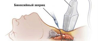
The thymus belongs to the endocrine glands and is of utmost importance. A fair question is, what is it for and what is it? The thymus is responsible for immune status. Due to the importance of the organ, an ultrasound of the thymus gland may be necessary - the procedure is painless and does not cause any harm to the body.
The thymus is located at the top of the retrosternal space.
Enlargement of the thymus gland is common in children. This deviation does not require medical intervention because it usually goes away on its own by age 5. But if there was an increase in childhood, then when the size of the gland normalizes, it will still need preventive control.
When is it prescribed?
It happens that ultrasound of the thymus gland is prescribed for babies under 1 year of age. Indications for such examination in infants are:
- severe dysbacteriosis;
- pronounced and difficult to treat diathesis;
- low-grade fever of unknown etiology;
- skin rashes that are not related to diathesis;
- enlarged adenoids and lymph nodes;
- excessive weight gain;
- other disorders of the immune system.
Older children, as well as adult patients, should examine the thymus gland using ultrasound if:
- There is a tendency to constant colds, which easily turn into various complications from sinusitis to pneumonia.
- There is a noticeable vascular network on the chest.
- There is a clear predisposition to all types of allergies.
- The patient quickly gets tired, feels general weakness and lethargy, and also complains of vague pain in the chest area.
- Worried about shortness of breath, increased sweating, and the skin is paler than usual.
- The thymus gland is enlarged.
- Heart rhythm disturbances appeared.
- There are various signs of decreased immunity.
How much does the examination cost?
Ultrasound diagnostics of the thymus in paid clinics will cost from 1000 to 2000 rubles. The cost depends on the pricing policy of the establishment. In government institutions, ultrasounds are performed only on the direction of a doctor. In this case, the examination is carried out free of charge.
Ultrasound of the thymus gland is a safe procedure that, if indicated, is prescribed at any age. It does not have a negative effect on the body and does not require special preparation.
Have you encountered ultrasound of the thymus gland? Did the procedure help in making the diagnosis? Share your experience in reviews and articles on social media. networks. Be healthy.
What are they watching?
At the end of the ultrasound of the thymus, the doctor analyzes the data obtained. The doctor checks whether the size of the gland and its functional parameters correspond to normal values. In addition, it is determined whether there are pathological changes in the organ and anomalies of its development.
Important! If a malfunction of the thymus occurs in adults, the consequences will be immune disorders, or rather incorrect formation of T-lymphocytes.
Parameters that are determined during ultrasound examination:
- thymus size - this parameter is of paramount importance, it depends on the patient’s age and is determined by the doctor using a special table;
- contours of the gland and its structure;
- the presence of inclusions that have a different structure than the rest of the glandular tissue.
Important! Ultrasound of the thymus is indispensable in identifying pathologies of the gland and diagnosing its diseases.
What determines the size of the gland?
The thymus gland actively develops and works in childhood, after which it gradually involutions. On average, by the age of 25-35, adipose tissue remains in its place. Consequently, normally the size of the thymus progressively decreases with age.
Also, the size of the organ is affected by the course of pregnancy and the newborn period, the presence of chronic diseases of the endocrine and immune systems, frequent acute respiratory viral infections, and allergic reactions. Physiological weight of the organ depending on age:
- children under one year old – 13.3 g;
- children from 1 year to 5 years – 23 g;
- at 6-10 years old – 26 g;
- from 11 to 15 years – 38 g;
- up to 20 years – 25 g;
- at 21-35 years old - 20 years.
Pathologies
The main diseases of the thymus gland, determined by ultrasound:
- enlarged thymus syndrome;
- thymomegaly – a significant increase in the size of the gland;
- thymoma is a concept that unites all types of neoplasms of the thymus;
- T-cell lymphoma – belongs to the group of hematological diseases;
- myasthenia gravis is a neuromuscular disease characterized by pathologically rapid muscle fatigue;
- MEDAC syndrome is characterized by a whole complex of symptoms.
Timomegaly
The most common pathology of the thymus is its enlargement or thymomegaly. The reasons for its occurrence in adults and children have not yet been clarified. It is believed that the disease is provoked by a number of factors, among which malnutrition, prematurity and birth injuries play an important role .
Clinical manifestations of thymomegaly are expressed in the following symptoms:
- Shortness of breath, cough, noisy, difficult breathing - occurs due to the fact that the enlarged thymus gland puts pressure on the trachea and superior vena cava, and also compresses the vagus nerve.
- Long-term, often complicated infectious diseases, which are a consequence of reduced immunity.
- Poor appetite - occurs due to a failure in endocrine and metabolic processes.
Other diseases
In newborns, in addition to thymomegaly, the following may be detected:
- hyperplasia – neoplasms of the thymus gland;
- hypoplasia – decreased production of T lymphocytes due to congenital disorders.
With hyperplasia of the 2nd degree, it is impossible to do without the intervention of a surgeon, but thymomegaly does not require special treatment. Such patients are only shown observation and a gentle regimen, as well as maximum protection from possible infections and diet .
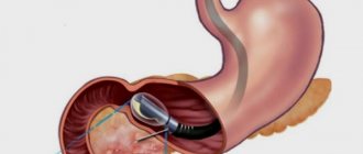
It happens that during an ultrasound, the doctor discovers a tumor - thymoma, which is characterized by a hypoechoic structure. Against the background of healthy tissue in adults and children, tumors are clearly visualized. Most often, neoplasms of the thymus gland are not malignant, but require mandatory treatment.
Structure of the thymus
We have already found out where the thymus is located. We will consider the structure of the thymus gland separately. This small-sized organ has a pinkish-gray color, soft consistency, and lobular structure. The two lobes of the thymus are completely fused or tightly adjacent to each other. The upper part of the organ is wide, and the lower part is narrower. The entire thymus gland is covered with a capsule of connective tissue, under which there are dividing T-lymphoblasts. The bridges that extend from it divide the thymus into lobules.
The blood supply to the lobular surface of the gland comes from the internal mammary artery, thymic branches of the aorta, branches of the thyroid arteries and the brachiocephalic trunk. Venous outflow of blood occurs through the internal mammary arteries and branches of the brachiocephalic veins. The growth of various blood cells occurs in the tissues of the thymus. The lobulated structure of the organ contains the cortex and medulla. The first appears as a dark substance and is located on the periphery. Also, the cortex of the thymus gland contains:
- hematopoietic cells of the lymphoid series, where T-lymphocytes mature;
- hematopoietic macrophages, which contain dendritic cells, interdigitating cells, typical macrophages;
- epithelial cells;
- supporting cells that form the blood-thymus barrier, which form the tissue framework;
- stellate cells – secrete hormones that regulate the development of T cells;
- “nanny” cells in which lymphocytes develop.
In addition, the thymus secretes the following substances into the bloodstream:
- thymic humoral factor;
- insulin-like growth factor-1 (IGF-1);
- thymopoietin;
- thymosin;
- Thymalin.
Norms and decoding
The main goal of ultrasound examination of the thymus is to assess its size and calculate the volume and mass of the organ.
Important! If the gland is significantly enlarged, then this can lead to the development of false croup.
In children, a healthy thymus appears on the ultrasound machine screen as a formation with average echogenicity. Normally, its contours are clear and its structure is uniform. The peculiarity of the thymus is that it is clearly distinguished from the surrounding tissues and structures of the chest cavity; the reverse side of the gland has smooth boundaries.
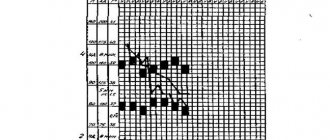
Table 1. Norms of thymic index
During the procedure, specialists determine the dimensions of the length, width and thickness of the lobes. Then, taking into account the data obtained, the doctor calculates the volume of each lobe and the total volume of the gland, for which the following formulas are used:
Share volume = X*Y*Z*0.523
Where X is the length (in cm), Y is the width, and Z is the thickness of each lobe.
The volume of the thymus in infants and older children is the sum of the volumes of the right and left lobes. After which the mass of each lobe and the total mass of the gland are calculated, and the thymic index is determined. To calculate mass, use the same formula as to calculate volume, only the coefficient of 0.523 changes to a coefficient of 0.704. The mass of the gland is equal to the total mass of the two lobes.
Table 2. Dimensional parameters of the thymus in different years of life
The thymic index in children is determined by the formula:
TI = Gland mass/Child weight* 100%
The resulting index is compared with standard tables, after which conclusions are drawn about compliance with the standards.
Table 3. Thymus weight in relative values per 1 kg of body weight
Conclusions:
From the 3rd to the 25th centile - indicators are below average, the size of the thymus gland is smaller than normal, which does not correspond to age standards.
From 25 to 75 centile – normal values;
From 75 to 97 centile – indicators are above average. The size of the thymus indicates an enlargement of the thymus gland, that is, thymomegaly of 1, 2 or 3 degrees.
Further actions of parents if a child has a pathology
If nonspecific thymomegaly of grade 1-2 is detected, the child does not need any drug treatment; he is only monitored by ultrasound until the indicators normalize.
In case of grade 3 thymomegaly, the baby is sent for consultation to a pediatrician, immunologist and endocrinologist. As a rule, vaccination is postponed for some time.
If the ultrasound doctor detects a pathology in the child, he will definitely describe this in the study protocol, and also refer him for a consultation to a pediatrician, who will decide on further examination and treatment tactics.
How do they do it?
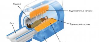
The study of the thymus gland in children and adults is carried out using a special ultrasound device. Scanning is performed using an ultrasonic sensor with a certain frequency. The patient is advised to stand throughout the procedure.
Research methodology:
- During an ultrasound, the width of the thymus gland is determined, for which the sensor is placed on the upper third of the sternum in a transverse position. Sometimes it becomes necessary to move it downwards (usually when the gland is significantly enlarged).
- In the same way, the thickness of each of the thymus lobes is determined; they are measured by placing the sensor longitudinally in turn on the left and right sides of the chest.
The examination of young children is carried out in a lying position, from 9 months to 1.5 years - in a sitting position, and from 1 year and 6 months, like adults, - in a standing position.
During ultrasound diagnostics, you need to calm the baby down and try not to change his position or interfere with the doctor.
Crying and active movements of the child will interfere with accurate recording of the size of the gland. In general, the procedure takes no more than 10 minutes.
Ultrasound of the thymus gland in children: indications for performance, how it is done, what it shows - MEDSI
Table of contents
- What is the thymus gland?
- For what purpose is an ultrasound of the thymus performed?
- Indications for use
- What can an ultrasound reveal?
- How is it carried out?
- Preparation
- Decoding the results
- Is it possible to examine the thymus in other ways?
- Advantages of carrying out the procedure at MEDSI
Ultrasound examination (ultrasound) is a procedure that is safe for a child of any age. It allows for accurate diagnosis quickly and painlessly. In this case, X-ray radiation is not used, which is harmful to the child’s body. The principle of its operation is based on the effect of different reflections of high-frequency sound waves from different types of organs and tissues.
What is the thymus gland?
The thymus gland (thymus) is an important part of the body responsible for the production of T-lymphocytes for human immunity. It is located in the upper region of the chest (in the upper mediastinum).
The size and weight of the gland changes throughout a person’s life:
- At birth she weighs 15 grams and is about 5 cm long
- By the end of puberty and throughout the main period of adult life, its weight is 20–37 grams, and its length is 7.5–16 cm
- By old age (about 75 years old) her weight decreases to 6 grams
The thymus gland has a gray-pink tissue color, and in old age it is yellowish.
For what purpose is an ultrasound of the thymus performed?
The doctor prescribes an ultrasound of the thymus gland for children when there are complaints about the child’s constant illnesses (colds, acute respiratory infections, etc.) and low immunity. This is necessary to determine the causes of regular ailments.
Ultrasound examination reveals a pathological increase in the size of the thymus - thymomegaly. As a result of its appearance, the immune system is weakened, and the child begins to regularly become infected with various diseases from other children and adults. In this case, allergies, gastrointestinal upset, and high fever may occur against the background of a common cold.
Indications for use
Ultrasound of the thymus is prescribed for symptoms such as:
- The child gets sick regularly (up to 10 times a year)
- The common cold often becomes more severe and is accompanied by complications (pneumonia, bronchitis, sinusitis)
- The temperature often rises to 37.5 and lasts for a long time
- Excessive regurgitation, excessive and heavy breathing
- The appearance of bronchial asthma, allergic reactions that are not eliminated even with a more proper diet
- For general weakness, chronic fatigue, chest pain
- If the network of blood vessels on the chest is clearly visible
- If your heart rhythm is abnormal (arrhythmia)
- With enlarged lymph nodes, tonsils or adenoids
What can an ultrasound reveal?
Ultrasound of the thymus gland in children helps to identify the following types of diseases and disorders:
- Timomegaly
- Pathologies of thymus development
- Aplasia (underdevelopment of the gland)
- Neoplasms (benign and malignant)
- Bernard-Horner syndrome (autosomal disorder)
- Nezelof's syndrome (appears due to the fact that the thymus gland cannot cope with its functions)
- DiGeorge syndrome (characterized by low T cell production)
Ultrasound examination will also allow you to determine parameters such as:
- Change in size (it should change within age norms)
- Correctness of organ tissue structure
- Contours (normally should be clear and uniform)
How is it carried out?
The process of performing an ultrasound scan of the thymus gland is quite simple:
- The child takes the body position necessary for the study:
- Until the age of one year, he should be in the arms of his parents
- From a year - he sits or lies down on the couch
- If the child is a teenager, then he must stand
- Clothes are removed from the upper body
- Gel is applied to the chest to improve sound conduction
- The doctor moves the sensor along the upper chest
- The image is displayed on the screen, and from it the specialist draws conclusions about the condition of the organ
The whole procedure takes about 10–15 minutes.
Preparation
Before performing an ultrasound examination of the thymus, no complex preparation is required:
- It is necessary to warn your doctor if you are taking various medications.
- You should find out the exact weight of the child and inform the doctor about it
- Clothing should be chosen that is easy to remove before the procedure.
- It is recommended to explain to the child what kind of examination is ahead and how it will be carried out so that he does not get nervous
Decoding the results
To decipher the results, the doctor must know the exact body weight of the child, since the determination of the normal volume and weight of the thymus gland (their compliance with age standards) depends on this value.
Normally, the weight of the gland is 0.3% of the total body weight. If this value is exceeded, a diagnosis of thymomegaly is made. It comes in three degrees, and its value is measured by a special CTTI index:
- I - with a CTTI index value from 0.33 to 0.37%
- II - with a CTTI value from 0.37 to 0.42%
- III - with an index value of more than 0.42%
Also, a sonogram (ultrasound result) can reveal changes in the gland such as: tumor, excessive tissue growth, etc.
Is it possible to examine the thymus in other ways?
Ultrasound of the thymus gland as a technique has been used relatively recently. Previously, X-ray examination was used to identify disturbances in the functioning and structure of the thymus gland. But such an analysis cannot be carried out frequently, unlike ultrasound, since X-ray irradiation is harmful to the child’s body.
If the results of an ultrasound scan of the thymus are not clear enough or contrast enough, the doctor may prescribe additional types of examination:
- Electrocardiogram (ECG)
- Heart scan
- Blood tests (general and biochemical) and urine tests
- Retroperitoneal scanning
Advantages of carrying out the procedure at MEDSI
- The MEDSI network of clinics conducts more than fifty different types of ultrasound examinations using modern expert-level devices (models Pro Focus 2202, Philips iU22)
- Interpretation of the survey results is done by experienced specialists who have high qualification categories and regularly improve their skills at major scientific conferences and seminars
- To make an appointment with a specialist, you need to call 8 (495) 7-800-500
- There are more than 20 clinics in Moscow, so it’s easy to find a convenient place for examination
- MEDSI clinics have comfortable waiting areas for children and parents