X-rays of the lungs can be done as often as the doctor prescribes. X-ray examination is accompanied by radiation exposure to the human body. The dangers of radiation have been confirmed by clinical studies.
There are different effects from the influence of chronic and acute doses. When performing an X-ray examination, low-dose radiation is generated. With frequent and prolonged exposure to the body, it leads to genetic mutations of cells.
An acute radiation reaction is accompanied by rapid death of organs and tissues. Doctors understand the difference between the benefits and harms of X-rays, so they prescribe X-rays of the lungs only when indicated.
The Ministry of Health clearly regulates the radiation safety of personnel and patients.
Advantages and disadvantages of fluorography
Every adult undergoes examination using this method at least once a year. Fluorography is a type of x-ray examination in which the image obtained by passing rays of the appropriate range through the patient’s chest is photographed.
The positive aspects of this survey are expressed in the following:
- Low cost of research. In every district clinic, any patient can undergo fluorography; all medical institutions are equipped with the appropriate equipment. With the introduction of digital technology, film was no longer needed for photographs. Therefore, examination costs have decreased even further.
- Speed of implementation. The shooting process takes two minutes. And you can learn about the results after some time, depending on the organization of work in the medical institution. In some clinics the result can be given out in half an hour, but in some you need to wait until the next day.
- Painless and no need to use any medications. The only unpleasant thing about this procedure is that you need to press your naked body against a cold metal plate. You also need to hold your breath when the nurse says. When examining using digital equipment, this will not be necessary.
- There is a high probability of detecting a disease in the human chest. This is why it is so important to undergo examinations every two years.
The disadvantages are minor:
- Use of radiation. But its dose is small, so there will be no harm to the body.
- Impossibility of accurate diagnosis. In the picture you can see the focus of the disease, but it is impossible to determine what kind of disease it is only by fluorography. For an accurate diagnosis, other studies and tests must be performed.
Indications and contraindications for undergoing
Fluorography is a mandatory part of the periodic medical examination of citizens.
It is prescribed to the following persons:
- all adults and adolescents over 15 years of age undergoing a mandatory medical examination;
- persons living with pregnant women and newborn babies;
- citizens who are HIV carriers.
A doctor may refer you for this examination if the following diseases are detected:
- inflammation of the lungs or pleura, that is, with pneumonia, pleurisy, etc.;
- pulmonary tuberculosis;
- diseases of the heart muscle and large vessels;
- cancer of the lungs and organs that are located next to them.
This type of examination is contraindicated for the following persons:
- Children under 15 years old.
- For pregnant women, X-rays can cause mutations in the child. In case of urgent need, it can be done after 25 weeks of pregnancy.
- Nursing mothers.
- Seriously ill patients who are unable to hold their breath for the required period of time.
- Persons who, for one reason or another, cannot be in an upright position while standing on their feet (wheelchair users, bedridden patients, etc.).
Possible health effects
Many people believe that it will be very harmful to health if they do fluorography twice in a row. This is sometimes required when the shot turns out to be unsuccessful. In this case, a repeat procedure is needed. But there will be no terrible consequences, because the dose of radiation received, even after two consecutive exposures, is several tens of times less than what we receive from surrounding natural sources. Modern technology uses a negligible dose of radiation.
Received radiation
Speaking about how often fluorography can be done, we note that the maximum safe radiation dose for humans is 500 mSv per year. The body receives irradiation of 3–4 mSv/g from external natural and man-made environmental sources. But he is exposed to this influence continuously throughout the year. Irradiation during photography is short-term and its harmful effects end immediately after the end of the shooting process, thus its harm is negligible. Let's analyze the radiation dose received during fluorography and x-ray:
| Examination method | Received radiation dose during fluorography, mSv per shot | Radiation dose for x-rays, mSv per shot |
| Film | 0,5–0,8 | 0,5–0,8 |
| Digital | 0,04 | 0,1–0,2 |
From the above figures, it becomes clear why you can undergo an X-ray examination of the chest organs twice throughout the year or in a row without harm to health. There is no noticeable damage to a person’s physical condition from this examination.
An interesting fact from everyday life. When smoking one cigarette, a person receives a dose of radiation that is equal to ten x-rays.
How can you neutralize the negative impact?
To remove ionizing radiation from the body, it is recommended to use certain medications. Here are some of them:
- Polyphepan;
- Activated carbon;
- Potassium orotate;
- Dietary supplements with calcium, iodine.
Polyphepan - 100 rub.
Potassium orotate — 96 rub.
Activated carbon - 20 rubles
Calcium magnesium plus zinc - about 1000 rubles
The following products will be useful after and before exposure to radiation:
- wine and grape juice with pulp;
- iodine-containing products - algae, fish, fruits;
- vegetable oil;
- honey;
- fresh milk and fermented milk products;
- dried fruits and decoctions from them;
- rice and oatmeal;
- quail eggs.
Principle of implementation
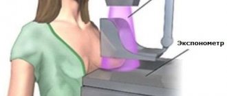
The operating principle of a fluorographic installation is similar to an X-ray one. Diagnosis occurs using a fluorograph - a special installation that is capable of producing 256 shades of gray. The load in different regions of Russia differs, but on average does not exceed 100 µSV. Due to this, an image of the internal organs is obtained.
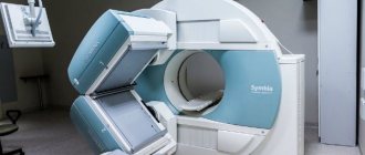
Scanning fluorograph (the safest and most modern diagnostic method)
During the examination, the patient is placed in a special area where X-rays pass through the body. They are unevenly absorbed by tissues of different densities, such as muscle tissue and bone. As a result, an image of the internal organs is obtained, which is displayed on a fluorescent screen. The image can be printed on film, but today digital devices that display it on a monitor are common.
The advantage of the new digital equipment is obvious; now the image is obtained much faster than before. In addition, the picture is viewed on a widescreen screen - this helps to examine the picture in detail.
The picture is saved in the database in DAICOM format or sent by e-mail to another institution, saved on an external memory device.
Digital examination puts much less burden on the patient, and also reduces the cost, since there is no need to use films. The study requires the installation of a special program, with the help of which the results are processed.
Example: digital fluorograph "ProScan" (scanning), when performing fluorography it emits 0.02-0.03 mSV (millisievert) or 20-30 µSV (microsievert). For comparison, in Moscow the natural background is 20 µSV. Such a device will not do any harm, even if you do fluorography twice in a row.
When and how often should you undergo the procedure?
The general recommendation of doctors is to undergo a fluorographic examination of the chest organs once every two years. As previously mentioned, without harm to your health, you can undergo this examination again and more often if directed by a doctor.
For an adult
There is no specific age gradation for the adult population according to the frequency of fluorography in the legislation. There is one general requirement for everyone - it must be done once every two years. When passing a medical examination during employment, for persons over 50 years of age, pensioners, students and citizens of any other categories, this very requirement applies.
Fluorographic examination
Certain professions
There is a certain circle of people whose profession, social status or health condition obliges them to undergo this examination 2 times a year:
- military personnel;
- health workers of tuberculosis medical institutions;
- maternity hospital workers;
- patients with pulmonary tuberculosis and those who have recovered from it;
- HIV carriers;
- citizens with drug addiction and mental illness;
- convicted and released after serving their sentence.
The following citizens are required to undergo fluorography once a year:
- patients with pulmonary, gastrointestinal, genitourinary diseases, diabetes mellitus;
- patients undergoing aggressive treatment, such as radiation therapy;
- people at high risk of disease - homeless people, displaced people;
- workers of children's and adolescent institutions, health and educational organizations.
For children
The procedure is contraindicated for children under 15 years of age. But as an exception, the doctor may order an X-ray to be taken if pneumonia, tuberculosis or another disease is suspected. In this case, fluorographic examination is necessary.
Teenagers over 15 years old, already at school, must undergo a medical examination every time at the clinic at their place of residence. Fluorography is included in the complex of this examination.
How long are the results valid?
Typically, fluorography is done for 12 months, so its result is valid for a year. For example, S.S. Savitsky was examined on March 22, 2020, and will be valid until March 21, 2017. For citizens who are required to check the condition of their chest organs more often, the results may be valid for 6 months. To determine at what time it will be necessary to take the scan again, you need to count the expiration date of the results from the date of examination.
Replay assignment
Typically, you should be tested again after the result expires. Another reason for prescribing repeated fluorography may be to monitor the course of the identified disease. For example, when treating pneumonia, the lungs are checked three times. The first - upon diagnosis, the second - after two weeks of treatment and the third - after a month in order to ensure complete recovery. When treating other diseases of the chest organs, the doctor, depending on the course of the disease, also prescribes repeat images.
Fluorographic image of the chest
Order to undergo fluorography
The obligation of the population to undergo fluorography is established by law. It is stated in the order of the Ministry of Health of the Russian Federation dated December 6, 2012 No. 1011 n “On approval of the Procedure for conducting a preventive medical examination.” It defines the sequence of the examination and the list of mandatory tests, including fluorography. By law, its frequency must be at least once every two years.
In addition, an enterprise or organization may issue orders that establish time limits and standards for mandatory fluorography. It may not be 24 months, but twelve. And for a certain range of professions - once every six months.
Sample order
Since June 18, 2001, the law “On preventing the spread of tuberculosis in the Russian Federation” has been in force in Russia. On its basis, a new order or instruction can be drawn up to undergo fluorography of the organization’s employees or residents of a certain area.
A sample of this document may have the following content.
ORDER
September 21, 2020
On employees undergoing a fluorographic examination
In order to detect diseases of the chest organs of workers
I ORDER:
All employees of the Mountain Lavender organization must undergo a fluorographic examination once a year, and a turner 3 rubles, a welder 5 rubles, a boiler room operator 4 rubles. – once every six months.
Responsibility for employees undergoing fluorography should be assigned to the heads of departments.
Is it possible to refuse the examination?
This aspect deserves special attention. So, we already know how often we need to undergo fluorography. But many people wonder if there is any way to legally avoid it. Despite the order of the Ministry of Health, no one has the right to force a person to undergo FGT. In addition, the following have the right to refuse the procedure:
- persons with limited physical abilities;
- people living in a region with poor environmental conditions.
However, there is no point in not undergoing examination without really compelling reasons. Tuberculosis is a very serious disease that spreads quickly and can lead to the development of an epidemic not only in the city, but also in the entire region.
Preparation and procedure
Virtually no preparation is required for the procedure. Before the examination, you need to undress to the waist, remove all jewelry, and put your long hair up.
Procedure for fluorography:
- Approach the metal plate, press your chest and shoulders against it.
- Hold the breath. But if you take a picture on digital equipment, then this is not needed.
- Go back and get dressed.
The process of undergoing fluorography is over. You will be notified when you can come for the finished result.
Decoding the results
Only a professional radiologist can interpret the image correctly. Depending on the type of disease, dark or light spots will be visible there. Modern fluorography makes it possible to identify serious diseases in their initial stages. Tuberculosis is characterized by dark spots in the upper part of the lungs in the form of small spots. If there is pneumonia, then dark spots of different sizes will be visible with blurred contours at the bottom of the lungs. With pleurisy, a solid dark spot is observed.
Contraindications
Conducts an examination of the chest organs in direct and lateral projections in a standing position.
There are no absolute contraindications to conducting a fluorographic examination. Relative restrictions:
- Claustrophobia - fear of closed spaces (only in devices with an X-ray protective cabin).
- A pronounced symptom of lack of air.
- Pregnancy. A procedure performed after 20 weeks, when vital organs have formed, is considered safe.
- Lactation period. If it is not possible to avoid the examination, then doctors recommend expressing milk after the procedure and feeding the child with the next portion.
- Children under 15 years of age. The unfinished formation of a child’s body can provoke the growth and development of cancer cells. In addition, immunity may decrease due to exposure to radiation, and increase the body's susceptibility to infections and viruses of various etiologies.
Fluorography has been used in medicine for more than 100 years; people began to fear not the procedure, but the fluorographic consequences.
Let us conclude that the danger of X-rays lies in film devices that have an overestimated radiation dose.
With digital devices, this dose is comparable to the natural background in Moscow, 0.02 mSV. Therefore, when undergoing fluorography, pay attention to the device. In developed countries, film fluorography is not used, since WHO has banned its use.
Video “Doctors order not to be lazy and do fluorography”
Information about the importance of fluorographic examinations can be found by watching a video report on the ont.by channel.
Do you have any questions? Specialists and readers of the HROMOSOMA website will help you ask a question
Was this article helpful?
Thank you for your opinion!
The article was useful. Please share the information with your friends.
Yes (100.00%)
No
X
Please write what is wrong and leave recommendations on the article
Cancel reply
Rate the benefit of the article: Rate the author ( 3 votes, average: 5.00 out of 5)
Discuss the article:

