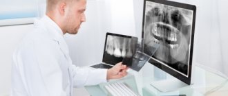
X-ray methods for examining an orthopedic patient
The main X-ray examination technique used in dental practice is radiography. Fluoroscopy is used much less frequently, mainly to determine the location of foreign bodies, sometimes in cases of traumatic injuries.
However, in these cases, transillumination is combined with preliminary or subsequent radiography. The anatomical features of the maxillofacial region (structure of the jaws, close arrangement of teeth in curved alveolar processes, the presence of multi-rooted teeth) determine the requirements for radiographs. Depending on the relationship between the film and the object of study, intraoral radiographs are distinguished (the film is introduced into the oral cavity) and extraoral (the film is located outside) . Intraoral radiographs are obtained on films wrapped first in black and then in wax paper to prevent exposure to saliva. For extraoral radiographs, cassettes with intensifying screens are used. The use of intensifying screens makes it possible to reduce exposure and thereby the radiation dose to the patient, however, the sharpness and structure of the image due to the fluorescent effect of the screens is worse than on intraoral radiographs. Intraoral radiographs, depending on the position of the film in the oral cavity, are divided into contact (the film is adjacent to the area under study) and bite photographs (the film is held by closed teeth and is located at some distance from the area under study). The structure of teeth and surrounding tissues is most clearly obtained on intraoral contact radiographs.
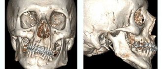
Methods of X-ray examination are divided into basic (intra- and extraoral radiography) and additional (tomography, panoramic tomo- and radiography, teleradiography, electroradiography, computed tomography, etc.). X-rays can reveal the presence of cysts, granulomas and impacted teeth. It makes it possible to diagnose benign and malignant tumors, traumatic injuries to teeth and jaws, the presence of foreign bodies in the maxillofacial area (bullets, projectile fragments, fragments of an injection needle, pulp extractor, root needle, bur, etc.).
Using radiography, you can clarify the diagnosis of apical or marginal periodontal lesions, differentiate chronic periodontitis (fibrous, granulomatous, granulating), establish the presence of osteomyelitis and other bone tissue disorders, diagnose periodontitis or periodontal disease and its stage depending on the degree of resorption of the walls of the tooth socket and alveolar process. Radiography facilitates the diagnosis of functional overload of individual teeth due to traumatic articulation or improper design of dentures. Radiography helps determine the severity of the process in periodontal diseases, the degree and nature of alveolar resorption (horizontal, vertical, funnel-shaped resorption, the presence of bone pockets), and determine the need for surgical or orthopedic treatment - with the help of splints and prostheses. This method facilitates the selection of the design of the orthopedic apparatus (removable, fixed) and supporting teeth.
BASIC AND SPECIAL METHODS OF X-RAY RESEARCH IN DENTISTRY
Methods of X-ray examination are divided into basic (intra- and extraoral radiography) and additional (tomography, panoramic tomo- and radiography, teleradiography, electroradiography, computed tomography, etc.).
The anatomical features of the maxillofacial region (structure of the jaws, close arrangement of teeth in curved alveolar processes, the presence of multi-rooted teeth) determine the requirements for radiographs. There are intraoral radiographs (the film is inserted into the oral cavity) and extraoral radiographs (the film is located outside). Intraoral radiographs are obtained on films wrapped first in black and then in wax paper on top to prevent the action of saliva. For extraoral radiographs, cassettes with intensifying screens are used. The use of intensifying screens makes it possible to reduce exposure and thereby the radiation load on the patient, but the sharpness and structure of the image due to the fluorescent effect of the screens is worse than on intraoral radiographs. Intraoral radiographs, depending on the position of the film in the oral cavity, are divided into contact (the film is adjacent to the area under study) and bite photographs (the film is held by closed teeth and is located at some distance from the area under study). Most clearly the structure of the teeth and surrounding
tissues is obtained on intraoral contact radiographs.
a) Intraoral bitewing radiography
Bitewing X-rays are performed in cases where it is impossible to obtain intraoral contact images (increased gag reflex in children), when it is necessary to study large sections of the alveolar process, to assess the condition of the buccal and lingual cortical plates of the mandible and the floor of the mouth. A film measuring 5´6 or 6´8 cm is inserted into the oral cavity and held with closed teeth. Bitewing radiographs are used to examine all teeth and all parts of the upper jaw, anterior teeth, and the anterior and posterior areas of the lower jaw.
b) Extraoral (extraoral) radiography
In certain cases, there is a need to evaluate parts of the upper and lower jaws, temporomandibular joints, and facial bones, the image of which cannot be obtained on intraoral photographs or is only partially visible. In extraoral photographs, the image of teeth and surrounding formations appears less structural. Therefore, such images are used only in cases where it is not possible to obtain intraoral radiographs (increased gag reflex, trismus, etc.).
c) Tomography
Tomography - layer-by-layer study - is an additional method that allows you to obtain an image of a certain layer of the studied area, avoiding superposition of shadows that complicate the interpretation of radiographs. Special devices are used - tomographs or tomographic attachments to an imaging table or a universal tripod. During tomoharphy, the patient is motionless, the X-ray tube and film cassette move in opposite directions (Fig. 9). The tube and the cassette holder are fixed at the ends of a metal rocker arm with a tomographic rod rotating around a horizontal axis. The position of the axis in each specific case changes taking into account the level (depth) of the body part being examined. When the patient is positioned horizontally on the X-ray machine table, the axis is set at the level of the layer whose image needs to be obtained. As a result of the synchronous movement of the tube and the film cassette, the image of all anatomical details is blurred, except for one layer lying at the level of the axis of rotation of the rod.
Tomography is used mainly to clarify pathologies of the upper jaw and temporomandibular joint.
The method makes it possible to assess the relationship of the pathological process with the maxillary pause, the floor of the nasal cavity, the pterygopalatine and infratemporal fossae, the condition of the walls of the maxillary pause, the cells of the ethmoidal labyrinth, and to detail the structure of the pathological formation.
d) Panoramic tomography
Panoramic tomography (orthopantomography) is a type of zonography. The practical use of panoramic tomography in dentistry began in 1949. An orthopantomogram produces a one-time image of the entire dental system as a single functional complex with virtually no angular distortions. The image on the film is slightly enlarged, and unequally in the central and lateral sections of the jaws. It should also be noted that the image of the anterior sections of the jaws and the projection of the cervical spine onto them are unclear.
e) Enlarged panoramic radiography
When performing enlarged panoramic radiography, the anode of a high-focus tube (focal spot diameter 0.1 mm) is inserted into the subject’s oral cavity, and X-ray film in a polyethylene cassette measuring 12 x 25 cm with intensifying screens is placed outside. The patient sits in a dental chair, the midsagittal plane is perpendicular to the floor, the occlusal plane of the jaw being examined is parallel to the floor. The tube is inserted into the oral cavity along the midline of the face to the level of the second molars (to a depth of 5–6 cm).
The focus of the X-ray tube is as close as possible to the object of study, the film is removed from the teeth by the thickness of the soft tissue. As a result, the image is enlarged by an average of 2 times.
f) Electroradiography
The scarcity of expensive silver, an integral part of photographic emulsion, dictates the need to search for materials for radiography that do not contain it. As a result, the method of electroradiography (xeroradiography) was developed and put into practice. The method is based on removing an electrostatic charge from the surface of a plate coated with selenium, followed by spraying colored powder and transferring the image to paper.
g) Computed tomography
The method allows you to identify the position, shape, size and structure of various organs, determine their topographic-anatomical relationships with nearby organs and tissues.
The method is based on the mathematical reconstruction of an X-ray image. The principle of the method is that after X-rays pass through the patient's body, they are recorded by sensitive detectors. Signals from the detector are sent to a computer (computer).
h) X-ray using contrast agents
The sialography technique for studying the ducts of large salivary glands consists of filling them with iodine-containing preparations. The study is carried out to diagnose mainly inflammatory diseases of the salivary glands and salivary stone disease.
Intraoral contact radiography
Dental X-rays can be obtained using any X-ray diagnostic machine. Special dental devices are most suitable for these purposes. The domestic industry produces devices 5D-1 and 5D-2. It should be noted that obtaining radiographs of teeth and craniofacial bones is more difficult than others due to anatomical features and the possibility of bones layering on top of each other, therefore, when taking contact intraoral photographs, it is recommended to direct the X-ray tube at a certain angle for the teeth of the upper and lower jaws, using the isometric rule: the central ray passes through the apex of the root of the tooth being removed perpendicular to the bisector of the angle formed by the long axis of the tooth and the surface of the film. Deviation from this rule leads to shortening or lengthening of the object, i.e. the image of the teeth turns out to be longer or shorter than the teeth themselves (Fig. 74).
To comply with the rules of isometry, it is necessary to use certain angles of inclination of the X-ray tube when photographing different areas of the jaws. To photograph individual teeth or groups of teeth, there are certain features of the position of the X-ray film of the oral cavity, the inclination of the X-ray tube, the direction of the central beam and the place of contact of the top of the tube with the skin of the face, which are described in the manuals on dental radiology.
In Fig. 75 shows a diagram of the projections of the apexes of the roots of the teeth on the skin of the face.
Interpretation of a dental radiograph
Only a dentist or radiologist can decipher and describe a dental image. The image shows the tooth: its root, internal canals, shape, anatomical features.
When assessing the image, the doctor performs the following actions:
- Examines the rigidity, density and uniformity of the bone structure. Evaluates the location of each element of the dentition.
- Determines the presence of signs of clearing or darkening, indicating the development of an inflammatory process, cysts, granulomas, neoplasms.
- Depending on what the dental x-ray shows, a diagnosis is made.
Carious formations in the picture look like light areas of various shapes with unclear boundaries.
The development of pulpitis is characterized by bone damage. The image shows a violation of its homogeneity in the interroot space. With the development of periodontitis, a granuloma appears in the area of the tooth root in the form of a rounded darkening with clear contours.
With periodontitis, the image shows a decrease in the density of the bone structure, a decrease in the height of the partitions between the elements of the dentition, and the formation of “pockets.”
Intraoral bite radiography
Bitewing X-rays are performed in cases where it is impossible to obtain intraoral contact images (increased gag reflex in children), when it is necessary to study large sections of the alveolar process, to assess the condition of the buccal and lingual cortical plates of the mandible and the floor of the mouth. A film measuring 5x6 or 6x8 cm is inserted into the oral cavity and held with closed teeth. Bitewing radiographs are used to examine all teeth and all parts of the upper jaw, anterior teeth, anterior and lateral areas of the lower jaw.
When radiography, the rules of projection are observed (the rule of isometry and tangent). The central beam is directed to the apex of the tooth perpendicular to the bisector of the angle formed by the long axis of the tooth and the film (Table 1). The patient sits in a dental chair, the film located in the bite is parallel to the floor of the office. The angles of inclination of the tube are given in table. 1.
Varieties
To obtain one image, two types of equipment are used - an outdated model of an X-ray machine or a digital radiovisiograph (its modern analogue).
In the first case, photographs are taken on film. The procedure is not comfortable for the patient; printing the image takes some time. The image turns out flat and loses quality, fading after one and a half to two years.
The second model of the device allows you to obtain photographs in electronic form of high quality and resolution. If necessary, the image can be expanded several times and a specific area can be studied in detail.
Images do not lose quality during long-term storage (for several years). In addition, the electronic device has 3 times less radiation exposure compared to the outdated model.
With modern equipment you can take two types of pictures:
- Interproximal - the condition of the coronal part is assessed. It is used for filling carious cavities with endomaterial, and for monitoring the correctness of prosthetics.
- Intraoral (done intraorally) - it is possible to determine the caries of adjacent units and the degree of adherence of the filling to the walls of the cavity after treatment.
This image is considered the most informative and effective way to diagnose dental caries and periodontal disease.
Extraoral (extraoral) radiography
In certain cases, there is a need to evaluate parts of the upper and lower jaws, temporomandibular joints, and facial bones, the image of which cannot be obtained on intraoral photographs or they are only partially visible. In extraoral photographs, the image of teeth and surrounding formations appears less structural. Therefore, such images are used only in cases where it is not possible to obtain intraoral radiographs (increased gag reflex, trismus, etc.).
Indications for use
Dental radiography is used for two main purposes: to diagnose and evaluate the quality of treatment performed.
Allows you to detect the following diseases:
- hidden carious lesion;
- pulpitis (inflammation of the neurovascular bundle of the tooth);
- cyst at the roots of the teeth;
- periodontitis (purulent inflammation of the tissue between the root of the tooth and the hole in which it is located);
- gum disease, in which bone tissue atrophies (periodontitis, periodontal disease);
- neoplasm.
Intraoral radiography also helps evaluate the results of procedures such as:
- root canal filling;
- treatment of periodontal diseases.
Study of dental x-rays
The tissues of teeth and jaws have different densities and thicknesses, so X-rays are absorbed to varying degrees. As a result, the X-ray image produces an image consisting of various shadows.
A normal dental x-ray (Fig. 76) shows:
- shadow of the enamel cover of the crown - 1;
- shadow of crown dentin - 2;
- clearing corresponding to the tooth cavity - 3;
- clearing corresponding to the root canal - 4;
- shadow of the tooth root, consisting of a shadow of dentin and an indistinguishable shadow of cement - 5;
- clearing corresponding to the lateral sections of the periodontal space - 6;
- dense strip of the cortical layer of the socket walls - 7;
- image of the interdental septum - 8.
The spongy bone tissue of the alveolar processes of the jaws appears in the photographs as a dense interlacing of dense bone beams intersecting in all directions and small light spaces filled with bone marrow. An x-ray of the upper jaw reveals a small-loop pattern; the lower jaw is characterized by a large-loop structure with a predominantly horizontal arrangement of bone beams. When assessing radiographs of the upper jaw, it is necessary to take into account its anatomical features, in particular the presence of air sinuses.
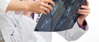
Each radiograph should be analyzed according to the following scheme:
1) determining the quality of the radiograph and the advisability of its use; the picture must be contrasty, clear, structural, without projection distortions;
2) identification of the upper or lower jaw on the image. For the upper jaw, the typical X-ray signs are the projection of the bottom of the cavities (maxillary, nasal) and a finely looped pattern of spongy bone, and for the lower jaw - the absence of a projection of the cavities and a largely looped pattern of bone;
3) determination of the anterior or lateral part of the jaws by the shape of the crowns of the teeth and the anatomical formations of this part in their x-ray image (especially in the absence of teeth). On intraoral radiographs of the upper jaw in the anterior section, as a rule, 7 main anatomical formations are projected, the bottom of the nasal cavity, the nasal septum, the inferior nasal turbinates, the inferior nasal passages, the anterior nasal spine, the intermaxillary suture and the incisive foramen (the latter is not always), and in the lateral section there are 3 main formations: the bottom of the maxillary cavity, the bottom of the nasal cavity, the zygomatic bone and behind the third molar (if an x-ray of the eighth teeth is obtained) additional 4 formations: the maxillary tubercle, the outer plate of the pterygoid process, the hook of the pterygoid process and the coronoid process of the mandible. On radiographs of the lower jaw, only the mental tubercle is projected in the anterior section and 3 formations in the lateral section: the mental foramen, the mandibular canal and the external oblique line;
4) detailed analysis of each tooth separately:
- crown assessment: size, shape, contours, intensity of hard tissues;
- tooth cavity: presence, absence, shape, size, structure; tooth root: number, size, shape, contours;
- root canal: presence, absence, width, if filling material is present - degree of filling;
- periodontal gap: width, uniformity;
- compact lamina of the alveoli: presence, absence, width;
- integrity violation;
- surrounding bone tissue: osteoporosis, destruction, osteosclerosis;
- interalveolar septa: location, shape of the apex, preservation of the compact end plate, structure;
5) determination of pathology in the area of the apical and marginal periodontium;
6) determination of pathology in the bone tissue of the jaws.
However, it is difficult to obtain two identical photographs of the same object taken at different times; the slightest deviation in the projection of the central beam onto the film gives a different picture of the x-ray image, which can lead to misinterpretation of the results of treatment measures. There are special devices and techniques for obtaining identical images of the teeth of the upper and lower jaws in the same projection.
Detailed analysis
The patient receives a traditional x-ray image from an analogue machine on photographic film. It is placed in a plastic or paper bag. The package indicates his last name, execution date and dental unit number.
In many clinics, the procedure is performed using digital devices. In this case, it is stored electronically on a computer, flash card or any other medium.
The taking of images should only be carried out by a qualified specialist who has undergone the necessary training. Quality largely depends on the correct implementation of the regulations adopted for these procedures.
Attention! The requirement to comply with the regulations also applies to the patient. If he wants to get a high-quality result, he must also follow the rules posted in front of the X-ray room.
The research data is reviewed by the attending physician. Based on them, the dentist receives detailed information about the condition of the tooth that is bothering the patient.
The following pathologies can be identified from the image:
- Pulpitis. appears at the bottom and center of the tooth. You may also find deposits in the root canal or dental cavity. This suggests that the disease has become progressive.
- Caries. If it is present, a change in the density of tooth enamel is noticeable. At the site of a carious lesion, the enamel will have a lighter appearance. If deformation of the tooth structure is visualized, then complicated caries occurs.
- Cyst. It can be traced in the form of an oblong figure with clear contours and a uniform structure. Its size and location are also well defined.
- Periodontitis. It is characterized by a sharp darkening in the upper part of the tooth root.
- Periodontal disease. At the same time, a decrease in bone tissue and signs of sclerosis and atrophy are visible.
This is interesting: Gingivitis in adults: complete information about the disease
In addition to a specific disease, the dentist can also detect concomitant dental anomalies (if any).
The examination will show the location of the tooth in the jaw, its inclination, and the presence of un-erupted neighboring teeth. This information can not only help in treating a specific disease. It can help prevent some problems in the future.
Tomography
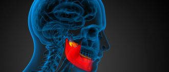
Tomography - layer-by-layer study - is an additional method that allows you to obtain an image of a certain layer of the studied area, avoiding superpositions of shadows that complicate the interpretation of radiographs. Special tomographs or tomography attachments are used. During tomography, the patient remains motionless; the X-ray tube and film cassette move in opposite directions. Using tomography, you can obtain an X-ray image of a specific layer of bone at the desired depth. This method is especially valuable for studying various pathologies of the temporomandibular joint, the lower jaw in the area of its angles (due to injury, tumor, etc.).
Tomograms can be obtained in three projections: sagittal, frontal and axial. Pictures are taken layer by layer with a “step” of 0.5-1 cm. The larger the angle, the greater the smearing and the thinner the selected layer. At a swing angle of 20°, the thickness of the layer under study is 8 mm, at 30°, 45° and 60° - 5.3 mm, 3.5 mm and 2.5 mm, respectively.
Tomography is used mainly to clarify the pathology of the upper jaw and temporomandibular joint. The method makes it possible to assess the relationship of the pathological process with the maxillary sinus, the floor of the nasal cavity, the pterygopalatine and infratemporal fossae, the condition of the walls of the maxillary sinus, the cells of the ethmoidal labyrinth, and to detail the structure of the pathological formation.
Layer-by-layer study with a small swing angle (8-10°) - zonography. In this case, the image of the area under study is clearer and more contrasting. Sonography at a depth of 4-5 cm in the frontonasal projection with the patient in an upright position is the method of choice for identifying effusion and assessing the condition of the mucous membrane of the maxillary sinus. The cut thickness is calculated to be 30 mm. To study the temporomandibular joint, lateral tomograms are performed in the position with the mouth open and closed. The patient lies on his stomach, his head is turned and the joint being examined is adjacent to the table top. The sagittal plane of the skull should be parallel to the plane of the table. The tomogram is carried out at a depth of 2-2.5 cm.
The scheme for measuring the parameters of the temporomandibular joint is shown in Fig. 77.
The width of the articular fossa at the base along line AB, connecting the lower edge of the auditory canal with the top of the articular tubercle; the width of the articular fossa is along the line CD, drawn at the level of the apex of the mandibular head parallel to the line AB; the depth of the articular fossa - along the perpendicular KL, drawn from its deepest point to the line AB, the height of the mandibular head (degree of immersion) - along the perpendicular KM, drawn from the highest point of the apex of the head to the line AB (almost always coincides with KL); width of the mandibular head - A1B1; the width of the joint space at the base in front is AA1 and in the back - B1B, as well as at an angle of 45° to the line AB from point K in the anterior section (segment a), in the posterior section (segment c) and in the upper section (segment b); angle of degree of inclination of the posterior slope of the articular tubercle to line AB (angle a).
Modern panoramic tomographs have separate programs for performing conventional orthopantomograms, zonograms of the temporomandibular joints, maxillary sinuses, the middle third of the face, the atlanto-occipital joint, orbits with the openings of the optic nerves, and the facial skull in a lateral projection.
Carrying out x-rays in children
Practice helps to conclude: radiography in children is carried out much more often than in adults.
This fact is explained by the fact that the baby teeth of young patients are especially susceptible to caries. In this case, the lesions are localized in those areas that cannot be seen in any other way. X-rays help identify defects in the process of eruption of distant elements of the dentition, ailments of dental and bone tissue, and effectively treat teeth and gums. The same diagnostic method is relevant if orthopedic manipulation is necessary for problems in the process of jaw formation in children.
The procedure is carried out for a child in a similar way. For children (children under 2 years old), radiography is prescribed only in extreme cases. For example, in case of injury during childbirth (to monitor the development of the dental system) or a child falling from a height (to assess the integrity of the teeth).
Enlarged panoramic radiography
When performing enlarged panoramic radiography, the anode of a high-focus tube (focal spot diameter 0.1 mm) is inserted into the subject’s oral cavity, and X-ray film in a polyethylene cassette measuring 12x25 cm with intensifying screens is placed outside. The patient sits in a dental chair, the midsagittal plane is perpendicular to the floor, the occlusal plane of the jaw being examined is parallel to the floor. The tube is inserted into the oral cavity along the midline of the face to the level of the second molars (to a depth of 5-6 cm). The X-ray film is pressed to the face by the subject himself, separately to the upper and lower jaws, and in this position the photograph is taken. Using this method, you can obtain a complete picture of all teeth in the form of a panoramic image with great sharpness and 2-fold magnification, and compared to conventional images, the patient’s radiation exposure is 25 times less.
X-rays of teeth
What is a visiograph and how does it differ from an x-ray?
This is one of the most common questions, like what is the difference between a car and a traffic light. It seems that both concepts have some kind of connection, but it is quite difficult to compare them. Also here. A radiovisiograph is a system that receives x-ray radiation, transforms it into digital form and displays the image on a computer screen.
Roentgen (who was Wilhelm Conrad) was a German physicist who gained worldwide fame for his discovery of short-wavelength rays with enormous penetrating power. The physicist himself called these rays X-rays (in English today they are called exactly that - X-ray), but now we often call them X-rays, and in everyday life simply “X-rays”. The unit of radiation power was also called the x-ray. Now it is clear that a visiograph and an x-ray are completely different things. If you compare the visiograph with anything, it’s with x-ray film, which it is universally replacing from all areas of medicine.
X-ray of teeth
Is it true that a visiograph is safer than a regular film photograph?
When asked about such a comparison, they mean the radiation exposure that the patient receives when using different techniques. In this sense, indeed, a visionograph is better, since its sensor is much more sensitive compared to film. Therefore, to obtain a high-quality image using a visiograph, you need significantly less shutter speed. To take a picture on film, the shutter speed is 0.5-1.2 seconds. To obtain the same image using a visiograph sensor – 0.05-0.3 seconds, that is, 10 times less. As a result, the radiation exposure received by the patient when using a visiograph is reduced to an insignificant minimum.
How many pictures can you take at one time? And in general, isn’t it harmful that when treating a large number of teeth, you have to take a lot of X-rays?
This is the most pressing of questions. This is an echo of the Chernobyl tragedy and life safety lessons that emerge in memory. But in our society there is still a very strong phobia towards everything that is even remotely connected in our heads with radiation. Any extra photo often raises questions about radiation sickness, or “will I glow in the dark?” Therefore, we will try to explain in more detail here. First from a scientific point of view.
To measure the amount of radiant energy applied to living tissue, various units are used - joule per kilogram, gray, rem, sievert, etc. In medicine, X-ray procedures typically measure the dose received by the entire body during one procedure—the effective equivalent dose, measured in sieverts. According to SanPiN 2.6.1.8-38-2003, when carrying out preventive medical x-ray procedures and scientific research, this dose should not exceed 1000 μSv (microsievert) per year. Moreover, here we are talking specifically about preventive studies, and not about therapeutic ones, where this bar is much higher.
What is 1000 µSv? Is it a lot or a little?
Remembering the famous cartoon, the answer is simple - depending on what you measure it in. 1000 μSv is approximately:
- 500 targeted images (2-3 μSv) obtained using a radiovisiograph
- 100 of the same images, but using good X-ray film (10-15 µSv)
So, apparently, even if we take 1 picture on a visiograph every day for the whole year, and also a couple of 3D computed tomograms per year, and the same number of orthopantomograms, then even in this case we will not get out beyond the limits of safe permitted doses.
there is no need to be afraid of receiving a significant dose of radiation during dental procedures . Even if you want to go beyond the permissible values, it is unlikely to be possible. To be clear, here are the doses needed to cause any serious health effects:
- 750000 μSv - short-term minor change in blood composition
- 4,500,000 μSv - severe radiation sickness (50% of those exposed die)
All these figures are incomparable in their significance with the doses we receive in everyday life. So even if, for some reason, several pictures are taken at once in a row, and the day before you were already “exposed” by doing an orthopantomogram, you don’t need to panic and run to the store to buy a Geiger counter or search on the Internet for “the first symptoms of radiation sickness.” To calm yourself down, it’s better to “detoxify” with a glass of red wine. There will be no point in this, but the mood will immediately improve.
Let us clarify that, for example, with one-stage basal implantation, control targeted images are taken before and after installation of implants (when installing 1-3 implants). And also after final prosthetics (7-8 days).
Is it possible to do x-rays for pregnant women?
We will not remind you that before pregnancy it is better to take care of your health in advance, including “preparing” your own teeth at the dentist. In order not to run away later with acute pain and doubts whether this or that manipulation will harm the development of the fetus... Therefore, you should leave the lyrics and pay attention to the facts and common sense. Without phobias, prejudices, speculations and myths. So, is it possible to do x-rays for pregnant women?
Here's what they write to us about this in the documents (SanPiN 2.6.1.8-38-2003):
7.16. Pregnant women are prescribed for X-ray examination only according to clinical indications. Studies should, if possible, be carried out in the second half of pregnancy, except in cases where it is necessary to decide on termination of pregnancy or the need for emergency or emergency care. If pregnancy is suspected, the question of the admissibility and necessity of an x-ray examination is decided based on the assumption that there is a pregnancy...
7.18. X-ray examinations of pregnant women are carried out using all possible means and methods of protection so that the dose received by the fetus does not exceed 1 millisievert for two months of detected pregnancy. If the fetus receives a dose exceeding 100 mSv, the doctor is obliged to warn the patient about the possible consequences and recommend terminating the pregnancy.”
In general, the conclusion from these two main points is simple and clear. It’s definitely not worth taking pictures in the first half of pregnancy, but in the second half - 1 mSv for a visiograph - this is practically unlimited.
I would also like to add that one often encounters bellicose stubbornness: an X-ray at the dentist during pregnancy is an absolute evil. It’s better, they say, to screw up a tooth, to cure crooked canals... there are a lot of teeth, pregnancy is more important. Moreover, such sermons are given not only by lay patients who have little understanding of the essence of things, but also often by dentists themselves who have forgotten their school physics course.
To resolve this doubt, we must understand that sources of ionizing radiation are not only found in medical offices. And it doesn’t have to be near Chernobyl (and now Fukushima) to receive some doses from the environment. After all, every second we are influenced by both natural sources (sun, water, earth) and man-made ones. And the doses received from them are much greater than those received from an x-ray of a tooth.
For clarity, we can give one simple example. As you know from a school physics course, the sun emits electromagnetic energy in a wide range, not only in infrared (heat), visible (light), ultraviolet (tan), but also in x-rays and gamma radiation. Moreover, the higher the earth's surface, the more rarefied the atmosphere and, therefore, the weaker the protection from sufficiently strong radiation from the sun.
After all, while “fighting” radiation at the dentist, the same people often calmly fly south to bask in the sun and eat fresh fruit. Moreover, during a 2-3 hour flight in a “healthy” climate, a person receives 20-30 μSv, i.e. the equivalent of approximately 10-15 images on a visiograph. In addition, 1.5-2 hours in front of a cathode ray monitor or TV gives the same dose as 1 picture... How many pregnant women sitting at home, watching TV shows and surfing the Internet, think about how many pictures they “took”? while watching another program, and then discussing it with friends on the forum and social networks? Almost no one, because people do not associate all this with ionizing radiation, unlike a picture in a doctor’s office.
And yet, dear expectant mothers, prepare for pregnancy in advance. For many people, visiting the dentist still remains stressful. And it’s not so much that anesthesia or x-rays can be harmful during this period, but what is important is your peace of mind and the absence of unnecessary worries (of which many already have more than enough during this period). You can study more information on this topic in our article “Can teeth be treated during pregnancy.”
What is the best protection to use if you need to take a photo while pregnant?
The number of aprons does not matter. In contact radiography, the apron essentially protects not from direct radiation, but from secondary, that is, reflected. For X-rays, the human body is an optical medium, like a glass cube for a flashlight beam. Point the beam of a flashlight at one of the faces of a large glass cube and, regardless of the thickness and direction of the beam, the entire cube will be illuminated. It’s the same with a person - you can swaddle him completely in lead and shine only on his head - at least a little, but it will reach every heel. So even under two aprons with a good lead equivalent, a pregnant woman will, at most, simply have a harder time breathing.
Is it possible to do x-rays for breastfeeding mothers? And if possible, then what about feeding the child after the procedure?
Can. X-rays are not the same as radioactive waste. By itself, it does not accumulate in the biological environment. If you irradiate a loaf of bread with a lethal dose, it will not mutate, get radiation sickness, or begin to “foul.”
X-rays differ from light rays only in wavelength and have a direct damaging effect only under certain conditions. If you shine a flashlight into a bucket of water and turn off the flashlight, the light won't stay in the bucket, right? The same is true in a protein-fat solution, which is what many biological fluids are (including breast milk) - the radiation passes right through. So, with such a load that is necessary to work with a visiograph, it is unlikely that anything will happen to the milk itself.
As a last resort, to reassure yourself, you can skip one regular feeding. Another thing is that the breast tissue itself during lactation is, of course, more susceptible to the harmful effects of radiation. But, again, we are talking about doses much more powerful than is necessary for digital radiography (of course, subject to all protective measures and without “shooting” 20 times anywhere).
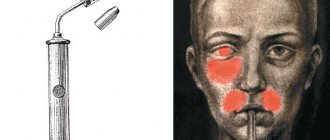
Electroradiography
The scarcity of expensive silver, an integral part of photographic emulsion, dictates the need to search for materials for radiography that do not contain it. As a result, the method of electroradiography (xeroradiography) was developed and put into practice. The method is based on removing an electrostatic charge from the surface of a plate coated with selenium, followed by spraying colored powder and transferring the image to paper. To carry out the method, a special electro-radiographic apparatus ERGA has been developed, consisting of two blocks: a charging block and a block for developing an x-ray image.
Where to get an x-ray, how much the procedure costs, video
The targeted radiography service is available in almost every specialized clinic. The cost varies around 400-450 rubles.
Some clinics provide the practice of booking several x-ray procedures (2-4) as part of dental treatment - the patient has the opportunity to save money.
A targeted dental image is a highly informative and safe procedure that allows you to identify a dental problem and monitor the effectiveness of the therapy. It is successfully carried out for both adults and children. X-rays are used with caution in pregnant women and infants.
Teleradiological examination in dental practice
The term “teleradiography” refers to performing a study at a large focal length, ensuring minimal distortion of the size of the organ being examined. The images obtained in this way are used to carry out complex anthropometric measurements, allowing one to assess the relationship of various parts of the facial skull in normal and pathological conditions. The technique is used to diagnose various malocclusions and evaluate the effectiveness of ongoing orthodontic measures. Teleradiograms are performed on a cassette with intensifying screens measuring 24×30 cm, the focal-film distance is 1.5-2.0 m. During the study, it is necessary to use a craniostat, which ensures fixation of the patient’s position and obtaining identical radiographs.
The complexities of the skull structure require radiographs to be taken in two mutually perpendicular projections - frontal and lateral. In practical work, in most cases, only teleradiography in a lateral projection is used. Determining on a teleroentgenogram the sizes of various lines drawn between certain anthropometric points and the magnitude of the angles between them makes it possible to mathematically characterize the characteristics of the growth and development of various parts of the skull in a particular patient. This is described in more detail in the chapter “Orthodontics”.
The dangers of dental x-rays
Digital radiovisiograph (without applying film)
In modern medical practice, when creating a targeted image, a digital radiovisiograph is used, which is characterized by a reduced level of radiation, in contrast to devices used as screening diagnostics. Radiation exposure at the time of examination for an adult is 7-15 μSV. For comparison, the natural background in Moscow is 20 µSV. (Use the “Dosimeter” at the end of the article)
This fact allows the equipment to be used to obtain a clear image in the treatment of preschool children without dangerous consequences for the child’s body.
The permissible annual dose for the population is 5000 µSV, and for regular visits to the dentist, a person may need no more than 100 spot images - this will total 1 mSV. There is no need to worry about the degree of harmfulness of such tiny radiation on the human body.
This is interesting: Purulent plugs in the throat: causes and treatment
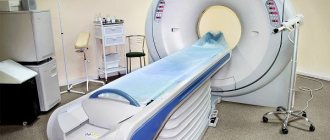
CT scan
The development and introduction into clinical practice of X-ray computed tomography (CT) was a major achievement of science and technology. The method allows you to identify the position, shape, size and structure of various organs, determine their topographic-anatomical relationships with nearby organs and tissues.
The method is based on the mathematical reconstruction of an X-ray image. The principle of the method is that after X-rays pass through the patient's body, they are recorded by sensitive detectors. Signals from the detector are sent to a computer (computer). A high-speed electronic computer processes the received information according to a specific program. The machine spatially determines the location of areas that absorb X-rays differently. As a result, a synthetic image of the area under study is recreated on the screen of a television device - a display. The resulting image is not a direct x-ray or tomogram, but is a synthesized image compiled by a computer based on an analysis of the degree of tissue absorption of x-ray radiation at certain points. CT slice thickness ranges from 2 to 8 mm.
The method expands diagnostic capabilities in recognizing traumatic injuries, inflammatory and tumor diseases, primarily of the upper jaw. As is known, significant difficulties are encountered during X-ray examination of this department. On CT, the cartilaginous disc of the temporomandibular joint may be visible, especially if it is displaced anteriorly.
Radiovisiography:
- Phosphoric: produced using film and reagents
- Digital: produced using a sensor, electronic matrix, block.
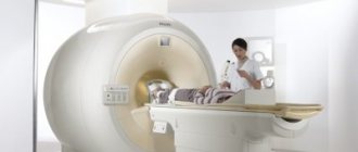
MRI – magnetic resonance imaging: a method that allows you to examine tissues (including soft ones) and organs using the physical phenomenon of nuclear magnetic resonance
Ultrasound is a research method using ultrasonic waves used to diagnose diseases of internal organs and soft tissues
EDI–electroodontodiagnosis: a method that allows you to assess the excitability of the dental pulp when it is irritated by electric current (Table 25)
Table 25 Indicators of electrodontodiagnosis of teeth with various pathologies
| EDI indications | Diagnosis | ||
| 2-6 µA | Intact pulp | tooth: | healthy |
| 6-12 µA | Caries | ||
| 20-80uA | Pulpitis | ||
| {amp}gt;90 µA | Periodontitis | ||
| Myography | -method | research | bioelectric |
| activity | muscles, | allowing | register |
contractile activity of muscles
Radiography using contrast agents
The sialography technique for studying the ducts of large salivary glands consists of filling them with iodine-containing preparations. The study is carried out to diagnose mainly inflammatory diseases of the salivary glands and salivary stone disease. Angiography is a method of contrast X-ray examination of the vascular system of arteries (arteriography) and veins (venography).
Did you like the article? Share with friends
0
Similar articles
Next articles
- Anthropometric examination of the jaws and dental arches
- Methods for determining chewing efficiency
- Methods for studying the general condition of the body of an orthopedic patient
- Contents and formulation of the diagnosis of an orthopedic patient
- Classification of materials used in orthopedic dentistry
Previous articles
- Instrumental and hardware methods for examining an orthopedic patient
- Questioning and examination of an orthopedic patient
- Tracheostomy
- Cellulitis of the mediastinal tissue (mediastinitis)
- Abscess, phlegmon of the posterior neck