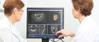
Dental CT is a type of modern x-ray examination. By performing it, a three-dimensional model of the area under study is obtained with maximum detail. A 3D image provides the doctor with a complete amount of information about the condition of the maxillofacial region and surrounding soft tissues.
During the procedure, the device creates a series of layer-by-layer images, which are digitized and turned into a three-dimensional model. The ability to change the image size and high accuracy minimize the risk of distortion of the result.
What is it used for in dentistry?
A three-dimensional image taken by CT allows dentists to examine in detail all the nuances of the problem that has arisen in order to make the correct diagnosis.
© apops/Fotolia
In implantation, three-dimensional models will allow you to select a high-quality prosthesis, all the components for its fastening and surgical intervention.
Modern CT devices allow you to perform research in various areas of dentistry :
- endodontology – one of the sections of the therapeutic field, work with the root canals of teeth;
- surgery – implantation, bone grafting, diagnostics, study of tumors;
- periodontology – study, prevention, therapy of periodontal tissues and their pathologies;
- orthodontics – analysis of causes, diagnosis, prevention, treatment of various anomalies of teeth and jaw bones.
In addition, CT allows dentists to see a real picture of what is happening with the patient’s teeth in order to prescribe the correct treatment or surgical intervention.
Which is better - CT or X-ray?
Computed tomography is considered one of the most informative diagnostic methods used in dentistry. Using this method, it is possible to obtain a multilayer image of the area under study. A 3D image is much more informative compared to a conventional X-ray, which only provides a planar image.
From the point of view of affordability, x-rays are preferable, but they do not always provide an accurate picture of the condition of the dental-maxillary system. As practice shows, if a CT scan is performed, then in 99% of cases it is possible to accurately determine the cause of toothache and other symptoms.
Diagnostic principle
To carry out CT scans spiral and multispiral tomographs are used , scanning soft tissues and transmitting a 3D image to the monitor.
Read how to treat tooth pulpitis in a new publication.
In this article we will talk about methods for treating flux at home.
We’ll talk about the symptoms of periodontal disease and its treatment with folk remedies at the link https://www.vash-dentist.ru/lechenie/desnyi/parodontoz/sposobyi-narodnyimi-sredstvami.html. Features of the course of the disease.
Modern tomography is a universal tool for high-quality diagnostics , which has great potential in the study of various areas of dentistry, as well as maxillofacial surgery:
- injuries;
- congenital developmental anomalies, defects;
- focal infections turning into chronic;
- implantations and planned operations;
- complex tooth extractions and prosthetics;
- control of the development of neoplasms in tissues and facial bones;
- clarifying diagnostics for drawing up a treatment plan.
CT capabilities allow for panoramic scanning of the entire oral cavity with saving 3D photographs on DVDs for further study by the attending physician.
In particularly difficult cases, specialists from various fields are invited - this is done in order to clarify the diagnosis, eliminate the possibility of errors, and a comprehensive treatment plan is developed.
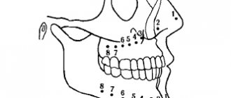
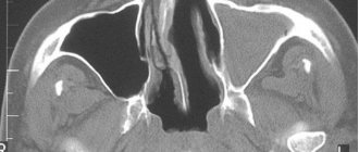
High-resolution and high-quality digital images make it possible to scan the area of interest to doctors in different planes:
- frontal – indicated in blue (see photo);
- sagittal or vertical, divides the object into left and right halves (red);
- axial – transverse, divides into upper and lower parts (green).
Such tomographic studies provide radiologists with complete information about the patient’s condition; in addition, it becomes possible to construct 3D models in various sections and perform more accurate measurements.
What consequences can there be if you don’t do a CT scan?
If the doctor does not receive detailed images before treatment, he may choose the wrong implantation method and artificial root model. The specialist may damage the nerve or sinuses. In the absence of this type of study, the risk of incorrect installation of implants increases, and accordingly, the risk of their non-engraftment or rejection also increases. Early failure of the entire system, improper distribution of the chewing load and discomfort, inflammation of the tissues surrounding the structure are likely. All these complications will not only undermine your health and make you nervous, but will lead to irrational use of funds (and implantation is not a cheap service), or to additional expenses.
On a note! Without a tool such as CT, the doctor will not be able to accurately plan all stages of the operation, which will inevitably lead to complications. So, for example, when choosing an implant of a smaller (than necessary) size, the load on the bone will be uneven. If the root is installed in the wrong area, the patient’s bite will be disrupted, and the structure will be non-functional.
Advantages
CT is already replacing the usual research methods using an X-ray machine, having certain advantages:
- Carrying out an examination with extensive information - the volumetric image shows not only soft tissues, but even the smallest foci of inflammation.
- High accuracy of reproduction of the final result, no need for decoding. The clarity of the image allows you to detect all the nuances of the structure, the ability to enlarge the image multiple times.
- 3D images make it possible to study an object not in one plane , as was the case with X-rays, but in three at once. Photos can be copied or printed for patient reference.
- Taking pictures in scale: 1:1 , which are then easy to enlarge and examine the area of interest from all sides.
- The procedure is highly safe – the radiation exposure is much less than with conventional x-rays, and the process is much faster.
- Obtaining a clear picture of the location of the implant in relation to the dental nerve.
The patient does not feel any discomfort during CT scanning: the procedure is performed in a standing or lying position, which depends on the modification of the device.
The radiation doses are so small that this method is also used for children of all ages.
With such short-term irradiation, doctors receive comprehensive information about the general condition of the jaw - all diseases of the oral cavity or teeth will be captured in photographs for subsequent diagnosis.
Visitor reviews
“When treating the roots of the tooth, the doctor decided to remove it. I had to go to another clinic for additional consultation, where a CT scan procedure was prescribed. This was the first time I did such a diagnosis, and thanks to it, the tooth was saved,” Victoria, Perm, February 2020.
“I had to do a dental computed tomography scan several times already. A quick, painless procedure that does not require additional preparation. The doctor immediately gives a transcript. I completely trust this research and consider it a worthy breakthrough in the development of medicine,” Vladimir, Moscow, December 2020.
In what cases is it prescribed?
When installing prosthetics and identifying various diseases of the oral cavity, the dentist prescribes a CT scan procedure.
The main indications for the use of this progressive method are:
- detection of unerupted teeth;
- identification of injuries, damages;
- study of jaw bone abnormalities or deviations from normal dental development;
- elimination of congenital malocclusion of irregular shape - before installing mouth guards or braces;
- detection of hidden cavities with the presence of caries, dental cyst or tumor;
- preparation for surgical interventions on the jaw and implantation;
- control during and after surgery.
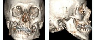
If you are planning implantation, then you cannot do without an accurate CT scan with subsequent printing of the results : the doctor must take very accurate measurements and do preliminary computer modeling.
Dental and maxillofacial surgery and otorhinolaryngology cannot do without high-quality diagnostics. In these cases, this method is also practiced.
Indications
The reason for a dentist to prescribe a computed tomography scan may be trauma to the jaw area. CT scans are also often performed to assess the condition of the dentition after an accident or other emergency situations. When using a CT scan, you can immediately see displacements of the jaw bones, cracks, damage to tooth roots, and more. Early diagnosis makes it possible to exclude further complications.
This picture is taken when teeth are positioned incorrectly. The doctor then correctly installs braces and performs other manipulations that promptly correct abnormal tooth growth. CT scan is a 3D image, thanks to which braces and aligners are made very accurately.
For complex operations and removals, this method comes in handy. After what he sees, the doctor will be ready for anything, and this increases the chances that the procedure will go without complications. It is advisable to do this both before and after surgery, since further therapy depends on what is seen in the photo.
In the image you can also see various formations appearing in the jaw area, so if doctors suspect a tumor, they can also resort to this type of diagnosis. For example, after injuries, a cystic formation often forms in the area of impact. If detected in time, it can be removed without serious consequences.
If the patient plans to have implants, then a dental CT scan should also be present. It is done to accurately measure the height and width of the alveolar processes.
It can be prescribed if an inflammatory process or other changes in the dental area are suspected.
On this topic
- Diagnostics
All about computed tomography of the jaw
- Maria Konstantinovna Tevs
- July 18, 2020
Some specialists resort to this method in order to see how well the canals are sealed, and also find out the number of roots in the tooth.
In case of inflamed gums, the periodontist will send you for a CT scan to see the whole situation from the inside and prescribe effective treatment.
A similar diagnosis is also carried out in case of detection of unerupted teeth.
After the doctor makes a diagnosis, an important role is played by the x-ray. Simple “photos” cannot fully convey the condition of the patient’s teeth. For a more accurate and high-quality examination, three-dimensional dental computed tomography was developed.
Attention! CT scan is appropriate for any inflammatory processes occurring in the jaw area.
Preparation
To perform a dental CT scan, all you need to do is pre-make an appointment with a dentist at the reception desk - no special preparation is required, you don’t need to take anything with you.
The patient, in a standing, sitting or lying position, rests the lower part of his face on a special stand in the CT machine and, at the doctor’s command, does not move for several minutes while the scan is performed.
People prone to claustrophobia before the procedure simply tune in and calm down - you need to understand that nothing terrible will happen in such a short time.
But, as a result, you will have high-quality information, and your attending physician will have the opportunity to make an accurate diagnosis and conduct high-quality therapy.
What treatment methods for alveolitis after tooth extraction are most effective?
In a separate article we will tell you why treatment of a fistula on the gums of a child using traditional methods is not recommended.
Here https://www.vash-dentist.ru/lechenie/zubyi/karies/prisheechnyiy-prichinyi-vozniknoveniya-foto.html we will talk about why the treatment of cervical caries of the anterior teeth requires a special approach.
Patients who cannot remain in one position for a long time should also remain motionless for several minutes - to obtain a high-quality result, you can be patient a little.
Even fidgety pediatric patients successfully undergo this easy procedure.
Contrast-enhanced tomography - what is it?
In order to identify hidden neoplasms in bone and soft tissue, vascular diseases, to assess the size of tumors and the location of metastases, contrast-enhanced tomography is performed. As part of the examination, a special iodine-based contrast agent is used. The drug is administered intravenously.
Important! Before carrying out such a procedure, the specialist must make sure that the patient does not have a possible allergic reaction to the contrast agent. Otherwise, a person risks encountering such unpleasant symptoms as swelling of soft tissues, itching, sore throat, hives, swelling of the larynx, breathing problems, nausea and vomiting.
The procedure with contrast is used to identify hidden tumors in bone and soft tissue
The drug can be administered before the procedure or during it. This technique allows you to detect and assess the extent of problems that cannot be identified without a contrast amplifier.
Sequence
Tomographs are bulky devices of various configurations, the principle of which is to smoothly rotate a special design around the patient’s head, with the help of which about 600 images are taken.
There are quite a few types of CT scanners, but they are all based on an absolutely painless scanning method.
Sometimes a secure head fixation is required, but this happens extremely rarely. The initial position of the patient (standing, sitting or lying) depends on the modification of the equipment.
When scanning with contrast is performed, a special substance is injected intravenously into the patient before the procedure begins. In this case, the doctor must first make sure that there is no allergy to similar drugs.
The session lasts no more than 5 minutes and does not require any special preparation of the patient..
Today, not only spiral, but also high-quality dental (cone beam) tomographs for special purposes are used, with the built-in function of an orthopantomogram - a panoramic image of the entire oral cavity.
Then the attending physician carefully examines the obtained images and makes a diagnosis. It is accurate CT that provides indispensable assistance to dentists in many matters, especially during surgical intervention after complex injuries.
How is a CT scan done?
A dental computed tomography machine resembles an orthopantomograph. The process itself is simple: the person stands or sits. The patient's neck and chest are protected with a special apron with a lead layer. The subject rests against the frontal support, which, together with the chin and temple supports, provides reliable fixation of the head; hands on the handrails; firmly holds a special contact segment with his teeth. Based on the required shooting mode and frequency, the patient must remain motionless in this position for 15-25 seconds, during which the device’s planar sensor will make a full revolution around the subject’s head. In these few seconds, up to two hundred photographs are taken in various projections.
It takes several minutes to process the received information and build a multidimensional model; the CT scan is recorded on disk. After 5-10 minutes, accurate data on the current condition of the teeth and jaws is ready, the doctor can, based on it, make a final diagnosis and prescribe treatment or monitor the therapy already completed.
Restrictions and contraindications
Minimal exposure to radiation waves still exists, so the frequency of the procedure must be decided individually, under the supervision of the attending physician and mandatory consultation with a radiologist.
All contraindications are advisory:
- Pregnant women should refuse a CT scan, because during the shooting there is, although minimal, radiation that is quite safe for the whole body, but still radiation that can have a negative impact on the subsequent development of the fetus.
- Nursing mothers are not prohibited from carrying out similar procedures, but we must remember that for 2 days after the tomography it is necessary to refrain from breastfeeding the baby.
- This type of diagnosis is not recommended for patients with claustrophobia - fear of limited space: the moving part of the tomograph that takes pictures can cause a panic state.
- People who cannot remain still even for a short period of time should also avoid having a CT scan.
- CT scanning using a contrast agent is contraindicated even mild diabetes
About the advantages of using dental computed tomography when planning implantation, watch the video:
Leading experts advise not to do CT scans more than twice a year, and a break of at least 12 months is needed between scheduled studies. It is necessary to take into account the types and doses of radiation, individual characteristics of the body, diagnosis, as well as circumstances surrounding the procedure.
Is it possible to do a CT scan with dental implants?
Unlike magnetic resonance imaging, computed tomography allows for the presence of implants and dentures in the mouth. The material from which the structure is made matters.
Modern implants consist of inert metals (titanium, zirconium) and ceramics. They do not create interference and do not distort the final model with spots and shadows. Previously used “simple” metals change the appearance of the image, but a qualified specialist takes this into account when decoding.
When answering whether it is possible to do a CT scan with dental implants, let’s say yes, but be sure to notify the x-ray technician about the presence of fixed structures in the mouth.
Sign up for the study
Price
The final cost of a CT scan is influenced by various factors:
- first of all, it is a tomograph ; the newer its modification, the higher the price. Today, the latest devices with a fairly wide range of capabilities already exist, but many clinics in the Russian Federation still have outdated equipment.
- often the price is influenced by the status of the clinic where the CT scan is performed: in Krasnodar there is a medical center where tomography is performed free of charge, other institutions are focused on costs ranging from 250 rubles to 3 thousand.
In Moscow, the cost of a CT scan in public clinics is 1-6.4 thousand, and in private clinics - from 4 to 8 thousand rubles. - The pricing policy is also determined by the quality of the images. In many private medical institutions, CT results are recorded on DVDs and then issued to the patient.
This innovation is not practiced in public clinics.
Types of computed tomography
Tomography for dental implantation can be performed using different devices. And here it is important to understand that they can produce images of different quality.
Step-by-step tomography
Today, this type of research is no longer carried out anywhere in innovation and metropolitan centers, because it uses old-generation devices that produce images of poor quality and strong radiation exposure, which, as some researchers claim, exceeds even the annual permissible norm of 1000 μSv by 1-1.5 times (and this is for 1 complete study, and in the process dental implantation requires at least three of them).
Multislice tomography
MSCT, unlike the first type, is one of the highest quality types of research. It is carried out on a spiral tomograph and allows you to see all the hidden processes in detail. This type of research cannot be done in dentistry, because The devices installed here are simpler than those required for MSCT1. However, professional doctors send their patients to specialized centers to conduct this type of research in cases where the clinical situation is complex and it is necessary, for example, to carry out zygomatic implantation.
Cone beam examination
This is the most common and optimal type of CT scan today. The resulting images are of good quality and high clarity, which allows them to be used with maximum benefit for working out and planning all stages of treatment. At the same time, cone beam devices have a low radiation dose, so they can be used for diagnostics as many times as necessary.
On a note! Specialists load the obtained data into the software. By the way, the capabilities of software from different manufacturers may also be different: some provide more opportunities for a comprehensive analysis of the data obtained, while others provide less. Some doctors believe that the best today are Sirona, Picasso.
Anatomy of the dental system
The organs of the dental system perform the most important functions - breathing, speech, and digestion. It includes:
- Skeleton, consisting of several bones, including the nasal, maxillary and submandibular, palatine.
- Salivary glands, thanks to the secretion of which the initial stage of digestion is ensured.
- Teeth whose main purpose is to chew food.
- Organs for grasping food and sound pronunciation (lips, tongue, palate, cheeks).
- Musculature.
- Temporomandibular joints (TMJ).
The difficulty of diagnosing diseases of the dentofacial system encountered in dentistry is due to the complexity of its structure and the relationships of all elements. In addition, the structure of the dental system determines the inaccessibility of all its elements for visual inspection. In this regard, great importance is attached to technological methods in dentistry, primarily CT of the jaw, or MRI.
Price
The cost of a dental CT scan depends on many factors. This includes the number of teeth being examined, the type of apparatus, and the need to transfer a digital image to film or recording on a medium.
The average prices for cone beam tomography of teeth are as follows:
- CT scan of a segment of 3 teeth - 1500 rubles,
- CT scan of the jaw on one side - RUB 2,700,
- CT scan of the upper and lower jaw on one side - RUB 3,700,
- CT panoramic — 4500 rub.
A complete tomogram of the teeth of both jaws and sinuses ranges from 5,000 to 5,500 rubles. If a dental CT image is performed on a simpler device, the cost of all services will be 1000–1100 rubles lower. Additional recording of results on flash media is estimated at 100 rubles.
It is obvious that CT dental diagnostics is a simple but effective procedure. The quality of the research largely depends on the completeness and accuracy of the transcription. Without dental tomography, it is very easy to make a mistake, which will then have to be difficult and time-consuming to correct. That is why CT examination should not be neglected. Otherwise, all material costs for treatment may be in vain.
Author: Elena Medvedeva, doctor, especially for Karies.pro
Take a 3D CT scan of teeth: what advantages does the technique give to patients?
Compared to other diagnostic methods, CT of the jaw has a number of advantages, including:
- 1. Speed of implementation. A CT scan can be done in just a few minutes and also quickly obtain all the data necessary for analysis and treatment planning.
- 2. CT eliminates the possibility of obtaining inaccurate and blurry images.
- 3. The CT method is absolutely safe for the patient’s health (provided that modern tomographs are used in the examination, with minimal radiation doses).
- 4. You can do a CT scan for one or two jaws at once, a separate area of the oral cavity. The ability to select the area of study is convenient for both doctors and patients.
All data obtained after a dental CT scan is easily stored on any information medium and this will allow them to be transferred to another specialist if additional consultation is necessary.
The most popular questions about CT scans in St. Petersburg
How often can I have a CT scan?
Radiation doses when using modern tomographs are minimal, so if you need a repeat CT scan, you do not have to worry that the procedure will cause harm to your health.
Is it painful to have a CT scan?
Computed tomography is performed quickly and does not cause any pain or discomfort during the procedure.
Is it possible to do a CT scan during pregnancy?
There is no exact data on the effect of CT on the fetus during pregnancy, so pregnant women can undergo this study only in cases of extreme necessity and only after agreeing on the procedure with the doctor monitoring the pregnancy. It is definitely not recommended to undergo a CT scan in the first trimester of pregnancy.
Where to get a dental CT scan in St. Petersburg?
The CT service is offered by different clinics in St. Petersburg, and when choosing a dentist for diagnostics, it is important to find out what equipment will be used in the procedure. Choose those clinics in St. Petersburg that have modern and high-tech equipment that allows you to conduct accurate research!
How to choose a clinic? It’s simple - enter a request on the Internet - do a CT scan of teeth in St. Petersburg, addresses and prices, and the system will give you a list of dentists offering the service. You will need to call clinics and find out what equipment is used to perform CT scans.
Where can I get a dental CT scan in St. Petersburg inexpensively?
The desire to save on dental services is quite understandable, but you should not chase excessively low prices for CT scans in St. Petersburg. An underestimated cost of a service may indicate that the research is being conducted on outdated equipment. Therefore, it is more correct not to ask the question about the price of CT scans in St. Petersburg, but to look for a clinic where the study will be carried out with the highest quality and safety for your health.