Peculiarities
- To adequately evaluate the vessel, angiography is performed with contrast.
- In non-selective angiography, contrast is injected into the cubital vein.
- The injection rate is 4-5 ml/sec, the drug is injected automatically.
- This examination method is not indicated for all patients.
Advantages
- High spatial resolution.
- Possibility of quantitative measurement of lumen narrowing.
- Assessment of vessels in combination with adjacent structures.
- Can be performed if there are metal objects in the body.
- Possibility of assessing vessels hidden on ultrasound by bone structures.
Flaws
- Contrast required.
- High radiation exposure.
- Metal artifacts (clips) that make visualization difficult.
- Possibility of allergic and anaphylactic reactions.
- Complications after venipuncture.
- Toxic effects on the kidneys.
Comparison of restrictions
Having figured out how MRI and MSCT tomographs differ, it becomes clear that certain limitations and contraindications appear. This greatly influences the choice of examination method. A magnetic resonance imaging scan of the brain should not be performed if the patient:
- Have implanted pacemakers, hearing aids or insulin pumps. They react to magnets and lose their working rhythm.
- The patient's weight is too high. It does not allow you to sit comfortably in the narrow tunnel of the device and causes damage to the retractable table.
- The injured person is in critical condition. During scanning, you have to remain motionless for at least 30 minutes, which is impossible in case of severe injuries accompanied by unbearable pain.
- Pregnancy in the 1st trimester.
- Implanted metal implants and brackets on teeth that distort sections.
- MRI is not recommended for neurological disorders. The narrow tunnel and sharp clicks when the coils operate are difficult for patients with claustrophobia, so they require choosing a different method.
When comparing the difference between MSCT and MRI, one should not forget about the powerful X-ray radiation of the first method. Despite all the positive aspects, it remains dangerous in terms of the degree of impact on tissue. Additional stress on internal organs is created when using a contrast agent, therefore contraindications include:
- pregnancy in any trimester;
- mental problems (claustrophobia);
- diabetes;
- thyroid dysfunction;
- renal failure;
- lactation.
The MSCT procedure is not recommended for muscle spasms: the patient does not control movements, so the sections are blurred, and the accuracy of the diagnosis is not guaranteed.
Options
MSCT angiography is divided into selective and non-selective arterio- and venography.
- In the first case, a diluted drug is injected into the vessel (artery or vein) that needs to be contrasted.
- In the second - into the vein of the upper limb.
The contrast agent enters first into the pulmonary circulation, then into the aorta and smaller arterial vessels throughout the body.
Usually only one or two adjacent zones are examined, because The scanning speed is low, and the device usually cannot keep up with the blood flow.
Contrast agents, scan scale and type
For MSCT angiography, contrast agents containing iodine are used.
- The active substance may be in ionic or non-ionic form.
- The first group includes urografin, the second - iohexol, omnipaque, yopamiro, ultravist, yomeron, etc.
- Non-ionic drugs give the least number of complications.
- Ionic preparations are now practically not used, because give many serious complications.
- Each drug contains a different amount of iodine. The most common iodine concentrations are 300-400 mg/ml.
- For selective arterio- or venography, substances with lower concentrations are used: 100-150 mg/ml. This concentration leads to a decrease in the number of artifacts from dense matter in the lumen of the vessel.
Attention: if you are allergic to iodine, contrast is contraindicated!
The scanning scale depends on:
- from the area of pathological changes,
- previously performed studies - ultrasound, MRI.
If the affected area is known, the scale is limited only to it and a few centimeters before and after.
If the extent and localization of pathological changes are not clear, the scanning area reaches half a meter or more, for example, when examining the vessels of the thigh.
Spiral tomography is used to evaluate vessels, because step-by-step is too slow and the tomograph cannot keep up with the blood flow.
On average, to study an area 10 cm long, it takes 10-30 seconds, depending on the width of the spiral turn. The time for stable contrasting of the arteries of the neck, for example, is 10-15 seconds.
Therefore, it is necessary to compare the speed of the device with the available time for vascular contrast.
CT angiography can be performed on tomographs with at least 16 rows of detectors. With such devices, blood vessels up to 1 mm wide can be distinguished, but only the lumen of arteries 2-3 mm wide can be characterized.
For coronary angiography, a computed tomograph with a large number of detector rows is suitable - from 32. The optimal one is 256 or 512.
In general, the more detectors, the higher the resolution and the smaller vessels can be assessed. However, radiation exposure increases accordingly.
Keep in mind: the more slices, the higher the resolution.
Study areas
| Zone | Borders |
| Head | From the base of the skull to the coronal suture |
| Neck | From the upper thoracic outlet to the base of the skull |
| Rib cage | 10 cm below the diaphragm to the superior thoracic aperture |
| Stomach | From the level of the ilium to 2 cm above the diaphragm |
| Pelvis | 2 cm below the symphysis pubis to the level of the iliac crests |
| Lower limbs | From the level of the aortic bifurcation to the toes |
| Upper limbs | From the level of bifurcation of the brachiocephalic trunk to the fingers |
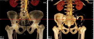
What does the photo show?
MRI images are thin sections presented as multiple images of the scanned area. The brain and its parts are shown in different projections, so it is difficult to miss tumors, bleeding or veins blocked by a blood clot. The diagnostician analyzes the contrast shades and determines the malignancy or benignity of the neoplasm.
Using MSCT, a computer creates a three-dimensional model of the patient’s skull and brain, allowing one to track anatomical abnormalities in the bone structure. The model clearly shows blood vessels of any size, their plexuses and pathologies. The image recreates the structure of the brain matter with inflammatory foci, hematomas and areas of tissue necrosis. When drawing up a surgical plan, a three-dimensional image allows you to anticipate nuances and complications.
Preparation
Preparation depends on the scanning area. General point:
- blood tests for creatinine, urea;
- determination of renal function.
Before examining the heart vessels:
- it is necessary to stabilize the heart rhythm;
- consult a cardiologist;
- take with you the data of previously performed examinations - MRI, ultrasound;
- Warn staff of previously observed allergic episodes and local reactions to contrast.
Methodology
- The patient lies down on the tomograph table, and a catheter is inserted into the vein of the elbow.
- First, marking scans are performed, then the area where arterial density will be monitored (zone of interest) and the boundaries of the scanning area are marked on them.
- Then a marking transverse scan is performed. The artery section is highlighted because Density changes will be monitored there.
- Then the “trigger” value (in Hounsfield scale units) is set - the trigger. Typically this parameter is 100-200 units.
- The injector is then started and begins to inject contrast into the vein.
- At this time, the tomograph takes scans of the area of interest and notes changes in density in it.
As soon as the density exceeds the set value, scanning begins with preset parameters.
Note! The trigger is set during research:
- cerebral vessels - to the common or internal carotid artery;
- pulmonary circulation - to the pulmonary artery;
- neck and head - on the aortic arch or one of its large branches.
Depending on the scanning area, the parameters presented in the table may change.
| Parameter | Characteristics |
| Patient position | With your feet or head towards the tomograph, on your back or stomach |
| Scanning direction | Caudal (to the feet), cranial (to the head) |
| Area of interest | The lumen of the artery is slightly above or below the scanning area |
| Scanning area | Anatomical area assessed |
| Trigger | Density value, above which the tomograph starts scanning |
| Injection rate | Typically 4-5 ml/sec |
| Contrast volume | 50-150 ml |
Duration
The study and preparation takes 10-30 minutes. The vast majority of time is spent on preparation; the scanning itself usually lasts 10-30 seconds.
Please note: data collection takes only a short time. It will take much longer to prepare for the procedure.
List of indications for examinations
MRI is the best method for scanning soft structures. It is better than MSCT if there is a need to identify the following pathologies:
- scanning the structure of the brain and spinal cord, individual hemispheres;
- the presence of tumors, cysts, gliomas or fibromas;
- the cause of impaired blood supply to the brain after a stroke, injury;
- pathology or displacement in the cervical spine;
- blood clots or aortic aneurysm supplying the brain;
- inflammatory foci during infections;
- detection of the cause of epileptic seizures, loss of consciousness or disorientation of the patient;
- early diagnosis of sclerosis and Alzheimer's disease.
MRI of the brain is often recommended for young children who develop hearing loss, pathologies of the nervous system due to birth injuries, or cerebral palsy. Using this method, neurologists monitor the patient's condition after chemotherapy or surgery to remove a tumor, making a decision on further therapy.
Vascular pathologies are better visible with multislice MSCT of the head with the addition of a contrast agent. The drug penetrates into the smallest blood vessels and highlights the boundaries of malignant tumors and metastases on sections. Indications for the procedure include:
- any benign neoplasms;
- oncological tumors at an early stage without metastasis;
- angiopathy due to diabetes mellitus;
- severe traumatic brain injury;
- damage to the bone tissue of the skull;
- growths on the arch;
- aneurysm;
- blockage of the bloodstream of an artery by blood clots;
- foreign bodies in the medulla;
- viral meningitis;
- assessment of the patient's condition after acute ischemic stroke;
- infectious foci in the brain;
- malformations.
MSCT is used when it is necessary to perform an operation requiring craniotomy. It helps to identify in the patient the destruction of the masticatory or temporal joint, lesions of the maxillary sinuses, which often become the source of a dangerous infection.
Complications
Complications in most cases are associated with the administration of contrast.
These include:
- allergic reactions - skin rashes, swelling of the respiratory tract;
- anaphylaxis is an emergency condition requiring immediate assistance.
During the administration of contrast, the following are possible:
- feeling of warmth;
- metallic taste in the mouth;
- cough.
In addition, at the site of contrast injection, the following may occur:
- phlebitis;
- thrombosis
Kidney failure may also develop. Risks from radiation exposure are low.
It is not recommended to perform MSCT - in young children, in pregnant women and during lactation - if this study can be replaced by another without damage.
Review of contraindications
MSCT or MRI are safe diagnostic techniques, but in certain cases they can cause harm to the patient. Magnetic resonance imaging is not prescribed for patients who have electronic devices installed in their bodies, such as a pacemaker. The magnetic field can cause disturbances in their operation, which will lead to undesirable consequences for the patient’s health and may even threaten his life.
The multispiral technique is not recommended for pregnant women and breastfeeding mothers. Diagnostic testing is not recommended for children under 7 years of age. The annual radiation exposure should be no more than 5 mSv per year.
MSCT is unacceptable for renal failure, asthma and decompensated heart failure. Patients suffering from diabetes mellitus are advised to stop taking medications containing metformin 2-4 days before diagnosis (preliminary consultation with a doctor is required).
Alternatives
| Method | Advantages | Flaws |
| Traditional angiography | High resolution, ability to visualize small vessels | Radiation exposure, need for contrast, invasiveness |
| MRI angiography | A developing diagnostic method, no contrast required, no radiation exposure, ability to evaluate adjacent structures | Duration of the study, artifacts from vascular pulsation, impossibility of performing in the presence of magnetic metal objects in the body |
| Ultrasound | No radiation exposure, high resolution of modern sensors, ability to assess the direction of blood flow (color Dopplerography) and the presence of blood flow (power Dopplerography) | Inability to visualize vessels shielded by bones and gas |