Nakhodki
A CT scan of the neck can detect traumatic changes in bones and joints and evaluate:
- location of the fracture;
- direction of displacement of fragments;
- damage to adjacent neural structures.
Fractures of the cervical vertebrae are dangerous - possible compression or damage to the spinal cord or spinal nerves.
You can also evaluate the relationships in the joints of the craniovertebral junction.
Violation of the relationship between the odontoid process and the atlas is often observed in children, but is not always a consequence of injury.
Patients with degenerative spinal disease have:
- decreased height of intervertebral discs,
- osteosclerosis of the endplates of the vertebral bodies,
- osteophytes (bone “spikes”, “spurs”).
Signs of arthrosis of the facet joints and uncovertebral arthrosis are also detected. The latter is due to the fact that the vertebrae “sit” on top of one another, and the posterior sections of the body of the overlying vertebra are pressed against the uncinate processes of the underlying one.
Osteochondrosis is also characterized by straightening the physiological curve of the spine - it becomes straight.
The next group of findings are anomalies in the development of vertebrae.
- Wedge-shaped or butterfly-shaped vertebrae may be detected, leading to kyphoscoliosis.
- Fusion of the vertebrae in the anterior and (or) posterior sections is also possible (Klippel-Feil disease).
- Ossification of longitudinal ligaments and joints is often observed (ankylosing spondylitis, Forestier's disease).
Anomalies of the craniovertebral zone are more common. These include:
- Non-fusion of the odontoid process with the body of the axis (odontoid bone);
- invagination of the odontoid process into the cranial cavity;
- non-closure of the arches of the atlas, etc.
Tumors of bones and soft tissues of the neck. For tumors, a contrast study is required to evaluate:
- intratumoral vascular network;
- better delineation from adjacent structures.
Most often, MSCT of the neck is performed in patients with squamous cell carcinoma:
- larynx;
- pharynx;
- tonsils;
- language;
- with metastatic cervical lymphadenopathy.
The method allows you to evaluate:
- neck anatomy;
- determine the location of the tumor and its size;
- invasion;
- secondary foci.
It is necessary to distinguish multiple cystic formations of a different nature from tumors. Most often these are congenital cysts.
Highlight:
- gill slits cysts;
- thyroglossal duct cysts;
- nasopharyngeal duct cysts, etc.
Cysts can be confused with necrotic lymph nodes or a neck abscess.
Contrast is indicated for differential diagnosis with tumors.
Inflammatory lesions. Most often, abscesses are found in the neck area, caused by an inflammatory process in the area of the teeth, tonsils, and salivary glands.
- The abscess looks like a fluid inclusion with a thick wall that accumulates contrast. It manifests itself as swelling and “clouding” of adjacent tissues.
- Diffuse diffuse inflammation (phlegmon) looks like widespread tissue swelling with fluid leaks.
Vascular pathology. To identify vascular disorders, such as occlusion, compression, thrombosis, embolism, developmental anomalies, aneurysms, MSCT of the neck vessels with contrast is required.
A worthy alternative to CT is ultrasound. Ultrasound can accurately assess:
- degree of narrowing of the vessel;
- its progress;
- presence of anomalies.
But the ultrasound does not show:
- vessels of the aortic arch;
- intracranial segments;
- segments of arteries located in bone canals.
Options
Computed tomography must be performed using different modes depending on the suspected pathology.
- In case of injuries, a study is performed without contrast with reformatting in three planes in the bone window.
- For degenerative diseases - examination in the bone and soft tissue window.
- For tumors and pathologies in the vessels, contrast is added.
In the first case, contrasting is two- or three-phase, in the second - arteriography and venography.
Standard research
By “standard” we mean research:
- in spiral or step mode without contrast;
- with construction of reconstruction in three planes;
- using bone and soft tissue modes.
Applicable:
- for injuries,
- with osteochondrosis,
- for vertebral anomalies;
- as an indicative method.
MSCT
Multislice tomography is performed on tomographs with several rows of detectors in a spiral mode.
Devices with one row of detectors are now almost never found, as a result of which all studies outside the step-by-step mode are considered multispiral.
MSCT scans an object with 4-helices in one revolution of the tube, while the rotation speed is 0.5 seconds faster than the MSCT.
Multispiral research is characterized by:
- higher speed;
- less radiation exposure;
- lower resolution.
CT with contrast
Contrasting is shown:
- for neck tumors;
- for vascular pathology (angiography).
For tumors, a standard two- or three-phase study is performed.
- An iodine-based contrast agent is injected at medium speed into a vein of the upper limb.
- The neck is then scanned in several phases.
The data obtained allows us to evaluate:
- tumor vascularization;
- metastases;
- more clearly separate the neoplasm from healthy tissue.
CT angiography
- Angiography is performed by injecting high-velocity contrast into a vein in the elbow and then performing a single-phase scan of the neck.
- Unlike a standard contrast study, scanning begins earlier.
- Accumulation of contrast in the parenchyma does not yet occur; only vascular structures are visualized, the lumen of which is filled with a radiopaque substance.
How to prepare for the study, and what preparation will there be in the clinic?
Before undergoing this type of examination, the patient should perform the following activities:
- Obtain a referral for a CT scan of the cervical spine from an appropriate specialist. For example, if there is a suspicion of cancer, such a referral is given by an oncologist. When studying the condition of the larynx and nasopharynx, an ENT doctor refers for examination, for the thyroid gland - an endocrinologist, etc. Many medical institutions do not accept a patient for examination without an appropriate referral.
- Extract from the patient's medical record.
- Results of previous examinations (if any).
This manipulation does not require any specific preparation. However, in the case of contrast, the patient must refrain from eating and drinking for several hours before the procedure. Otherwise, the gag reflex may be triggered.
Among other things, the use of a contrast agent has a number of contraindications, so you need to donate blood in advance to determine the level of creatinine and urea.
In order not to distort the results of the study, all jewelry must be removed.
Parts of clothing can also interfere with the scanning process, so it is best to bring a hospital gown with you.
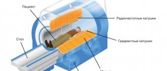
Device for scanning the neck and cervical spine
Indications and contraindications
Indications:
- neck injuries, wounds;
- foreign bodies of the esophagus, respiratory tract;
- developmental anomalies of the cervical vertebrae;
- abnormalities of the craniovertebral junction;
- bone tumors;
- soft tissue tumors;
- congenital cysts of the neck, for example, gill slits;
- ectopia of the salivary glands, thyroid gland;
- other developmental anomalies;
- vascular pathology - aneurysms, malformations, vascular abnormalities;
- postoperative control.
Contraindications:
- allergy to contrast;
- increased “fragility” of veins;
- impaired renal function;
- pregnancy;
- childhood.
The first three points are related to the administration of contrast, the last two are relative contraindications.
Methodology
First of all, you need to remove all metal objects from the scanning area - earrings, chains, brooches. Their presence leads to the appearance of artifacts on tomograms, which greatly complicate the interpretation of the results by a doctor.
- The patient lies on the tomograph table on his back.
- If contrast injection is planned, a catheter is installed in the vein, connected to a machine that injects the drug.
- Then marking scans are performed - straight and lateral. The operator marks the scanning area on them and starts data collection.
- If contrast is planned, data are collected in one or more phases after the start of drug administration.
- The received data is processed and transferred to a workstation, where a doctor views it and issues a conclusion on it.
Stages of performing computed tomography of the neck and cervical spine
When contrast is performed, a dye is injected into the patient a few minutes before the scan begins.
If the object of study is the digestive tract, contrast is administered orally .
For all other cases, bolus contrast - a small amount of iodine-containing X-ray contrast agent enters the body through an installed catheter in a vein throughout the study.
In this case, certain side effects may occur: nausea, dizziness, perspiration. All this goes away in just a few minutes.
If these pathological conditions are pronounced, this should be reported to the doctor.
In general, at the time of scanning, the patient is in a separate room, and communication with medical staff is maintained thanks to a special intercom.
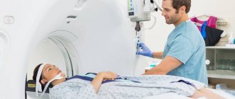
At the very beginning of the procedure, the nurse helps the patient lie down on a retractable table, which, after connecting the system, automatically enters the tomograph, where layer-by-layer sections of the area under study are made. At the time of scanning, the person should not move or even swallow saliva.
Computed tomography of the cervical spine takes, on average, 5-10 minutes . The scanning process itself lasts no more than a minute , which is very convenient for examining people who are in serious condition.
After the diagnosis is completed, the patient is sent home. He is notified in advance that the first day after the procedure, urine and feces may change color. This phenomenon is a consequence of the removal of the contrast agent.
CT or MRI: which is better?
Each method has its own purpose. MRI is better for patients with soft tissue lesions, computed tomography for bone lesions.
MRI is an ideal method for visualizing intervertebral discs and nerve roots in adult patients without any radiation exposure. It is more correct to use it in patients with osteochondrosis. If there are contraindications, the method may well be replaced by computer diagnostics.
In any case, a specialist must make a decision.
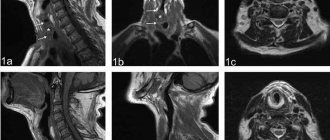
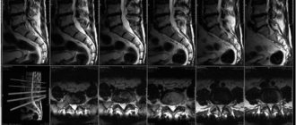
Neck CT results – what does a neck CT scan look like?
During this procedure, the tomograph takes several black and white images. Only the most successful of them are given to the patient.
For an additional fee, you can also request a disk/flash drive on which the entire scanning process will be displayed.
In addition, based on the information received, the radiologist makes a written conclusion .
The patient can discuss all questions regarding the examination with the doctor at the time the results are received.
Price
In 2020 in Russia, the cost of computed tomography ranges from 1,500 to 10,000 rubles.
- The standard procedure costs 1500-4000 rubles.
- With contrast - up to ten thousand.
Layer-by-layer examination of the neck is an advanced, fast and accurate method for diagnosing various diseases.
- The method is used for both outpatient and inpatient patients.
- The method is characterized by multiple advantages.