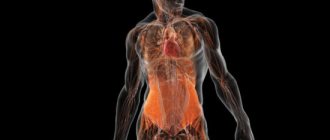
The mediastinum is a complex of anatomical structures in the chest cavity. The mediastinum is divided into inferior and superior; anterior, middle, posterior. The organs of the mediastinum include the thyroid gland, respiratory tract, esophagus, blood vessels, muscles, nerve trunks, heart, and lymph nodes.
Mediastinal pathology can be divided into diffuse lesions - mediastinitis and local space-occupying formations - tumors. Computed tomography is used to diagnose pathology, allowing the attending physician to quickly decide on a tactic.
Classification of mediastinal lesions
Mediastinal masses can be divided into cystic and soft tissue. Cystic have a density corresponding to fluid. Solid (soft tissue) formations are more dense, their structure is homogeneous or heterogeneous: with cavities, calcifications, partitions.
Cystic formations
| Education | Characteristics |
| Bronchogenic cyst | A congenital disorder of the development of the bronchial tree, most often found in children. Bronchogenic cysts are asymptomatic unless they cause compression of the airways. Discovered by accident. |
| Esophageal duplication cyst | Congenital pathology of the anterior germinal intestine, manifested as a cystic formation of a homogeneous structure and density near the wall of the esophagus. |
| Neuroenteric cyst | Hypodense formation near neural structures (spinal cord, spinal roots). It occurs due to incomplete resorption of the neuroenteric canal. |
| Pericardial cyst | Rare benign cystic formation of the anterior and middle mediastinum. On CT, it appears as a well-circumscribed fluid collection near the pericardium. |
| Meningocele | A developmental anomaly characterized by the release of the meninges and neural structures into the surrounding soft tissue. Often located within the chest. |
| Lymphangioma | Occurs mainly in children as a result of impaired development of the lymphatic system. The formation of anastomoses between lymphatic and venous vessels is disrupted. On CT it looks like a cystic formation. |
| Cystic teratoma | A volumetric formation containing organ and tissue components of varying degrees of maturity (skin and its appendages, fat, teeth). In cystic teratoma, the fluid component predominates. |
| Thymus cyst | Thymic cysts account for 1-3% of volumetric formations of the anterior mediastinum. They are divided into acquired and congenital. Acquired ones are more often single-chamber, developing from the remnants of the thymopharyngeal duct. Congenital, predominantly multilocular, arise due to damage to the thymus. |
| Cystic tumor (lymph node) | Due to impaired blood flow, necrosis often occurs in the center of a rapidly growing node. A hypodense zone is detected in the center of the lymph node or tumor, which does not accumulate contrast. The “rim” on the periphery is intensely contrasted. |
| Thoracic duct cyst | A cystic formation with a fluid density, with smooth edges, adjacent to the wall of the thoracic duct, filled with lymph. |
| Pseudocyst | Pancreatic pseudocyst protruding into the mediastinum through the hiatus of the diaphragm or the foramen of Morgagni. On CT it appears as a thin-walled cystic formation associated with the pancreas. |
Soft tissue formations
| Thymus tumors (thymoma, cancer, lipoma, carcinoid, etc.), hyperplasia | Thymoma is one of the most typical tumors of the anterior superior mediastinum. It looks like a soft tissue formation between the sternum and large vessels. In 10-15% of cases it contains cysts and calcifications. Invasive thymoma grows into the tissue or neighboring organs, resembling cancer. After contrast administration, an uneven increase in the density of the solid component is determined. Concomitant mediastinal lymphadenopathy is often found. Thymolipoma is a rare benign tumor represented by mature adipose tissue. A malignant variant of lipoma is liposarcoma. Carcinoid is the most common neuroendocrine tumor of the thymus with a heterogeneous structure and density. |
| Tumors of the thyroid gland and parathyroid glands, goiter | Adenoma is a benign tumor of follicular cells. The method of choice is ultrasound. Thyroid cancer (mostly papillary) can be assessed on CT. A nodular space-occupying formation with compression or invasion of neighboring organs, unevenly accumulating contrast, is determined. Multinodular goiter is also best assessed by ultrasound. If suspicious nodes are identified, a biopsy is indicated. Tumors of the parathyroid glands are better distinguished on ultrasound. |
| Other neoplasms | Tumors of the pericardium, heart, nerve sheaths (schwannoma, neuroma), esophagus (cancer, leiomyoma). Enlarged lymph nodes (with metastatic lesions). |
How is the examination carried out?
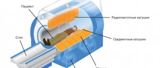
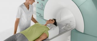
Diagnosis of mediastinal diseases using magnetic resonance imaging is carried out as follows:
- the patient is placed on a special table on which a tomograph is installed (it is a cylindrical chamber);
- fix the chest of the person being examined using special rollers and belts;
- then the table with the patient moves inside the cylindrical chamber (tomograph) and the diagnosis begins.
All data obtained during the MRI process is recorded on the monitor screen.
During the procedure, a person should not move, because any movements can cause distortion of the information received. The CT scan machine produces loud noises that may cause discomfort to the patient. Therefore, in such cases, the examinee may be given soundproof headphones. The tomograph itself is also equipped with a two-way communication device, and if the patient is bothered by something, he can contact the doctor at any time.
The procedure lasts from 15 to 30 minutes. The time it takes can increase to one hour if the tomography is performed with intravenous contrast enhancement.
What does tomography of organs and mediastinum include?
MRI of the mediastinal organs includes the ability to examine:
- Hearts. Using the procedure, the doctor can evaluate the condition of the heart muscle (myocardium), blood vessels, valves and chambers of the internal organ. In addition, tomography makes it possible to detect areas of myocardial scarring (in the event of a heart attack).
- Lungs. MRI allows you to determine the presence of modified areas in the organ and its pleura, as well as determine the extent of the pathological process.
- Soft tissues, bone and cartilaginous structures of the thoracic region. The examination can provide the doctor with information about the condition of the ribs, vertebrae, and fatty tissue of the chest.
- Blood vessels, arteries and nodes of the body's lymphatic system. Using magnetic resonance imaging, you can detect the presence of diseases of the lymphatic system and anomalies in the structure of blood vessels and arteries (aneurysms).
Feelings during the procedure
During the diagnostic examination, the patient may be concerned about the following:
- a metallic taste in the mouth or a rush of blood to the extremities (if a contrast agent is used);
- increase in body temperature;
- a burning or cold feeling in the area where the contrast agent was injected;
- discomfort from prolonged standing still.
Despite possible discomfort, the procedure is easily tolerated and does not cause pain in the person being examined.
Using Contrast Enhancement
A contrast agent is used in cases where it is necessary to increase the accuracy of the diagnostic procedure. If contrast enhancement is unavoidable, then before the MRI the patient is given an intravenous injection of a special drug that contains gadolinium.
Contrast enhancement allows the doctor to establish the exact location of tumor tumors and their boundaries.
Cost of MRI
The cost of magnetic resonance imaging in both public and private medical institutions starts from 4,500 rubles and above.
Other pathological changes
In addition to tumors and cysts, the following are found in the mediastinum:
- inflammatory changes - abscess, mediastinitis;
- lymphadenopathy of reactive or infectious nature;
- gas inclusions - pneumomediastinum;
- wounds (penetrating through the chest or esophagus);
- foreign bodies, such as swallowed bones, perforating the esophagus;
- anomalies in the development of the heart, blood vessels, and respiratory tract;
- vascular disorders - wall dissection, atherosclerosis, aneurysm;
- pathological changes in the heart - aneurysms, changes in size;
- changes in the esophagus - diverticula, stenosis, Shatsky rings;
- pathological changes in the respiratory tract: pharynx, larynx, trachea, bronchi;
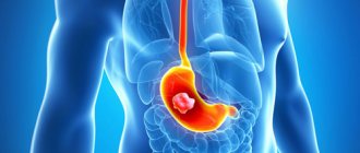
- diaphragmatic hernia with prolapse of the stomach into the mediastinum.
To kid
There are no age restrictions for MRI of the mediastinum.
To perform an MRI, a prerequisite is immobility throughout the entire examination. Children may find it difficult to lie still, so anesthesia may be considered.
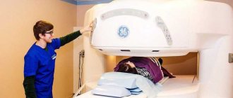
Children may be afraid to be in the confined space of a closed-type CT scanner. In this case, an examination can be performed using an open-type tomograph. The parent will be able to stay next to the child and hold his hand.
Research methodology
- If a CT scan of the mediastinum is performed without contrast, the patient is placed on the tomograph table, having first removed all metal objects.
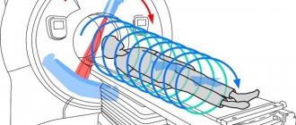
- The table then slides into a ring, inside which the X-ray tube rotates.
- In helical tomography, the table moves slowly inside a ring and the tube rotates around the patient, delivering radiation to the patient. The attenuated radiation is perceived by detectors on the other side, then converted into cross sections on the screen.
- If contrast is administered, a catheter (a thick needle with a wide lumen) is first installed in the vein. The catheter is connected to an injector, which pumps contrast at high speed.
Consider: all metal objects on the chest, incl. decorations need to be removed. Women also need to remove their bra.
CT scan
How to prepare
Special preparation, if no contrast is introduced, is not needed. As before any other study with contrast, for example, angiography of the cerebral arteries, it is necessary to evaluate the level of creatinine and urea in the blood serum.
The doctor should be warned about previous allergy episodes.
What to take with you
You need to take with you:
- previous conclusions, disks (films);
- direction;
- opinions of other doctors;
- blood test results for creatinine and urea.
This list is subject to change. It is better to call the clinic first and find out what else you need to take with you.
Contraindications to CT of the chest and mediastinum
It is prohibited to perform a CT scan during pregnancy. The gestational age is not important. The radiation from the device can have a teratogenic effect on the fetus, that is, provoke the appearance of congenital pathologies.
CT scanning is not performed in cases where the patient’s weight exceeds the permissible load on the equipment. Domestic clinics use devices with a maximum capacity of 200 kg.
There are certain limitations for computed tomography of the chest using contrast solutions:
- individual intolerance to contrast components, which can provoke the development of a severe allergic reaction;
- breastfeeding, as the contrast may pass into the milk. The substance is removed from the body within 48 hours;
- renal failure. Deterioration in the function of removing substances from the body can lead to poisoning and allergies;
- the patient's serious health condition: this means shock, coma, various severe injuries. A CT scan with contrast will only worsen the severity of a person’s condition.
MRI indications
MRI of the mediastinum is performed during differential diagnosis or at the stage of additional examination, if deviations from the norm have previously been identified.
MRI of the mediastinum is indicated to confirm:
- heart attack, other damage to the myocardium or heart sac;
- congenital defects;
- aneurysms and other vascular lesions;
- inflammation of the lymph nodes or other tissues in the mediastinum;
- blood clots in the vessels of the heart and lungs;
- cysts;
- neoplasms;
- spread of metastases;
- location and severity of injury.
MRI has no quantitative or age restrictions. This method is used in the process of examination and treatment for children or adults (except for pregnant women).