- Large fruit - body weight 4001 - 5000 g.
- Giant fruit - more than 5000 grams.
- Low fetal weight - less than 2500 g.
- Very low fruit weight - less than 1500 g.
- Extremely low fruit weight - less than 1000 g.
What should the baby's weight be?
This question worries many expectant mothers. Some simply for the sake of idle curiosity, others for the purpose of determining the tactics of childbirth, etc. Therefore, modern obstetrics necessarily involves determining the weight of the fetus in various ways. Both regular arithmetic calculations and automatic methods for determining your baby’s birth weight come to the rescue.
When and how does the research take place?
Routine ultrasound examinations are carried out three times: at 12, 20, and 32 weeks. The normal course of pregnancy gives reason to carry out fetometric diagnostics during the same period.
The procedure is carried out in two ways:
- transvaginally - a vaginal sensor is inserted into the vagina.
- transabdominal - the contents of the uterus are viewed through the external abdominal wall.
During the examination, the doctor takes measurements of the fetal organs on the monitor screen, then makes a diagnosis about the correct development and formation of individual organs.
Additionally, fetometric analysis is usually carried out in the following cases:
- the mother's condition is of concern to the gynecologist;
- there is a suspicion of a violation of intrauterine development of the fetus.
By month
The monthly pregnancy calendar will become your guide to all 40 weeks of pregnancy.
Every expectant mother will be able to leave her reviews, observations, share experiences and useful information here. Together we will watch how quickly our babies grow and develop! And give advice on the right lifestyle, diet and safe treatment of all sorts of ailments. Each month of pregnancy is marked by serious changes in the development of the child: he achieves greater success in physical and mental development, the baby’s brain grows rapidly, muscle mass strengthens, vital organs and systems are formed, and even the first natural skills! The pregnancy calendar by month will familiarize you in detail with these events at each stage.
Personal pregnancy calendar
Every woman can keep a personal pregnancy calendar. Moreover, it is welcomed and recommended. After all, now you are in a very special state. Many sensations are new to you, and can even cause worries and fears - the body of a pregnant woman undergoes enormous changes, both in the physiological sense and in the psychological sense.
The pregnancy calendar by week and month is compiled with an attempt and purpose to explain each of them.
You can keep a personal pregnancy calendar in a separate notebook, which you decorate in accordance with your mood, tastes, experiences, as well as whether you are expecting a boy or a girl. Such an album will be an excellent start to a family chronicle or a chronicle of the life of your heir.
Key points of fetometric research
The key data of a fetometric study are the following indicators:
- DB - thigh length;
- BPR - biparietal size;
- DP - shoulder length;
- KTP - coccygeal-parietal size;
- DN - nasal bone length;
- LZR - fronto-occipital size;
- OG - head circumference;
- DG—tibia length;
- AB—abdominal circumference;
- TVP is the thickness of the collar space.
A decoding of the designations of the studied parameters is provided, since the fetometric data is written in the table in Latin.
A video about the stages of ultrasound was presented by the 1st Medical Quarter of Crede Experto on Taganka.
Child's weight
By the 12th week, the baby’s body weight is normally only 19 g; by the middle of pregnancy, the baby will weigh about 345 g, and by the 32nd week - almost 2 kg.
If you promptly pay attention to the problem with the discrepancy between the fetal body weight and the standard, and take preventive measures, then it will be possible to correct the situation relatively easily. The rate of weight gain is greatly influenced by genetic factors. The gynecologist makes sure that the dynamics are positive.
KTR (CRL, coccygeal-parietal size)
KTR (Latin analogue of CRL) means coccygeal-parietal size, that is, the height of the child. It is calculated from the crown to the end of the coccyx.
If this indicator differs slightly from the norm, then the fetus is not in danger. An increase in CTE over several weeks by the same value indicates that the fetus is relatively large in size.
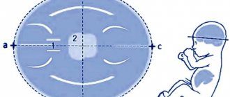
BPD (BPD, biparietal and fronto-occipital head sizes)
The letters BPD indicate the width of the fetal head. This is the maximum distance between the parietal bones. The size can be determined by taking measurements along the smallest axis of the circle between the child's temples. BDP allows you to determine the exact duration of pregnancy.
The parameter of biparietal fetal head size (BSD) helps to identify developmental abnormalities already during the first trimester. The data obtained characterize the state of the fetal nervous system.
The LZR or fronto-occipital size is calculated between the most distant points of the forehead and the back of the head.
OG (chest circumference)
The volume of a child's chest is determined by calculating the diameter of its circumference. A size that does not correspond to the norm should not cause any particular concern; most likely, this is a genetic feature. Perhaps the baby will simply be born large. It is necessary to take into account the physical characteristics of the mother and father.
AB (abdominal circumference)
Abdominal circumference is measured at 20 and 32 weeks of pregnancy. The parameter is calculated along the line of the liver, stomach and umbilical vein. When the difference in indicators exceeds the permissible norm, the doctor will diagnose intrauterine growth retardation. However, to confirm it, the size of the coolant is calculated in relation to other parameters - the size of the head, femur, and BPR. If most indicators are normal, then this indicates a delay in the development of the asymmetric form.
DB (femur length)
If a discrepancy in the thigh length is detected, this also does not indicate the presence of pathology. Much depends on individual characteristics. For example, when the length of the femur and shin bones is longer than normal, it means that the parents of the unborn baby or other relatives have a long leg.
PMP (PVP)
PVP is the estimated body weight of the fetus. During an ultrasound examination, there is a possibility of an error in weight. To eliminate errors, this figure is calculated using various medical formulas.
Calculation methods:
- Zhordania (Lebedeva) - PMP = height of the uterine fundus × abdominal circumference.
- Bublichenko - PMP = 1/20 of a woman’s weight.
- Lankowitz - PMP = (height + maternal weight + abdominal circumference + uterine fundus height) × 10.
- Jones - PMP = (height of the uterine fundus - 11) × 155. The value 11 is a conditional coefficient for a pregnant woman weighing up to 90 kg.
- Yakubova - PMP = (abdominal circumference + uterine height) × 100/4.
Calculations are made after 38 weeks of pregnancy.
The Family TV channel presented a video about performing an ultrasound in the third trimester.
The importance of estimated fetal weight in the choice of obstetric tactics
The estimated weight of the fetus has been determined, but how to correctly interpret the results obtained, how to most reliably determine the weight of the fetus. It is clear that if the baby’s weight is higher than 5000 g, then a planned caesarean section is indicated. However, there have been cases of such large babies being born with good outcomes. Sometimes, in order to remove the shoulder girdle, the collarbones had to be forcibly broken. This leads to a sharp reduction in the shoulder girdle. If the child’s weight ranges from 4000 g to 5000 g, then it is necessary to approach individually to resolve this issue. It is imperative to take into account the woman’s height, evaluate her pelvis and many other factors.
A fetus measuring less than 2500g should be considered premature, or born prematurely, as a variant of deviation from the normal physiological course of pregnancy.
We have discussed how to calculate the estimated weight of the fetus. Now it is necessary to discuss the main issues that lead to some degree of distortion of reality. This could be obesity, structural features of the fetus, etc. In the case where the fetal weight is less than normal, you should think about delivery by cesarean section. It should be taken into account that the child’s weight can be less than normal. This doesn't mean anything. However, you should be very careful when choosing delivery tactics. Such childbirth can also be carried out through the natural birth canal, but it is necessary to monitor the progress of the fetal head.
In conclusion, it should be noted that the weight of the child plays a large role in determining obstetric tactics for childbirth. The approximate weight of the fetus can be calculated in various ways, but the final decision on the weight of the fetus is determined based on a comprehensive analysis of the results obtained, since they depend on many factors. The final determination of fetal weight can be determined after birth by weighing the newborn baby. This is how you can evaluate the correctness of your own calculations.
Fetal fetometry norms by week
Table of approximate norms of fetal development by week.
| Gestational age | Weight, g | KTE, cm | OG (GDK), mm | DB, mm | BPR, mm |
| 11 | 11 | 6,8 | 20 | 7 | 18 |
| 12 | 19 | 8,2 | 24 | 9 | 21 |
| 13 | 31 | 10,0 | 24 | 12 | 24 |
| 14 | 52 | 12,3 | 26 | 16 | 28 |
| 15 | 77 | 14,2 | 28 | 19 | 32 |
| 16 | 118 | 16,4 | 34 | 22 | 35 |
| 17 | 160 | 18,0 | 38 | 24 | 39 |
| 18 | 217 | 20,3 | 41 | 28 | 42 |
| 19 | 270 | 22,1 | 44 | 31 | 44 |
| 20 | 345 | 24,1 | 48 | 34 | 47 |
| 21 | 416 | 25,9 | 50 | 37 | 50 |
| 22 | 506 | 27,8 | 53 | 40 | 53 |
| 23 | 607 | 29,7 | 56 | 43 | 56 |
| 24 | 733 | 31,2 | 59 | 46 | 60 |
| 25 | 844 | 32,4 | 62 | 48 | 63 |
| 26 | 969 | 33,9 | 64 | 51 | 66 |
| 27 | 1135 | 35,5 | 69 | 53 | 69 |
| 28 | 1319 | 37,2 | 73 | 55 | 73 |
| 29 | 1482 | 38,6 | 76 | 57 | 76 |
| 30 | 1636 | 39,9 | 79 | 59 | 78 |
| 31 | 1779 | 41,1 | 81 | 61 | 80 |
| 32 | 1930 | 42,3 | 83 | 63 | 82 |
| 33 | 2088 | 43,6 | 85 | 65 | 84 |
| 34 | 2248 | 44,5 | 88 | 66 | 86 |
| 35 | 2414 | 45,4 | 91 | 67 | 88 |
| 36 | 2612 | 46,6 | 94 | 69 | 89,5 |
| 37 | 2820 | 47,9 | 97 | 71 | 91 |
| 38 | 2992 | 49,0 | 99 | 73 | 92 |
| 39 | 3170 | 50,2 | 101 | 75 | 93 |
| 40 | 3373 | 51,3 | 103 | 77 | 94,5 |
Tables of norms were created based on world average statistical data on pregnancy and fetal development.
Methods for determining fetal weight
Finding out how much the fruit weighs and how many centimeters it is in length is another task. Each pregnancy proceeds individually, so even the weight gain of the expectant mother does not mean anything - moreover, some women lose weight almost the entire first trimester, while the child can grow in full accordance with the norms. It is absolutely impossible to get close to this baby with a ruler and scales - at the same time, you need to measure him as accurately as possible, because in the early stages we are talking about grams and millimeters. There are two ways to solve this problem - using ultrasound or special formulas that use the body parameters of a pregnant woman.
According to the measurements of the expectant mother
To roughly estimate the weight of the fetus, special formulas are used - they were derived by various doctors based on numerous measurements of the weight of already born children and the parameters of their mothers before birth. Their advantage is that it is enough to know only a few parameters of a woman - the height of the uterine fundus (UFH), the abdominal circumference (AC), sometimes the woman’s body weight is also required. There are two downsides - firstly, they are used only in the later stages of pregnancy, after 32 weeks. And secondly, they provide very approximate data, the probability of error is high, so you should not rely on the obtained value for any further actions.
Knowing the abdominal circumference and the height of the uterine fundus, you can approximately calculate the weight of the fetus
The most well-known formulas for calculating the weight of a baby:
- OJ*VDM;
- (coolant + VDM)/4*100;
- (VDM - K) * 155 (if the pregnant woman’s weight is less than 90 kg, the K parameter is taken equal to 11, if it exceeds - 12);
- (OB + GMR + height of the pregnant woman + body weight of the pregnant woman) x 10.
Using ultrasound
When a doctor conducts an ultrasound examination of the fetus, he simultaneously takes measurements of the child’s parameters - the length of some bones, skull circumference, abdominal girth, chest diameter, and many others. A special formula based on these values calculates the weight of the fetus. It is in this way that the result that is closest to reality is obtained, since such a calculation does not depend on high or low water levels, the number of kilograms gained during pregnancy, the position of the embryo in the uterus - everything that affects the size of a woman and can make the result of the above formulas incorrect.
Using an ultrasound of the fetus, you can determine its weight and height by measuring key parameters of the embryo
Video: Ultrasound anatomy of the fetus
The role of fetometry in assessing fetal development
The parameters and dimensions of the fetus obtained during fetometric analysis allow the doctor to more accurately determine:
- child health (for example, intrauterine growth retardation);
- mother's condition;
- date and outcome of the upcoming birth.
By changing the size of individual organs, the development of syndromes can be detected:
- Down;
- Patau;
- Edwards;
- Smith-Lemli-Opitz;
- Miller-Dicker;
- Williams;
- Angelman.
Deviation from the norm
For a normal pregnancy and a successful birth, the baby’s weight is very important. Using various formulas and fetometry, not only the weight of the fetus at a given moment is determined, but also its estimated weight at birth.
Normally, a child at birth should weigh from 2.5 to 4 kg. Deviations from the norm in any direction indicate the presence of any pregnancy pathologies. The fastest growth in fetal weight is observed in the last trimester, starting from the 30th week. The gestational age at the time of birth is also important. Childbirth from 38 to 42 weeks is considered within normal limits.
If, even during pregnancy, doctors note too rapid or slow growth in the weight of the fetus, the doctor will warn about the possible consequences.
What is dangerous about deviation of fetal weight from the norm:
- A large fetus can lead to premature birth. Rapid growth in a child's weight is not an indicator of a successful pregnancy. The greater the deviation from the norm, the greater the likelihood that labor will begin ahead of schedule.
- A large fetus is an indication for a planned cesarean section. Natural childbirth in this case may be too traumatic for mother and child if the size of the child's head and the mother's pelvis do not correspond to each other.
- A low-weight fetus may indicate developmental pathologies, hereditary diseases, etc. The woman is further examined and the ultrasound is repeated several times.
- Lack of weight may also indicate fetoplacental insufficiency. If a baby doesn't get enough nutrients from the placenta, he or she gains weight more slowly.
Useful video: how to find out a baby’s weight before birth
With a low-weight fetus, they also say malnutrition, but at the same time determine its type. If the fetus is completely symmetrically small, then they speak of insufficient nutrition, incorrect gestational age or other pathology. If the child’s head size is normal, and the internal organs are not sufficiently developed, the cause of this condition may be the mother’s poor lifestyle (smoking, alcohol) or an intrauterine infection.
During pregnancy, most deviations from the norm are corrected. Therefore, women are advised to listen to the doctor and not refuse hospitalization.
Source: DiagnozLab.com
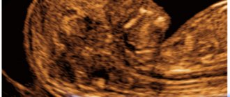
Photo gallery
The photo shows ultrasound images taken in different trimesters of pregnancy.
12th week of pregnancy, ultrasound image
32 week of pregnancy, ultrasound image
20th week of pregnancy, ultrasound image
Technique for calculating weight using ultrasound diagnostic data
Weight calculations are carried out by medical professionals. With the ultrasound data, the patient should visit her gynecologist. It is impossible to determine body weight in the first trimester. At this time, all the doctor can determine is:
- diameter of the fertilized egg;
- coccyx-parietal distance;
- Head circumference.
The listed data is not enough to determine weight. That is why weight is not established in the first trimester.
It is possible to determine a child’s weight using ultrasound data only if the following fetometric indicators are available:
- embryonic gestation period of a baby in weeks;
- fronto-occipital size;
- Head circumference;
- BPR;
- abdominal circumference;
- length of the femur.
Additionally, information about the size of the shin bone, humerus and forearm may be required. Such indicators are measured after the 19th week. Ultrasound diagnostics allows you to most accurately determine the baby’s weight. Small errors are possible if the baby is not presented correctly.
It is worth understanding that the baby’s weight according to ultrasound and at birth may differ. No calculator or formula can give 100% correct results. However, in most cases the error does not exceed 100–200 g.
Knowing the fetometry data of the child, you can calculate the weight yourself. To do this, you will need to use specialized calculators into which all available data is entered. However, in this case one cannot be sure of the correctness of the result obtained.
Self-calculation of weight is needed only for self-development. Without ultrasound diagnostics, determining body weight is impossible.
Video
Rumiya Farkhutdinova, a specialist at the 9 Months clinic, talks about the importance of fetal fetometry.
Do you have any questions? Specialists and readers of the HROMOSOMA website will help you ask a question
Was this article helpful?
Thank you for your opinion!
The article was useful. Please share the information with your friends.
Yes (100.00%)
No
X
Please write what is wrong and leave recommendations on the article
Cancel reply
Rate the benefit of the article: Rate the author ( 1 vote(s), average: 5.00 out of 5)
Discuss the article:
Determining the expected date of birth (DAD)
So, to calculate when to expect the baby to be born, you need to add 280 days (or 9 months and 7 days) to the first day of the last menstruation. It is a little easier to count in reverse order - from the date of the first day of the last menstruation, you need to subtract 3 months and add 7 days. In this case, do not forget to add one year. This is a workout for the brain.
There are many services on the Internet where, by entering the start date of your last period, you will receive an accurate calculation of the expected due date.
In fact, not many women give birth on this scheduled day. It is considered that the child was born at term if the birth occurred from the 38th to the 42nd week of the obstetric period. So your baby has a lot of choice when exactly to be born.
Management of childbirth based on weight
Weight is determined in order to select the most appropriate technique for pregnancy and delivery. After an ultrasound, hypertrophy can be diagnosed. Small size of the child or malnutrition may be a consequence of:
- acute toxicosis;
- maternal hypotension;
- anemia in a expectant mother;
- early aging of the placenta;
- fetal hypoxia;
- smoking and alcohol addiction in the mother;
- intrauterine infection.
If there is a violation, the woman is recommended to adhere to a diet and give preference to a balanced and healthy diet. Additionally, the woman is prescribed a set of diagnostic examinations.
From this video you will learn how the baby's weight changes during pregnancy:
Hypertrophy or excess fetal body weight can be a consequence of:
- diabetes mellitus in the mother;
- chromosomal disorders in the expectant mother;
- hereditary predisposition;
- maternal malnutrition.
It is important to determine weight to determine appropriate delivery tactics. If there is a deficiency or excess weight of the expectant mother, a caesarean section is prescribed. This measure is necessary and allows you to avoid possible injuries.
Each case is considered individually. The doctor takes into account the age of the woman in labor, the likely presence of diseases, the woman’s physique and the history of earlier pregnancies.
Source: infouzi.ru
Let's sum it up
It's no secret that the health of a child depends entirely on the mental balance and health of the mother. In order for a mother to worry less about her baby, she must be sure that his development is proceeding normally.
Therefore, it is extremely important for her to determine how to calculate the weight of the fetus, which method of calculation is more reliable. Thanks to state-of-the-art equipment, calculating mass using ultrasound is the most accurate method. Today, such an examination is quite accessible and has many advantages.
The most important thing is not to worry about trifles. And may you have a healthy and strong baby!
Source: www.syl.ru