- Mom's health
Medicine has advanced a lot in recent years. If just ten years ago, during the entire pregnancy, women took only a few tests and underwent an ultrasound scan 1-2 times, now expectant mothers are under the constant close supervision of doctors and undergo thorough examinations. And, of course, this is not the whims of doctors, but an urgent need, because modern diagnostic methods make it possible to detect various anomalies and developmental defects in the fetus already in the early stages of pregnancy.
Hypoplasia of the nasal bone in the fetus - what is it?
The length of the fetal nasal bone is one of the most important indicators of its formation, and it can be determined by ultrasound.
So if your doctor has prescribed this procedure for you, do not refuse. The harm from ultrasound during pregnancy is nothing more than a myth, but there can be a lot of benefits from this examination. The nasal bones are quadrangular, elongated bones that are visible as early as –11 weeks of pregnancy. If the length of the nasal bone in the fetus is less than it should be at this stage of pregnancy, then in this case they speak of hypoplasia of the nasal bone. If the fetus's nasal bone is completely absent, the condition is called aplasia.
Reasons for an extraordinary examination
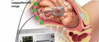
An unscheduled ultrasound at 17 weeks is performed if the doctor suspects a discrepancy between the expected gestational age and the date of delivery. An alternative may be the symptoms and complaints presented by a woman:
- nagging pain in the lower abdomen;
- the presence of abnormal vaginal discharge for this period (blood, ichor, profuse leucorrhoea);
- persistently high blood pressure;
- severe swelling of the lower extremities and face;
- ongoing toxicosis.
If the listed symptoms are present, an ultrasound procedure is performed transvaginally for a detailed assessment of the condition of the cervix and the area where the cervix and the body of the uterus (cervical canal) meet. The planned ultrasound at this time is performed:
- during complicated pregnancy;
- in case of detection of serious pathological abnormalities in the primary screening indicators.
In case of multiple embryonic pregnancy, additional examination is carried out at the discretion of the doctor.
Why do you need to know the length of the fetal nasal bone?
Hypoplasia of the nasal bones in the fetus or their aplasia is considered one of the most important signs (markers) of some chromosomal abnormalities, for example, Down's disease, Edwards, Turner, Patau syndromes and some others.
True, in the very early stages, the very presence of nasal bones in the fetus is much more important than their size. And you can determine the size no earlier than the 12th week of pregnancy.
Here are a few examples as an example:
1. Below on the ultrasound we see a normal nasal bone. 3 clear lines can be distinguished.
2. Hypoplasia of the nasal bone. Ultrasound done at 12 weeks of pregnancy. Length - 1.4 mm (below the normal limit).
Reasons for the development of pathology
Future parents may be interested in the question of why the fetus may be diagnosed with nasal hypoplasia and what is the relationship between the length of the nasal bone and genetic defects. Previously, the possibility of developing nasal hypoplasia in an embryo did not worry doctors too much. However, recent studies have shown the relationship between deviations in the length of the nasal bone from the norm and chromosomal abnormalities. As statistics show, based on the results of thorough research, 80% of nasal hypoplasia was present in a child diagnosed with Down syndrome at birth.
As a result, the conclusion was that nasal hypoplasia, noticed during an ultrasound, is a symptom accompanying congenital abnormalities in the embryo. The factors that cause it are numerous and varied, but the following can be particularly highlighted:
- Taking pharmaceutical drugs, including antimicrobial substances, at the initial stage of pregnancy.
- Abuse of smoking and alcoholic beverages.
- Overheating of a pregnant woman, lasting for a long time.
- Predisposition to pathology at the level of heredity.
- Severe pathologies at the initial stage of pregnancy.
- Infectious pathological conditions, including toxoplasmosis, rubella, influenza and others.
- Injuries or bruises received during pregnancy.
- Intoxication of the body under the influence of hazardous chemicals.
- Gamma radiation directed at a pregnant woman.
- Environmental factors that negatively affect the female body.
As we see, congenital nasal hypoplasia in the fetus may not manifest itself as a natural result of hereditary predisposition, but develop as a result of an incorrectly formed lifestyle or as a consequence of the influence of negative factors. Usually pregnant ladies do not focus their attention on them.
Norms for nasal bone size by stage of pregnancy
The size of the fetal nasal bones depends on the stage of pregnancy. For example, at 12–13 weeks of pregnancy, the length of the nasal bone is only 3.0 mm. At -21 weeks it increases to 5.5 - 5.7 mm, and by the 35th week of pregnancy it reaches 9.0 mm.
In order to correctly and accurately assess the parameters of the nasal bone obtained during an ultrasound examination, the doctor must have extensive experience and high qualifications. In addition, it is necessary that the ultrasound be performed on a modern device - otherwise, obtaining reliable results is very doubtful.
Gnomik.ru recommends Rmh24.ru Refrigerator repair shop We repair refrigerators of all brands. Free visit of a specialist and diagnostics!
Use promo code: Gnomic.ru
and get a guaranteed 10% discount
We work in Moscow and the region.
Call
Call a specialist
Nasal bone hypoplasia and other indicators
Many parents, hearing from a doctor that the size of their unborn child’s nasal bone does not correspond to normal values, panic. In fact, it is too early to worry - it is simply impossible to accurately determine Down syndrome or any other chromosomal abnormality in a fetus just by the size of the nasal bones.
In order to confirm or refute the alleged diagnosis, a pregnant woman is recommended to undergo a second ultrasound - with a different specialist and on a different device.
If, upon repeated examination, it is determined that the size of the baby’s nasal bone does not correspond to those that should be at this stage of pregnancy, then the woman is usually recommended to undergo additional examination - amniocentesis, i.e., sampling a small amount of amniotic fluid for subsequent genetic testing. analysis.
Nasal bone on ultrasound: normal at 12 weeks and fetal NK hypoplasia (small nose)
Advances in modern medicine make it possible to carefully monitor pregnant women and identify those who are at risk for possible chromosomal abnormalities of the fetus. Thus, ultrasound examination of the fetal nasal bone (NB) is an important diagnostic tool for identifying possible anomalies.
Why is it necessary to measure the fetal nasal bone?
The length of the nasal bone is an indicator of the normal development of the fetus. There are two pathological conditions – hypoplasia and aplasia. Hypoplasia is a decrease in its length, and aplasia is the absence of this bone.
Both of these conditions indicate the presence of pathologies in the fetus associated with chromosomal abnormalities. This indicator is detected through ultrasound.
Its normal values indicate intrauterine development without deviations.
If it is clear that the indicators are small, i.e. If there are deviations in the direction of decrease, then this condition is a sign of hypoplasia. An obvious pathology and deviation is the absence of bones, which indicates the absolute underdevelopment of this organ and a gross anomaly. This happens rarely, in exceptional cases.
The nasal bone is visualized by ultrasound already at 10-11 weeks. Deviations in this indicator are considered a sign of certain diseases caused by chromosomal abnormalities. These are conditions such as Down syndrome, Edwards syndrome, Patau syndrome, etc.
In the early stages of pregnancy, the presence of this bone in the fetus is important, and measurement of its size for diagnostic purposes should be made no earlier than 12-13 weeks.
With hyperplasia or aplasia detected on ultrasound, one can suspect not only abnormal intrauterine development, but also Down syndrome.
In this case, it is necessary to conduct an additional examination after 2 weeks to monitor the situation.
At 10-11 weeks, the doctor checks the presence of the nasal bone in the fetus, and by 12-13 weeks, deviations in its development can be detected. They indirectly indicate chromosomal abnormalities. The study is carried out by comparing the obtained data with normal indicators
In addition, in such a situation, specialists compare the results of the first screening with existing standards using a special table.
When deciphering, it should be remembered that, first of all, the fact that the fetus has a nasal bone is important, and measurement indicators at this stage are not so informative, since chromosomal abnormalities for this marker appear somewhat later.
However, we should not forget that the conclusion about chromosomal abnormalities leading to the birth of a child with serious illnesses is a serious diagnosis that requires careful verification and detailed analysis.
Norms of indicators at different stages of pregnancy
The growth and development of the fetus occurs in accordance with certain patterns and standards, confirmed by science and life. This also applies to the parameters of the nose of the unborn child:
- So, the norm at 12 weeks is considered to be a length of 3 mm;
- at 20 weeks the bone should be from 5.7 to 8.3 mm;
- at 35 weeks - at least 9 mm.
How accurately this indicator will be determined depends on the equipment of the ultrasound machine and on the professionalism and qualifications of the doctor conducting such a diagnosis. These normal indicators are the starting point for deciphering and studying the results of prenatal screening.
The information obtained during the examination is the basis for further monitoring of the condition of the fetus, in order to identify possible developmental anomalies.
If during an ultrasound examination it is impossible to determine the presence of NK and thickening of the collar zone is noted, then doctors note a high degree of probability that the child will be born with congenital defects or with Down syndrome.
If the doctor detects bone hypoplasia or cannot find any signs of their presence at all, additional studies are carried out. For example, when thickening of the nuchal zone is detected, the development of Down syndrome can be diagnosed with a high probability
Determining this parameter is so important that some parents, having learned about this possibility, decide to terminate the pregnancy.
Therefore, it is important to undergo the appropriate examination in a timely manner and make sure that the unborn baby is healthy and does not have various developmental disorders.
The presence or absence of hypoplasia indicates normal or pathological intrauterine development of the child. Only an appropriate specialist can make a diagnosis and determine the likelihood of such a risk.
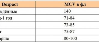
To determine hypoplasia, the following table is used, which presents the norms of the indicator at different stages of pregnancy. Based on this table, which shows the average, minimum and maximum values, doctors summarize the data obtained from ultrasound and give the appropriate conclusions:
Duration, week Average value, mm Minimum value, mm Maximum value, mm
| 12 — 13 | 3,1 | 2 | 4,2 |
| 14 — 15 | 3,8 | 2,9 | 4,7 |
| 16 — 17 | 5,4 | 3,6 | 7,2 |
| 18 — 19 | 6,6 | 5,2 | 8 |
| 20 — 21 | 7 | 5,7 | 8,3 |
| 22 — 23 | 7,6 | 6 | 9,2 |
| 24 — 25 | 8,5 | 6,9 | 10,1 |
| 26 — 27 | 9,4 | 7,5 | 11,3 |
| 28 — 29 | 10,9 | 8,4 | 13,4 |
| 30 — 31 | 11,2 | 8,7 | 13,7 |
| 32 — 33 | 11,4 | 8,9 | 13,9 |
| 34 — 35 | 12,3 | 9 | 15,6 |
What are the causes of this pathology?
Expectant mothers may have a natural question as to what causes the appearance of such a pathology, and how it is related to the definition of genetic anomalies and defects.
As a result of numerous studies in this area, it turned out that such hypoplasia was found in 80% of children born with Down syndrome.
This gave rise to the conclusion that such a pathology detected on ultrasound is a sign of congenital abnormalities in the child.
Factors that can cause such a deviation are quite diverse. Genetic scientists identify the following:
- taking potent medications in the early stages of pregnancy, including antibacterial drugs;
- excessive consumption of alcohol and tobacco products;
- prolonged overheating of a pregnant woman;
- hereditary predisposition;
- severe illness in the early stages of pregnancy;
- infectious diseases such as rubella, toxoplasmosis, influenza, etc.;
- injuries and bruises of a pregnant woman;
- poisoning with dangerous chemicals;
- gamma radiation from a woman during pregnancy;
- environmental factors that have a harmful effect on the body of a pregnant woman.
During pregnancy, smoking or even inhaling other people's tobacco smoke can be one of the main causes of birth defects in the fetus.
So, such a congenital pathology can be the result not only of a hereditary predisposition, but also appear as a result of an incorrect lifestyle and the influence of unfavorable factors, to which pregnant women most often do not pay due attention.
At the same time, these factors directly affect the development of the unborn child. Diagnostic methods of modern medicine used in obstetrics make it possible to detect such abnormalities in the early stages of pregnancy and inform parents about them.
Fetal malformations - reliability of ultrasound
Each person is individual and, naturally, this individuality can be traced even at the stage of intrauterine development. Therefore, the sizes of body parts, including the nasal bone, may differ from the table values.
It is possible to speak confidently about the presence of chromosomal abnormalities in the fetus only when not only underdevelopment of the nasal bones is detected, but also hypoplasia of the fetus itself - short arms and/or legs, too small sizes of some internal organs.
Therefore, if during the next ultrasound it turns out that the size of your baby’s nasal bones is smaller than normal, this does not mean that there is a genetic pathology. It is quite possible that this is simply an individual feature of the fetus and your baby will be born quite healthy and with a charming snub-nosed “button”. In any case, only your doctor can correctly interpret all the results obtained and make the correct diagnosis. Trust him.
What to do if a child develops incorrectly?
In some cases, even if the child’s skeleton is formed with a delay, this can be corrected, and by the time the baby is born everything will be fine.
As a rule, a pregnant woman is recommended:
- nutritious diet rich in microelements and calcium;
- giving up bad habits: smoking, alcohol, drugs;
- special preparations containing certain substances necessary for the fetus and placenta;
- sets of physical exercises;
- physiotherapy;
- if necessary, the infection is treated if it is the cause of malnutrition.
Unfortunately, some pathologies of the development of the musculoskeletal system are incompatible with life. True, such pathologies are extremely rare - approximately 2-3 cases per 10 thousand newborns.
If such a serious congenital disease is detected in the early stages, then the woman has the opportunity to terminate the pregnancy. In the later stages, some pathologies of skeletal development are indications for cesarean section and emergency medical care for the born child.