Features of the procedure
Ultrasound usually begins with an assessment of the fetal head, and the following parameters are determined:
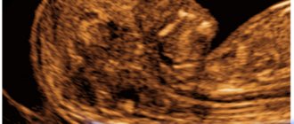
- Biparietal size of the fetal head (BPR)
is the distance from the outer surface of the upper contour to the inner surface of the lower contour between the parietal bones (Figure 1, line bd). - Fronto-occipital size (FOR)
is the distance between the outer contours of the frontal and occipital bones (Figure 1, line ac).
Figure 1 – Scheme for measuring biparietal and fronto-occipital dimensions:
1 – cavity of the transparent septum, 2 – visual thalamus and cerebral peduncles,
bd – biparietal size, ac – fronto-occipital size
Measurement of BPR and LZR is carried out with strictly transverse scanning
at the level of brain structures: the cavity of the transparent septum, the cerebral peduncles and the visual thalamus, as shown on the right side of Figure 1. When measuring, the doctor evaluates the structures of the brain, the integrity of the bones of the skull, and the presence of tumors.
The biparietal and fronto-occipital dimensions increase in proportion to the duration of pregnancy, by the end of which the growth is not as pronounced as in the first and second trimesters (see table).
Dimensions of the fetal head by week of pregnancy
| Gestation period, weeks | Fronto-occipital size (LZR), mm | Biparietal size (BPR), mm | ||||
| Percentiles | Percentiles | |||||
| 10 | 50 | 95 | 10 | 50 | 95 | |
| — | — | — | 13 | 17 | 21 | |
| — | — | — | 18 | 21 | 24 | |
| — | — | — | 20 | 24 | 28 | |
| — | — | — | 23 | 27 | 31 | |
| — | — | — | 27 | 31 | 35 | |
| 41 | 45 | 49 | 31 | 34 | 37 | |
| 46 | 50 | 54 | 34 | 38 | 42 | |
| 49 | 54 | 59 | 37 | 42 | 47 | |
| 53 | 58 | 63 | 41 | 45 | 49 | |
| 56 | 62 | 68 | 43 | 48 | 53 | |
| 60 | 66 | 72 | 46 | 51 | 56 | |
| 64 | 70 | 76 | 48 | 54 | 60 | |
| 67 | 74 | 81 | 52 | 58 | 64 | |
| 71 | 78 | 85 | 55 | 61 | 67 | |
| 73 | 81 | 89 | 58 | 64 | 70 | |
| 77 | 85 | 93 | 61 | 67 | 73 | |
| 80 | 88 | 96 | 64 | 70 | 76 | |
| 83 | 91 | 99 | 67 | 73 | 79 | |
| 86 | 94 | 102 | 70 | 76 | 82 | |
| 89 | 97 | 105 | 71 | 78 | 85 | |
| 93 | 101 | 109 | 73 | 80 | 87 | |
| 95 | 104 | 113 | 75 | 82 | 89 | |
| 98 | 107 | 116 | 77 | 84 | 91 | |
| 101 | 110 | 119 | 79 | 86 | 93 | |
| 103 | 112 | 121 | 81 | 88 | 95 | |
| 104 | 114 | 124 | 83 | 90 | 97 | |
| 106 | 116 | 126 | 85 | 92 | 98 | |
| 108 | 118 | 128 | 86 | 94 | 100 | |
| 109 | 119 | 129 | 88 | 95 | 102 | |
| 110 | 120 | 130 | 89 | 96 | 103 | |
Biparietal size of the fetal head
To monitor intrauterine development during pregnancy, 3 ultrasound examinations are performed. During the ultrasound, fetometry is performed - measurements of the child’s body parameters.
Biparietal, or large transverse, size (LTS) is one of the indices by which the gestational age is determined. Among them, using the BPR, the deadline can be most accurately determined. This indicator is most informative from 3 to 7 months. BDP is measured using ultrasound examination.
The BDP is represented by the distance from one parietal bone to the other (between the temples). We can say that the BPR is the width of the child’s head.
As pregnancy progresses, the head of the unborn child grows, and, accordingly, the BPD increases. Specialists in the field of obstetrics have developed BPR standards. At the end of pregnancy, the rate of increase in this indicator decreases slightly.
Determining the gestational age using BDP is informative only up to the 22nd week; within this period, the accuracy of the determination reaches 5–10 days. But starting from the 28th week, the reliability of this method in terms of determining the period is questionable.
Moreover, after 38 weeks, the shape of the baby’s head changes, and the compliance of the BPR with established standards will depend on its configuration. For example, if the child's head has an elongated (dolichocephalic) configuration, then the BPD will be reduced compared to the norm.
BPD is considered one of the main indicators of child development, since the enlargement of the skull can be used to judge the enlargement of the brain, and, consequently, the development of the fetal nervous system.
In addition, BDP in combination with abdominal circumference and thigh length makes it possible to determine possible body weight. This indicator is very important before childbirth, since taking it into account, among others, determines the method of labor and obstetric care.
What is BPR and how is it measured?
Biparental head size is the distance between the two temples, measured along the minor axis, in other words, the width of the head, or its diameter . As already mentioned, this indicator is determined during an ultrasound scan as part of the second (after 20 weeks) and third (after 30 weeks) screenings. To do this, the doctor measures a line connecting the two contours of the temples and passing strictly above the thalamus.
This value is very important, as it indicates the full development of the brain and nervous system, and makes it possible to answer the following questions: are there any disturbances in the development of the child, does the expectant mother need to be prescribed additional drug therapy, will she be able to give birth through natural delivery, or you will have to resort to a caesarean section if the size of the baby’s head significantly exceeds the width of the birth canal, etc.
Fronto-occipital size
The fronto-occipital dimension (FOR) is represented by a segment connecting the frontal and occipital bones. This is a line that is perpendicular to the BPR.
LZR is measured by scanning brain structures in a transverse direction. In addition to measurements of the BPR and LZR, the specialist notes the structure of the brain, skull bones (their integrity), and the presence of neoplasms.
Having measured the parameters, a cephalic index is derived, equal to the quotient of dividing these indicators, expressed as a percentage. This index gives an idea of the shape of the baby's head.
Both BPR and LZR increase proportionately during intrauterine development, then closer to childbirth their growth slows down.
What to do if the BPR and LZR are not normal?
If the BPD and LZR differ slightly from normal indicators, then this is most likely a developmental feature of a particular child.
When the size of the head exceeds the norm, it is necessary to evaluate other parameters (abdominal circumference, length of limb bones). If a large fetus
(over 4 kg), then all sizes will be increased by 1 or more weeks. In addition, children do not always grow proportionally, so the head may be larger relative to other sizes, but after 2-3 weeks it will level out.
An increase in BPR and LZR may also indicate the presence of pathology: space-occupying formations of the brain or skull bones (tumors), the presence of cerebral hernias, hydrocephalus
(excessive accumulation of cerebrospinal fluid in the ventricular system of the brain). In case of large formations or hernias, termination of pregnancy is suggested, since such deviations are often incompatible with life.
Hydrocephalus, as a rule, is a consequence of intrauterine infection, so a course of antibiotic therapy is carried out, and if there is no effect of treatment and the head grows in size, pregnancy is terminated. If hydrocephalus does not increase, pregnancy can be prolonged under ultrasound control over time. After birth, the child may need bypass surgery (implantation of a special tube into the brain with the other end in the abdominal cavity to drain excess fluid), which significantly complicates future life.
A decrease in the size of the fetal head may indicate underdevelopment or absence of anatomical structures of the brain (cerebral hemispheres, cerebellum). This is an indication for termination of pregnancy at any stage. It is necessary to look at the proportionality of the sizes of the head and other parts.
With reduced BPD and LZR, especially in the third trimester, intrauterine growth retardation syndrome
, which requires immediate correction, as it can lead to fetal death. The correction is carried out with drugs that improve uteroplacental blood flow, increasing the delivery of nutrients to the fetus (Actovegin, pentoxifylline, chimes), under mandatory ultrasound control after 7-10 days.
Additionally Summarizing the above, we can conclude that BPD and LZR are important parameters for assessing fetal development, establishing the anatomy of brain structures and excluding the diagnosis of FGR.
What to do if the BPR deviates from the norm?
A deviation from the norm, which is detected once, does not have much significance if it does not differ from the normal indicator within 2-3 lines in the table.
To suspect any developmental disorder in a child, it is necessary to record a change in the BDP several times in a row. For this purpose, ultrasound fetometry is carried out dynamically, usually at intervals of 1-2 weeks.
Increase in head size
Macrocephaly or large head
1. The reasons for a large head size may be hereditary factors. If a large head size is detected in one of the parents, then no special measures are taken.
In this case, the attending physician must determine the possibility of independent vaginal birth or choose a cesarean section as a method of delivery.
- If the large size of the fetal head is not associated with hereditary characteristics, then it is necessary to exclude developmental anomalies.
2. With hydrocephalus, there is an expansion of the internal cavities of the brain, which contain brain fluid - cerebrospinal fluid. The volume and pressure of the fluid increases - the size of the head increases.
The causes of hydrocephalus can be a violation of the development of the fetal brain, infection for a period of 13 to 27 weeks, tumors, injuries, hypoxia, spina bifida (non-fusion of the vertebrae, as a result of which the spinal cord does not have a full-fledged bone frame).
Detection of hydrocephalus before the period of fetal viability (3rd trimester) raises the question of termination of pregnancy. If the fetus is already at a viable age, then karyotyping, tests for viruses, a detailed ultrasound, and consultation with a neurosurgeon on the treatment of the child after birth are performed.
- If pressure in the ventricles increases, it is necessary to carry out early delivery and further surgical elimination of the problem.
3. Macrocephaly can be observed with disorders of the development of bone and cartilage tissue. This disorder is thanatoform dysplasia, a congenital disorder of skeletal development that is associated with a lack of ossification.
An enlarged head, its shape in the form of a trefoil, can be detected on an ultrasound in the middle of the 2nd trimester. Another pathology is achondrogenesis, in which the production of cartilage tissue is impaired, also leading to the development of macrocephaly.
- This condition is a defect incompatible with life.
Hyperplastic development or large fetus
1. In this situation, heredity may also be the cause, and the increase in fetal size may be due to the family constitution. It is necessary to find out what body weight mom and dad were born with, as well as determine their body type.
- The large size of the fetus influences decisions on labor management tactics.
2. The second reason for a large fetal mass may be gestational diabetes, or diabetes mellitus in pregnant women. It is necessary to conduct urine and blood tests for sugar. On an empty stomach, in the blood, the level of this indicator in pregnant women is lower than usual and amounts to 5.0-5.8 mmol/l.
There should be no sugar in the urine. If changes are detected in the tests, as well as for all pregnant women between 24 and 28 weeks, it is necessary to conduct an oral glucose tolerance test with 50 g. glucose. The test is carried out without prior preparation, at any time. After taking the glucose solution orally, the amount of sugar in the blood is determined after 1 hour.
If this indicator is more than 7.8 mmol/l, further examination by an endocrinologist is necessary. Treatment for gestational diabetes involves following a special diet. If blood sugar continues to be elevated, insulin injections are prescribed.
- Any tablet preparations for lowering sugar during pregnancy are contraindicated.
Reducing head size
Microcephaly or small head
1. As with large head size, a small fetal head may be associated with small head sizes in the family. This feature does not require treatment.
2. Spina bifida is a developmental disorder of the spine, which can be indirectly suspected when the head shape changes to the “banana” or “lemon” type.
Refers to those defects in which termination of pregnancy is recommended if the fetus has not yet reached a viable age. In other cases, it is necessary to inform parents about the possible manifestations of this disorder after the birth of the child, such as impaired movement and sensitivity of the lower extremities, chronic pain, fecal and urinary incontinence, and curvature of the spine.
- If pregnancy continues, it is necessary to determine the date and method of delivery.
3. Intrauterine infection, which leads to damage to the embryo at a period of 3-12 weeks, can lead to the formation of malformations with a decrease in the size of the head. If the infection occurs at a period of 28 weeks or more, the infection is severe and spreads, that is, it becomes generalized - fetal development is delayed and an associated decrease in head size and BPD occurs.
If an infectious disease is suspected in the mother, a number of studies are carried out aimed at identifying the pathogen: PCR, detection of antibodies in the blood, microscopy of smears, bacterial cultures on nutrient media.
Treatment depends on the type of pathogen and trimester of pregnancy. From 14-16 weeks, placental insufficiency is prevented against the background of general treatment.
- If you become ill with rubella before the 16th week and if malformations are detected, it is recommended to terminate the pregnancy.
Hypoplastic development or small fetus
1. Hereditary low weight in children in the family, asthenic type of constitution in parents. Special measures are not taken during normal pregnancy.
2. A decrease in body size can be due to genetic diseases, such as triploidy, trisomy on the 13th pair of chromosomes - Patau syndrome, on 18 - Edwards syndrome, or on 21 - Down syndrome, which lead to early symmetrical growth retardation of the fetus: that is, a discrepancy with the gestational age has both the head and body of the fetus.
In each case, the trip is individual. Children who are born alive have severe malformations of organs and their life prognosis is unfavorable - most die in the first year.
The exception is children with Down syndrome, who, with the necessary treatment and social support, are able to have a fairly high quality of life.
Osteogenesis imperfecta is a genetic developmental defect that leads to a group of diseases that impair the formation of bone and cartilage tissue. Has several types.
When performing an ultrasound, only 2 types can be detected: shortened, curved bones and multiple fractures are recorded; deformation of the head occurs when the sensor presses on the soft bones of the skull. Osteogenesis imperfecta type 2 is not compatible with life.
BDP is important when calculating the cranial index (CI, Y), which indicates the arrest of fetal development, its intrauterine death and the approximate time the dead fetus remains in the uterus.
- Cranial index = BPR/LZRx100. If CHI is 83 or more, this is the norm.
4-5 days after the death of the fetus, destruction of the internal structures of the brain occurs and a significant decrease in the BPD: if CHI = 64-74, then the death of the fetus occurred less than 3 weeks ago, if CHI = 64, then the fetus is not viable for more than 3 weeks.
Reducing BPR and LZR
If the BPR and LZR indices are below normal, then the following may be observed:
- underdevelopment of parts of the fetal brain;
- absence of some parts of the brain;
- when the head is proportional to the entire body in comparison with normal values, intrauterine growth retardation syndrome is diagnosed.
In the first two cases, termination of pregnancy is necessary, regardless of the period. In the latter case, timely correction is required, since without it intrauterine fetal death is possible. For treatment, medications are prescribed that activate uteroplacental circulation, which improves fetal trophism (chimes, actovegin, pentoxifylline). After 7–10 days from the start of treatment, ultrasound diagnostics are performed again.
The diagnosis of developmental delay is made based on ultrasound results. Intrauterine growth retardation is considered to be a deviation of the established fetal weight from the standards presented in the tables of the ratio of fetal weight and its age by gestational age. Pathology is considered to be weight less than the 10th percentile of the tables.
There are cases when even with such a diagnosis there is no need to worry, since the child is simply small in size (the child’s parents, for example, may be short and light in weight). Then low weight is considered a peculiarity of the baby, which does not indicate reduced activity or mental and physical development. In this case, no special treatment is carried out either during pregnancy or after childbirth.
If this is not the case, then such a delay can lead to developmental deviations and even intrauterine death. With IUGR, it can be assumed that the nutrition of the fetus is impaired, so it does not receive the necessary nutrients and oxygen. Violation of fetal trophism is caused by:
- chromosomal abnormalities;
- smoking, alcohol or drug addiction of the mother;
- maternal hypertension, anemia or cardiovascular disease;
- improper attachment of the placenta;
- multiple births;
- taking certain medications without the advice of a specialist;
- long pregnancy (more than 42 weeks);
- maternal malnutrition;
- toxoplasmosis, rubella, syphilis and some other infections.
Increase in BPR and LZR
If during ultrasound diagnostics the dimensions of the head are determined to be larger than normal values, this is interpreted as follows:
- a proportional increase in all parts of the fetal body indicates that it is a large fetus (all parameters of its body are increased by 1 or more weeks). A fruit is considered large if its weight exceeds 4 kg;
- the head can grow faster than the rest of the body, so the situation is considered normal when within 14 to 21 days the parameters of the fetus become proportional;
- an increase in size may indicate pathologies: tumors of the brain or skull bones, brain hernias, hydrocephalus (accumulation of cerebrospinal fluid in the ventricles of the brain).
If tumors or hernias are diagnosed, termination of pregnancy is necessary; such disorders usually lead to fetal death. If hydrocephalus is diagnosed, an intrauterine infection is usually suspected. Therefore, antibiotics are prescribed for hydrocephalus. If hydrocephalus progresses, the pregnancy should be terminated. If progression of hydrocephalus is not observed, further pregnancy is carried out under the control of ultrasound diagnostics. After childbirth, shunting is possible to drain excess cerebrospinal fluid.
If deviations of the BDP and LZR from the norm are small, then they are considered developmental variations.
Determining the parameters of the fetal head is very important, since most often during childbirth it is the first to pass through the birth canal. Since the baby's head is dense and large in comparison with the rest of his body, it is with it that the main difficulties of the baby passing through the birth canal are associated. After the head has passed, the birth canal is ready for the passage of other parts of the baby's body.
Therefore, studying the size and shape of the child’s head is extremely important from the point of view of preparation for childbirth and prognosis of its course.
The fetal BDP exceeds the norm - what does this mean?
In some cases, the index exceeds the permissible norms. In this case, the attending physician is obliged to determine other parameters of the embryo (such as abdominal circumference, thigh length, etc.) in order to make sure that there are no pathologies. And if other parameters exceed the norm by at least one or two weeks, it means that the expectant mother has a large fetus. But if these indicators are within acceptable limits, then it is likely that the child is simply developing in leaps and bounds, and all parameters will soon level out.
As for significant deviations of the BPD from the norm, they often indicate serious problems in development. For example, an increased index may occur with a tumor of the cranial bones or the brain itself, as well as with hydrocephalus and cerebral hernia. In each of the listed cases (the exception is hydrocephalus), women are advised to immediately terminate the pregnancy, since such pathologies are, unfortunately, incompatible with life. But hydrocephalus is treated with antibiotics or (if treatment does not produce any results) an abortion is performed.
Note! If the BPD in the embryo is too low, nothing good should be expected either - this often indicates underdevelopment of the brain or the absence of certain of its components (right, left hemispheres, or both at once, the cerebellum, etc.). In such cases, the fetus is aborted regardless of the gestational age.
In the last trimester of pregnancy, low biparietal size indicates delayed fetal development. This syndrome is treated with special drugs (such as Actovegin, Chimes, etc.), which stimulate blood flow in the uterine cavity and placenta.
Average fetal fetometry values
| Week of pregnancy | Height, mm (KTR - coccygeal-parietal size) | Weight, g | Chest diameter, mm |
| 11 | 6,8 | 11 | 20 |
| 12 | 8,2 | 19 | 24 |
| 13 | 10 | 31 | 24 |
| 14 | 12,3 | 52 | 26 |
| 15 | 14,2 | 77 | 28 |
| 16 | 16,4 | 118 | 34 |
| 17 | 18 | 160 | 38 |
| 18 | 20,3 | 217 | 41 |
| 19 | 22,1 | 270 | 44 |
| 20 | 24,1 | 345 | 48 |
| 21 | 25,9 | 416 | 50 |
| 22 | 27,8 | 506 | 53 |
| 23 | 29,7 | 607 | 56 |
| 24 | 31,2 | 733 | 59 |
| 25 | 32,4 | 844 | 62 |
| 26 | 33,9 | 969 | 64 |
| 27 | 35,5 | 1135 | 69 |
| 28 | 37,2 | 1319 | 73 |
| 29 | 38,6 | 1482 | 76 |
| 30 | 39,9 | 1636 | 79 |
| 31 | 41,1 | 1779 | 81 |
| 32 | 42,3 | 1930 | 83 |
| 33 | 43,6 | 2088 | 85 |
| 34 | 44,5 | 2248 | 88 |
| 35 | 45,4 | 2414 | 91 |
| 36 | 46,6 | 2612 | 94 |
| 37 | 47,9 | 2820 | 97 |
| 38 | 49 | 2992 | 99 |
| 39 | 50,2 | 3170 | 101 |
| 40 | 51,3 | 3373 | 103 |