The availability of ultrasound diagnostics allows a woman who has just learned about her pregnancy not to speculate about the due date, but to find out exactly how her baby is developing. Naturally, when visiting an ultrasound room, a woman roughly assumes what time the doctor will name, but it happens that her expectations are not met.
How often does this happen, how can we explain the situation when the term according to ultrasound differs from the period according to menstruation, or the size of the fetus differs from the standard at a specific period?
How much can you trust ultrasound?
Ladies who really want to get pregnant want to know about a miracle happening in their lives as early as possible, literally in the first days of the delay. Of course, you can always buy a pregnancy test. But even ultra-sensitive tests can only accurately give a specific answer to the question “has pregnancy occurred?” They are able to answer yes or no, but more precise methods will be needed to set the deadline.
Positive pregnancy test
During a manual examination of a pregnant woman, an obstetrician-gynecologist may notice that the uterus is loose and slightly enlarged. But this phenomenon is also observed before menstruation. To accurately clarify the situation, you will need an ultrasound examination.
Ultrasound can accurately determine pregnancy when the level of hCG (human chorionic gonadotropin) in a woman’s blood exceeds one thousand units. In this case, the doctor is already able to see the fertilized egg in the uterine cavity (in case of multiple pregnancy - two or three fertilized eggs). The longer the period, the more complete the picture the study shows - the doctor will note the presence of not only the fertilized egg, but also the yolk sac in it, or even see the embryo and its heartbeat, and will be able to measure the CTE of the embryo (the distance from the tailbone to the crown).
If your menstrual cycle is more than 30 days, the gestational age according to ultrasound and menstruation may differ significantly.
The following situation often arises: a woman comes for an ultrasound, the doctor asks about the date of her last period, conducts a study, and then announces that the pregnancy most likely turned out to be frozen - the embryo and its heartbeat are not visualized, a fetal egg is visible, the size of which is smaller than it should be be. The doctor may be wrong if a woman had late ovulation. For a significant proportion of women, the menstrual cycle is not the standard 28 days, but 33, or even 35-40. This means that conception did not occur on days 14-16 of the cycle, but a week or two later, and the woman simply came for the ultrasound too early, so the fetus was not seen. Most likely, the pregnancy will not be frozen and the fetus is developing normally, it’s just worth repeating the study in a couple of weeks to make sure there is no mistake. It is also possible to explain the situation when pregnancy is not visible at all on an ultrasound: probably, the level of hCG in the blood is less than that at which pregnancy can be seen.
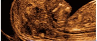
First ultrasound
According to doctors, pregnancy itself can be detected on the fifth day from the moment of fetal implantation, but for this the sensor will have to be inserted into the vagina. Until this moment, the embryo cannot yet be seen.
This type of examination cannot always be used to calculate the due date; if a woman is at risk of miscarriage, then inserting a sensor into the vagina is prohibited. Ultrasound diagnostics in the first weeks after conception is not carried out without special reasons. Indications for examination may include a suspected ectopic pregnancy or hydatidiform mole.
Even in the earliest stages of pregnancy, ultrasound does not harm the baby. In a situation where the doctor assumes that the embryo has stopped developing, there is no way to do without an ultrasound examination. A preliminary diagnosis can be made during examination of the pregnant woman. The size of her uterus does not change. At the fifth week, the embryo begins to experience its first heartbeats. And if pregnancy begins to be detected by ultrasound from three weeks, then ectopic pregnancy is diagnosed starting from the tenth day. Simultaneously with the transvaginal examination, a blood test for hCG is prescribed.
Can an ultrasound be mistaken about the gestational age? Despite the fact that ultrasound is one of the most modern methods, even here, during the examination, not entirely accurate results can be obtained. The error can be made either larger or smaller. This may be due to old equipment that does not allow accurately determining the size of the fetus.
Fetal ultrasound calculator online allows you to independently calculate the gestational age. For an accurate calculation, you need to enter several parameters into the calculator:
- Approximate gestational age based on the first day of the last menstruation
- One of three measured head sizes
- Femur length
- One of the values obtained by measuring the circumference of the chest or abdomen.
At the same time, it is worth noting that the result obtained is approximate, since each fruit develops at its own pace.
It is worth considering another important aspect of ultrasound examination. Why do doctors insist on early registration and early ultrasound up to twelve weeks? This examination primarily determines how the embryo was implanted. The second objective of the examination is to determine the duration of pregnancy from the moment of conception. At this time, all embryos develop almost equally, so the risk of error is minimal. During the same examination, it is determined how many fetuses are supposedly in the uterus, and the doctor can determine whether there is a threat of termination of pregnancy.
Differences in pregnancy terms according to menstruation and ultrasound
Obstetric gestational age is calculated by week
All obstetricians and gynecologists use the term “obstetric period” when determining the duration of pregnancy. This allows all gynecologists in the world to speak the same language. It is carried out in weeks, counting from the first day of the last menstruation. The first and second obstetric weeks are those weeks during which the egg is still maturing in the body, which will subsequently be released from the ovary during ovulation and meet with the sperm.
Thus, a woman who has a standard menstrual cycle of 28 days and comes to the doctor with a delay of one week will be diagnosed with pregnancy at exactly five obstetric weeks. At the same time, an ultrasound examination can set a slightly shorter period - three or four weeks. How justified is this and why does the deadline not match? This situation is normal, since ultrasound does not aim to determine the exact period in weeks, but to determine how many weeks from conception the fetus is developed according to its parameters (its gestational age). Understanding the difference between these two concepts is important. Therefore, a discrepancy of 1-2 weeks between the period according to menstruation and according to ultrasound in favor of menstruation (i.e., the period according to menstruation is longer, but according to ultrasound is less) is a common occurrence, there is nothing terrible about it.
Obstetric term
The beginning of pregnancy is the first day of the last menstruation. This method is called obstetric. It does not take into account the individual characteristics of a woman’s body, but is almost universal. Any doctor will use it.
The obstetric method has its own logic. The calculation of the period starts from the earliest stage of pregnancy - the beginning of egg maturation.
Using the obstetric method, the doctor will determine the expected date of birth (ED), as well as the period of maternity leave. In medicine, it is generally accepted that pregnancy lasts 280 days. These are the well-known 40 weeks or 10 lunar months.
Why 10 months and not 9? And why are the months lunar? Astronomy is to blame for this. The moon repeats its phases every 28 days (4 weeks). This is the lunar month. And if you count in calendar months, then only 9 of them really fit into a normal pregnancy.
How to determine the gestational age with an irregular cycle?
Some women suffer from hormonal imbalances and, as a result, irregular cycles. With pathologies such as polycystic disease, multifollicular ovaries, and excessive amounts of male sex hormones, menstruation may not occur for several months. However, ovulation with such disorders still occurs from time to time, which means that such women have almost the same chance of getting pregnant as others.
The girl remembers the date of her last menstruation
If pregnancy is not desirable for you, the use of contraception is mandatory, even if you suffer from irregular cycles and have not had a period for several months.
Often, women with irregular cycles do not consider it necessary to use protection, believing that they will definitely not be able to get pregnant. In this case, you can detect pregnancy in the third or even fourth month, because the absence of menstruation for several months becomes a kind of norm. Correct calculation of the obstetric term in such a situation is practically impossible, therefore, when determining the term, the doctor can rely solely on ultrasound data. If the fetus is not too large or too small, ultrasound can determine the gestational age of the fetus to within 2-3 weeks. The date of birth will also be determined using an unconventional method - by counting 40 weeks from the first day of the last menstruation, and relying on ultrasound data.
When is the last time an ultrasound is performed during pregnancy?
An ultrasound scan at the third screening is done at 30-31 weeks. As in the second study, the third ultrasound evaluates the condition of the uterus, placenta, umbilical cord, waters and the fetus itself. In addition, it is necessary to determine the presentation of the embryo - the position inside the uterus. Normally it is cephalic, that is, the fetus lies head down, with the top of the head towards the exit. It is important to establish the position of the placenta, the length of the cervical canal, and check the quality of the placenta.
The course of the third study does not differ from the course of the previous ones. An ultrasound is performed only on the abdominal wall.
In what cases is the term according to ultrasound ahead of the obstetric one?
Currently in Russia, all pregnant women are given the right to undergo free ultrasound examinations three times. They are usually prescribed between 11 and 14 weeks, between 18 and 22, and between 32 and 34. Each of these ultrasounds has its own goals (the first is to determine whether gross malformations of the fetus and genetic abnormalities have been detected, the second is to monitor the development of internal organs, the third is determining the condition of the placenta, the position of the fetus and its approximate weight). However, with each examination, the doctor determines how many weeks the fetus is developed. And often this period is ahead of the obstetric period. For example, a woman knows for sure that her pregnancy is 20 weeks, and the doctor indicated in the conclusion that the size of the fetus corresponds to 22 weeks. Why is this situation happening? There is no error in it. This can be explained as follows:
- The fruit is large. The large size of the fetus, which is ahead of its gestational age, is not a pathology. Just like all people, the fetus already in the womb has its own individual characteristics.
- The size of the fruit is slightly smaller than it should be. This may well be a variant of the norm; perhaps the baby is just small, especially if his parents are not tall and impressive in weight.
- Obstetric weeks are incorrectly defined. There are cases when bleeding, which a woman mistakes for her next period, is actually a threat of miscarriage during a pregnancy that has already occurred in the previous cycle. This phenomenon is popularly called “washing the fetus.” It turns out that the woman tells the doctor one date of her last menstruation, believing that conception occurred in this cycle, whereas in fact it happened in the previous one, and accordingly, the obstetric period will be longer.
Ultrasound screenings
Every pregnant woman should undergo a routine ultrasound, because this examination is included in the list of mandatory examinations, which are aimed at the timely detection of all kinds of pathological conditions, including malformations or arrest of fetal development. So, in which weeks is a planned ultrasound performed:
- first screening - 10-14 weeks;
- second screening - 20-24 weeks;
- third screening - 30-36 weeks.
The doctor will tell the pregnant woman a more precise date. It’s worth mentioning right away that none of the screenings should be skipped, because the range of diagnosed pathologies varies greatly in each case. In addition to identifying diseases, the gestational age, fetal position and other parameters are assessed, which are primarily necessary to determine the tactics of pregnancy and childbirth.
In order to understand how to correctly calculate the gestational age and determine whether there are discrepancies in terms of ultrasound, it is worth understanding what embryonic and obstetric terms are.
Before we begin the explanation, it is worth noting that an ultrasound shows a fairly accurate gestational age, but you need to understand that this is not its main purpose.
How often is ultrasound wrong?
There is always a possibility of error, both up and down, when determining the due date using ultrasound. Why does this happen and how many factors are to blame for this? There are several of them: outdated equipment, insufficient qualifications of a functional diagnostics doctor, confusion with the date of the last menstruation, the fetus was not immediately seen, the individual characteristics of the fetus (too large or too small). You can minimize the risk of an error occurring. It is enough to visit trusted medical centers where the level of qualifications of doctors and the quality of equipment is beyond doubt, and also to carefully monitor your cycle and know exactly the date of your last menstruation.
The duration of pregnancy is a significant indicator. It is used to evaluate how the fetus is developing and to find out the expected date of birth. There are many ways in which a pregnant woman can determine her due date (for example, by the date of her last period, by ovulation).
Ultrasound diagnostics (USD) deserves special attention. It is prescribed during the gestation period for several reasons. First of all, ultrasound is necessary to confirm the development of intrauterine pregnancy. Reasons for performing a scan include determining the duration of the gestation period.
With the help of ultrasound diagnostics, the duration of the gestation period can be determined as accurately as possible in the first trimester. In the following trimesters, the information received is not entirely correct. Errors arise due to the constitutional characteristics of fetal development, as well as due to existing and progressive complications in some women during pregnancy.
Accuracy in determining the gestational age using ultrasound
- In the period up to 8 weeks, the accuracy of determining the period is approximately 1-2 days . This is due to the very similarity of fetal development in these weeks.
- In the second trimester, timing is assessed by measuring the distances between anatomical landmarks, as well as the size of the head, abdomen, etc. Special attention should be paid to the coccygeal-parietal index (CPI). It is a segment drawn between the coccyx and the head end. KTE allows you to determine not only the length, but also the weight. To assess it, special tables are used, which include the possibility of deviation of the indicator due to the constitutional characteristics of the child.
- The same methods can be used in the third trimester. But when assessing the fetus in the second and third trimester, the constitutional features of a particular fetus should be taken into account.
8 weeks pregnant fetal size ultrasound photo
Particular attention should be paid to three-dimensional and four-dimensional ultrasound. The first allows you to create and evaluate a full-fledged 3D model, and the four-dimensional one allows you to evaluate everything in the same 3D, but, in addition, in real time. These methods allow, in addition to timing, to determine the presence of pathologies in the anatomy of the face, which are not always detected by conventional ultrasound.
How is the duration of gestation determined using ultrasound?
In the first 3 months, when it is impossible to see the embryo, specialists will determine the due date using ultrasound by the calculated SVD of the fetal egg - the average internal diameter. This parameter is determined using the algorithm below:
- the anteroposterior and longitudinal dimensions of the fetal egg are measured during longitudinal scanning;
- The width is measured during transverse scanning;
- The arithmetic mean is calculated from the obtained numbers.
At 5.5 weeks. the average internal diameter is characterized by values from 0.6 to 0.7 cm. Every day the embryo grows in a normally developing pregnancy:
- at 6 weeks the indicator in question already becomes equal to 1.1 cm;
- at 6.5 weeks - 1.4 cm;
- at 7 weeks - 1.9 cm;
- at 7.5 weeks - 2.3 cm;
- at 8 weeks - 2.7 cm.
When the embryo begins to be visualized, the indicator that allows you to find out the duration of the gestation period becomes CTE - a size called the coccyx-parietal.
Determination of CTE by ultrasound
It is determined by sagittal scanning. This parameter means the maximum distance from the coccyx to the outer contour of the head end:
- at 1 month and 3 weeks CTE is 0.81 cm;
- at 2 months - 1.48 cm;
- at 2 months and 1 week - 2.24 cm;
- at 2 months and 2 weeks - 3.12 cm;
- at 2 months and 3 weeks - 4.21 cm;
- at 3 months - 5.11 cm;
- at 3 months and 1 week - 6.32 cm;
- at 3 months and 2 weeks - 7.67 cm.
In the second and next trimesters, the duration of pregnancy is determined by various fetometric indicators.
Specialists can take into account the size of the fetal head in circumference, biparietal size, the average diameters of the abdomen and chest, the size of the abdomen in circumference, and the length of the femur.
What period does ultrasound show: obstetric or from the moment of conception?
Obstetricians-gynecologists use terms such as obstetric and gestational (embryonic) terms of pregnancy in their work. There is a slight difference between these concepts. By obstetric period we mean the number of weeks that have passed since the beginning of the last menstruation. The gestational (embryonic) period is the period that begins from the moment of fertilization of the egg.
The period determined by ultrasound is considered embryonic. In obstetric practice, the first concept is widely used. That is why, in order to avoid confusion, experts convert the gestational period to the obstetric period, adding 2 weeks to it.
If the period calculated according to ultrasound data exceeds the obstetric...
Theoretically, the gestational age is a couple of weeks less than the obstetric one. However, sometimes ultrasound diagnostics show something completely different. Some women note that their gestational age according to ultrasound is longer than the obstetric one
. This is a completely acceptable phenomenon.
The difference is explained by a decrease in the accuracy of determining the date as the fetus develops. The most accurate information is provided by an ultrasound scan performed in the first 3 months of pregnancy. During this period, all women develop the fetus almost equally, so errors in determining the term are minimal.
In the second trimester, the gestational age can be determined quite accurately based on fetometric parameters, but in the third trimester, errors already occur due to the fact that each fetus begins to develop individually and genetic factors influence it. The errors in some cases are ±3-4 weeks. In the last trimester of pregnancy, fetometry is recommended to be used not to clarify the duration of gestation, but to determine whether the size of the fetus corresponds to an already known period.
When can you do an ultrasound?
The question of how long an ultrasound can be done is initially incorrect. An ultrasound examination can be done when a pregnant woman wants it or when doctors need it. There are no reasons why ultrasound may be contraindicated, because it is a safe diagnostic method. But the question of when it should be performed is more precise. To determine whether pregnancy has occurred, an ultrasound examination can be performed as early as 5-7 days of delay.
But if the test shows two lines, and there is nothing on the ultrasound, then you shouldn’t be afraid. During this period, it is difficult to detect pregnancy on an ultrasound, and for this reason you should trust the test and wait a little.
Why is the deadline specified using ultrasound?
Post-term pregnancy is one of the problems faced by pregnant women. In this condition, the embryonic and obstetric periods are longer than the established values. Normal pregnancy lasts 38 embryonic or 40 obstetric weeks. Post-term pregnancy is considered a factor that increases the likelihood of complications during delivery and leads to increased rates of perinatal morbidity and mortality.
To prevent the consequences of post-term pregnancy, there are certain preventive measures. One of them is the exact determination of the gestational age based on the results of ultrasound diagnostics (it is advisable that pregnant women undergo scanning no later than 20 weeks). Determining the number of weeks also avoids unnecessary stimulation of labor.
Knowing the duration of the gestation period allows the doctor to determine whether the fetus is developing according to the norm and whether there are any deviations. Another reason why you need to know the exact number of weeks is the need for a woman to undergo screenings and take various tests at a certain time (if you take a particular test later or earlier, you can get an unreliable result).
In conclusion, it is worth noting that ultrasound scanning is a fairly simple way to determine the duration of pregnancy. The method provides the most accurate information in the first trimester. It is from the period calculated at the beginning of pregnancy that doctors base the future. It is also worth noting that many mothers are interested in the safety of ultrasound. Ultrasonic waves can cause harm. However, modern devices have minimal impact on the body, so the diagnostic method is considered safe for both the expectant mother and the fetus.
Many expectant mothers worry that, according to the results of an ultrasound examination, the gestational age suddenly turns out to be two weeks shorter or, conversely, longer than the period. Their worries are understandable. It is necessary to correctly calculate the expected date of birth. Because both late and premature births can significantly affect the health of the child.
However, before you panic, you need to figure out what calculation method the doctor used when conducting ultrasound diagnostics. And most likely it will turn out that everything is fine with the baby. You can just count it differently.
A shorter or longer period is associated with a different approach to its definition
Is the exact result so important?
Of course, parents expect the baby to arrive on time. And this period is determined by counting from the first days of the embryo’s life. If you know it exactly, you can determine the date of birth, and from here we have the opportunity to identify their possible delay. In general, in the early stages it is easier to find out the gestational age, because starting from the 28th week, the development of different embryos occurs differently.
It should be remembered: a woman in labor may need drug treatment, and some drugs cannot be taken during certain periods of pregnancy, so knowing the duration of pregnancy will help prescribe adequate treatment.
Let's add one more aspect here - legal. According to the law, a woman can go on maternity leave from the thirtieth week, the date is confirmed by the doctor. Therefore, ultrasound diagnostics is an important aspect of pregnancy monitoring.
Therefore, ultrasound is an important procedure that every expectant mother needs. This is determined, first of all, by the aspect of the physical health of the woman and the fetus. Ultrasound can also give erroneous results. Therefore, every caring woman reserves the right and opportunity to receive parallel consultation from another doctor.
How and why are the weeks of the cherished nine months counted?
And yet, why does the study suddenly set the period to be 2 weeks less or more than the monthly period, and how is it correct? There are several methods for this in medical practice.
The simplest of them is based on the fact that on average pregnancy lasts no more than 40 weeks or 280 days. This is the so-called “obstetric period”. This is how long it usually takes from the start of your last period to the birth of your baby. The first question that a future mother is asked at the antenatal clinic is: “When was the last time your period started?”
To find out the expected date of birth of the heir, you need to count three months ago from this day, and then add 7 days.
This “obstetric” formula was developed by the French gynecologist F.K. Negele. However, it is only suitable for women with a regular 28-day menstrual cycle. It is imperative to keep in mind that it is impossible to predict the specific date of birth. It is only assumed. This is a period of ± 10-12 days. After all, for each woman everything is strictly individual.
Determination of the embryonic period
Another method is used to calculate “embryonic life.” It is not counted by menstruation, but from the day of conception, which, as a rule, coincides with ovulation. A woman's egg matures by the beginning of the third week of the menstrual cycle. Modern doctors know that fertilization can occur after ovulation for another two days. The activity of male sperm lasts longer - four days. Thus, conception can occur within about six days. The “embryonic period” thus differs from the “obstetric period”, which is approximately fourteen days longer.
As a rule, during ultrasound diagnostics, more calculations are used based on the “obstetric period”, because it is easier to find out from patients when they had their periods than to find out the exact date of conception. Almost everyone finds it difficult to name it.
Other methods of determining the period are also used. For example, by the size of the uterus or by the movement of the fetus. However, these criteria are purely individual in nature for each woman in labor and, because of this, are less accurate. Indeed, with the same time intervals of gestation in different women, uterine parameters vary over a very wide range, which makes it impossible to estimate the period with weekly accuracy in each specific case.
Intrauterine movements of the fetus are also felt very subjectively, this is influenced by the sensitivity threshold, which is different for all women. So, for example, one expectant mother begins to feel the baby kicking from the inside from the eighteenth week, and another - only from the twenty-second. And this despite the fact that in reality the activity of the fetus manifests itself already from the second month, unnoticed by the mother.
How does the obstetric period differ from the embryonic period?
information As already mentioned, the obstetric period is determined by menstruation, and the embryonic period is determined by the date of conception. With a regular 28-day menstrual cycle, the first is longer than the second by an average of 14 days, since ovulation occurs in the middle of the cycle (and it is during this period that conception occurs). However, if a woman has an irregular menstrual cycle, the difference can be more than a month.
For example, if a woman ovulated on the 53rd day of her cycle, then the difference in this case is 53 days. This is possible with polycystic ovary syndrome, hormonal disorders, stress, and taking certain medications.
There are also cases when a woman had her last period before her previous pregnancy. Then she breastfed, and at that time there was lactational amenorrhea (lack of menstruation). With a decrease in the amount of feeding, her egg began to mature, and then, when it met a sperm during ovulation, pregnancy occurred. In this case, there is no need to talk about the obstetric period as such, because menstruation occurred more than a year ago!
Is pregnancy due date determined by ultrasound?
Quite often among expectant mothers there is a misconception that ultrasound determines the period of pregnancy and solves this problem exclusively. In fact, this study provides doctors with very different insights. The procedure helps to solve a problem that is relevant for monitoring the condition of the unborn baby, namely, with what period of waiting for the baby the information about the size and other characteristics of the fetus is currently comparable.
If the expected waiting period for the child is , and an ultrasound examination shows parameters characteristic of the characteristics of 19, then doctors will not think that the date of future birth has been determined incorrectly. They will come to the conclusion that the baby is developmentally delayed. This means that other tests are required to fully understand the causes of the problem. Also, setting the correlation with the duration of pregnancy allows you to estimate the growth rate. For the majority, they are normal, which is why the illusion arises that the ultrasound correctly showed how much time has passed since the day of conception.
It is also necessary to take into account the following fact: in the first 3 months, the ratio of the data obtained from an ultrasound examination of the fetus is mostly kept according to tables that are based on the “embryonic period”. an assessment is made (coccygeal-parietal size), and also calculated (the average diameter of the ovum). But later, that is, after three months of waiting for the baby, tables are used that are calculated according to the “obstetric period” data, and not the “embryonic period”, which, as we found out, is almost half a month less.
If the doctor who examined the patient did not initially add these 2 weeks, then later discrepancies appear between the periods according to ultrasound data up to 12 weeks and after. But in reality it turns out that there is no disagreement. You just need to add a couple of weeks to the result obtained during the first ultrasound examinations.
The purpose of ultrasound is to monitor the progress of pregnancy
Let us add that specific conclusions about the rate of fetal growth and its development can only be made based on combining data, including the date of the last menstruation, the date of conception, and ultrasound results over time.
Accurate determination of the moment of conception is of great obstetric importance for expectant mothers. Thanks to this, it is possible to name the date of delivery with precision down to the day, assess the condition of the fetus and whether its development is normal. The term can be assessed by ultrasound in each trimester of pregnancy. The technique is based on determining embryonic parameters and correlating these data with average values, in accordance with gestational time. As a rule, to determine the period in the I and II, III trimesters, different indicators are used for assessment, in accordance with the clinical recommendations of the Russian Association of Obstetricians and Gynecologists. This allows the procedure to be performed with greater precision.
It is important to note that in addition to the instrumental method of determining the gestational age, there are computational methods. They have a significant error (7-10 days), but allow the doctor to navigate the expected picture on the ultrasound. There are 2 main algorithms for determining gestational age:
- calculating the obstetric period is the time period from the beginning of the last menstruation to the present moment;
- determination of embryonic period - the time from the date of last ovulation to the present.
The embryonic period of pregnancy is always shorter than the obstetric period by 12-16 days. Computational methods are not an alternative to ultrasound diagnostics, but only complement it and help determine disorders of intrauterine development of the fetus. Ultrasound remains the most accurate.
Which ultrasound is better during pregnancy - 3D or simple?
The difference between 3D ultrasound during pregnancy and a regular one is only in the resulting image. During a normal examination, it is understandable only to the doctor, but the patient, as a rule, has difficulty examining the picture. Three-dimensional ultrasound allows you to see a clear three-dimensional image in color. High contrast and clarity allow you to examine the embryo in detail even at the earliest stages. If desired, a woman can get the first photo of the child printed on a printer from the doctor.
These types of ultrasound do not differ in terms of the level of impact on the child or other parameters - both are harmless to the fetus and painless for the mother. To get more positive emotions, it is better to choose a 3D ultrasound, but if it is not possible, you should not be upset.
Video: Ultrasound during pregnancy!
Attention!
This article is posted for informational purposes only and under no circumstances constitutes scientific material or medical advice and should not serve as a substitute for an in-person consultation with a professional physician.
For diagnostics, diagnosis and treatment, contact qualified doctors! Number of reads: 7614 Date of publication: 11/13/2017
Gynecologists - search service and appointment with gynecologists in Moscow
Determining the duration of pregnancy in the first trimester
An ultrasound sign that most accurately shows the period of conception in the first 13 weeks is CTR (coccygeal-parietal size). It is quite simple to determine - to do this, measure the length of the fetal body from the most protruding points of the sacrum and skull (parietal tuberosities). It is enough to use the transabdominal ultrasound method, as it is simpler. The use of an additional vaginal sensor is indicated only in case of atypical position of the fetus. Here are the normal embryonic development indicators, in accordance with the gestational age:
If there is a difference between the stage of gestation and the coccygeal-parietal size, the risk of developing intrauterine diseases significantly increases. Professor G.M. Savelyeva recommends that a deviation of a parameter by 2 weeks, up or down, be considered significant. This difference is taken as a “starting point” to eliminate the error of the ultrasound examination.
If the CTE is determined to be within 45-86 mm and the gestational age is 11-14 weeks, an additional diagnostic method is used to assess the condition of the embryo - measuring the nuchal translucency. This is the distance between the surface of the skin of the embryo and the inner surface of the soft tissues of the uterine cavity. The collar space is available for measurement in 96-100% of cases, since this area has good echographic properties. The difference between normal values is more than 5 mm; in 73%, it indicates the development of intrauterine or obstetric (in the future) pathology.
How else to determine the period?
If the obstetric period cannot be calculated in any way, doctors will use other methods of calculation. There are several of them:
- based on a gynecological examination;
- using ultrasound;
- by the first movement of the fetus;
- according to the size of the uterus.
In some cases, the doctor “looks after” all the signs in order to make fewer mistakes in calculating the duration.
Gynecological examination
An experienced gynecologist will be able to calculate the correct period based only on the size of the uterus. The doctor’s hands will accurately determine the boundaries of the uterine cavity. If the uterus is comparable in size to a chicken egg, the period is 4 weeks. And if it’s closer to a goose, then we’re talking about eight weeks.
This method works effectively if the pregnancy is less than 12 weeks.
Ultrasound
Ultrasound scanning these days allows you to effectively examine the fetus and even take some measurements. In the first trimester, the doctor will determine the size of the fertilized egg and compare it with traditional data. In the second and third, the doctor will measure the circumference of the chest, tummy or head. The last “measure” is considered the most accurate for determining the period.
In the first weeks of pregnancy, calculating the due date in this way gives a very accurate result. Later, future babies begin to differ greatly: some are larger, some are smaller. Just like in the life that awaits them in the future.
The baby is pushing!
The first movement of the fetus is another indicator. If a woman is preparing to give birth to her first child, she will feel his movements at 20 weeks. If the baby is the second, third, and so on, then the first movement is expected at 18 weeks. This is official medical data. And future kids do not at all think that they must follow them!
The fetus actually makes its first movements in the first trimester of pregnancy. But the unborn child is still so tiny that the mother does not feel anything for many weeks. But there are also exceptions.
Inna was expecting her second child. I was already skinny, but in the first weeks I still lost weight. With a height of 167 cm - 46 kg. And this is in the second trimester! The doctor shook her head disapprovingly and was worried. And Inna felt great. There was almost no nausea, and vomiting occurred occasionally. True, I always wanted oranges, and one red “handsome” one was always in my bag. And there were no problems at all.
The baby pushed at the seventeenth week. First once, and after a couple of hours - again. And the next day, and the day after, the woman experienced the same sensations. At the next appointment with the gynecologist, Inna named the date. The doctor shook her head once again, smiled and clarified - maybe it was gases? Inna laughed - she perfectly remembered the baby’s movements from her first pregnancy and definitely could not have been mistaken.
True, sometimes you can still get confused. If the expectant mother regularly suffers from flatulence, and is also expecting a child for the first time, the movement of gas through the intestines is sometimes mistaken by her for the baby’s movements.
When weeks are equal to centimeters
And one more way, which is related to the size of the uterus. More precisely, with its height. This method is available only to doctors. A pregnant woman lies down on the couch. The doctor takes a measuring tape or a special instrument - a pelvis gauge. Determines the upper and lower boundaries of the uterine cavity and takes measurements.
The height of the uterus in centimeters is the gestational age. That is, if the doctor measures 30 cm, then the gestational age is 30 weeks.
These four methods (usually in combination with each other) provide the most accurate determination of gestational age.
Determination of gestational age in the II and III trimesters
The accuracy of determining the gestational age after the first trimester is significantly reduced, since the fetus may have individual characteristics in the size of body parts and other fetometric indicators. According to the recommendations of Professor M.A. Kurtser, to estimate the date of conception, the optimal stage of pregnancy is up to 24 weeks. Therefore, during ultrasound examination it is important to take into account several parameters in combination. The difference between the indicators must be reduced to the arithmetic mean.
The most significant embryonic development indicators include:
- BDP (biparietal size) is the distance between the most prominent points of the parietal bones. To measure it, the diagnostician measures the width of the horizontal slice of the ultrasound image. It has greater diagnostic significance as an obstetric indicator;
- Fronto-occipital size - the length between the most distant points of the embryo’s skull in the sagittal plane;
- Head circumference;
- Chest diameter;
- Diameter and circumference of the abdomen;
- Length of the femur.
Average values taken from the clinical recommendations of Professor O.V. Makarov, are given in the fetometric table:
When assessing fetometric parameters, it is important to remember the possible error of ultrasound diagnostics and the slight individual variability of the fetus. Therefore, you should pay attention to significant deviations from normal values (3-5 mm).
Determination of gestational age in difficult diagnostic cases
In obstetric practice, there are situations when it is extremely difficult to determine the exact age of the fetus. As a rule, these are late gestation periods with a complicated pregnancy or intrauterine pathologies. In this case, it is almost impossible to correlate the ultrasound results with normal embryonic indicators, since they will not determine the real date of conception.
For successful obstetric manipulations, it is necessary to determine the approximate gestational age. For this purpose, less specific, but more independent echographic signs are used. The main criterion for their selection is that the parameter should reflect the course of gestation primarily from the side of the maternal body.
Clinically significant ultrasound signs are:
- the height of the uterus and its symmetry - the asymmetry of this organ disappears only after the 3rd month of intrauterine development of the fetus. This parameter can be determined not only by the ultrasound method, but also by palpation. It is more relevant for obstetric research;
- state of the placenta - there are 4 degrees of placenta development during gestation, which allow us to judge the true date of conception. Their distinctive features are quite complex, so the difference between them must be determined by a qualified ultrasound physician. Here are the time intervals for each degree:
- manifestation of fetal vital activity - the first sign is cardiac activity. It is first registered at 7-8 weeks. It is irrational to determine the gestational age based on heart rate in case of atypical pregnancy. The first intrauterine movements of the fetus are recorded at the 20th week.
Determining the duration of pregnancy is of great importance in obstetric practice. This procedure allows not only to predict the date of birth, but also to evaluate the development of the fetus. As a rule, for this purpose, fetometric parameters are measured using ultrasound. If it is impossible to determine the gestational age using standard methods, diagnosticians are forced to resort to additional diagnostic methods that are less accurate but more objective.