Almost every specific period during pregnancy requires careful monitoring and monitoring of fetal development. It is necessary to control its correct position, the formation of organs and limbs. Ultrasound occupies a special place in this observation. That is why it is so important to attend this procedure in a timely manner at all stages.
At 8 (10 obstetric) weeks, some pathologies of fetal development and abnormal course of pregnancy can be identified, so this stage can be considered a special case.
Why is it recommended to do this?
An ultrasound at 8 weeks of pregnancy is necessary to resolve several vital issues . First of all, it is necessary to establish whether the pregnancy is indeed intrauterine and what is the age of the fetus. The significance of this action is important because this period is the deadline when, without serious complications, an abnormally developing fetus can be removed if it is located outside the uterus.
In addition, the procedure solves the following problems:
- determine the number of embryos (if there are several);
- assessment of the viability of the unborn child (established by heart rate, activity, movement);
- determination of size, structure and relationship with reference standards for a given period;
- examine the organs of the expectant mother: uterus, fallopian tubes, ovaries;
- establish the exact location of the embryo (the correct position is at the back wall);
- examine the corpus luteum.
Important! If an ectopic pregnancy is suspected, an ultrasound scan is recommended earlier - in the fifth week.
Registration at the antenatal clinic
During your initial visit, you can choose any gynecologist in the antenatal clinic whom you trust, and he will manage your pregnancy . When registering, he will keep a large card in which he will collect information about previous attempts at conception and childbirth (if any). The gynecologist will also carefully examine you in the chair, take a smear for analysis and give directions for urine and blood tests.
Whether an ultrasound will be performed depends on the specific antenatal clinic. Usually, for registration, the results of an hCG test are sufficient, but with ultrasound it is recommended to wait until the first screening: it will be very soon, at 11–13 weeks.
How does pregnancy develop from the moment of conception?
From the moment of conception, a woman’s body, as a rule, undergoes some changes, including hormonal changes. Some women hardly feel this or even know that they are pregnant.
However, most often the physical condition of the expectant mother is assessed as follows:
- toxicosis: can persist for up to twelve weeks;
- change in the volume of the uterus: as a rule, the tummy becomes noticeable most often by the eighth week ;
- deterioration of skin condition due to the effects of hormones;
- breast enlargement;
- general deterioration of the body, fatigue;
- change in vaginal color and heavy discharge.
In the first days of pregnancy, a woman may also notice a drop in temperature, feel mild malaise, drowsiness and headaches. At this stage, this condition is considered normal.
What happens in the body of the expectant mother
There are no visual changes yet, only inside the female body
In the eighth week, the uterus continues to grow little by little, and the ligaments that hold it stretch. Because of this, slightly painful sensations in the abdomen may periodically occur, and urination may also become more frequent. In comparison, we can say that the uterus becomes the size of a grapefruit . This does not visually enlarge the tummy, but in tight clothes it already becomes uncomfortable.
The volume of blood circulating in the body increases by 50%, mainly in the vessels of the growing uterus. Add to this the dilated blood vessels due to the action of the hormone progesterone - and it’s clear why the heart rate increases and dizziness begins to bother you.
The breasts are already beginning to enlarge: the glands grow due to the action of the hormone prolactin. Breasts will continue to grow throughout pregnancy and will gain one or two sizes by the fortieth week.
At the eighth week, the mucous membrane of the softened cervix produces cervical secretion. The thick mucus that accumulates forms a plug that protects the growing fetus from possible infection from the vagina .
What will it show?
It cannot be said that at 8 weeks of pregnancy an examination is necessary. However, it can be very important if there is a threat of pathology.
The exact gestational age cannot be determined using ultrasound, but the reference structure and size of the fetus are compared with those available from already known data.
So, the following criteria may indicate complications:
- Bloody issues.
- Lack of embryonic heart activity.
- Empty fertilized egg (anembryony).
- Incorrect size, lack of fetal activity.
In addition, by doing an ultrasound at 8 weeks, you can determine the risks of miscarriage.
This can be determined by the following factors:
- too small or too large yolk sac;
- earlier reverse development of the sac (usually begins from the thirteenth to fourteenth week).
Reference! In order to obtain correct data about the condition of the expectant mother and child, it is necessary to determine the gestational age as accurately as possible and assess the condition of the woman’s body.
Recommendations regarding nutrition and taking vitamins
Hormonal imbalance can lead to a lack of appetite in the eighth week of pregnancy or strange food cravings. You shouldn't follow all of them. Don’t forget that your diet is shared with your child, so try to eat right. Eliminate processed foods, lean on vegetables and fruits, meat and fish with a small amount of fat in their composition. Replace the juices in the box with freshly squeezed ones.
It is important for an expectant mother to eat right
If you don't want to experience heartburn, reduce your consumption of spicy and smoked foods. And if you don’t want to suffer from constipation, lean on beets and prunes. And don't forget about fermented milk products. If you have been diagnosed with a common diagnosis during pregnancy - anemia - increase the number of animal products in your diet and those containing a lot of vitamin C: the latter will help iron be better absorbed.
How to prepare?
As a rule, transvaginal diagnosis does not require special preparation. It’s another matter if you have to wait in a huge queue, for which you need to be prepared.
So, you need to take into account several important rules:
- Before the procedure, the bladder must be empty;
- It is recommended not to drink liquids before the examination;
- You must have a special condom for the sensor with you (can be purchased at a pharmacy);
- There should be no gas in the intestines (it is desirable that it is also emptied).
In order to reduce flatulence, you can take appropriate medications. In addition, it is useful to take hygiene products with you: napkins and pads for every day.
This form of examination is usually carried out only in the early stages. Subsequently, examination through the abdominal wall is used.
How will an ultrasound be performed on a woman at 8 weeks?
Usually a woman undergoes 2 types of ultrasound, although for such a short period of time the transvaginal method is considered the optimal method of examination. It provides the most complete information about the development and condition of the fetus; if performed correctly, it does not pose a threat to the baby.
Before starting the transabdominal procedure, a conductive gel is applied to the woman in a lying position on her stomach, after which the specialist carefully moves the sensor in the desired directions. Next, a condom is put on another sensor, lubricated with gel and inserted into the vagina for transvaginal ultrasound. Upon completion of the procedure, the woman is given a research protocol.
The price for an ultrasound scan at 8 weeks will be 1000-3000 rubles, depending on the type of clinic. Usually, if there are clear indications, such diagnostics are performed free of charge under the policy in public medical institutions.
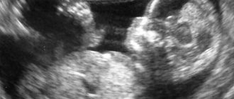
What can be seen in the photo of the fetus?
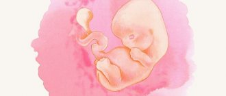
In the photo of the 8th week of pregnancy you can already see some outlines of the baby or babies (in case of multiple pregnancy). The tail of the fetus begins to disappear, fingers on the limbs gradually form, the intestines begin to form, the brain and nervous system begin to form. Eyelids appear and the visual apparatus develops.
In the photo of the embryo you can see the yolk sac, arms, legs and head of the embryo. But despite the fact that the testicles and ovaries are already developing at this point, it is still impossible to determine the sex of the unborn child.
In the first weeks of pregnancy, it is quite difficult to determine the presence of twins, but closer to the eighth week, two small black circles located in the uterus can already be detected on the monitor screen of the device. Here it is also possible to see the fingers and toes, the umbilical cord and also the two placentas.
Early problems
If the pain in the lower abdomen becomes constant and nagging, this is a serious reason to consult your doctor so as not to miss the threat of miscarriage. Periodically occurring slightly painful sensations are quite normal, because the uterus increases in size. Breast growth is also normal, but this hardly becomes a problem for the fair sex.
But what other troubles can await you in the eighth week:
- Persistent morning sickness. Usually the gynecologist prescribes Hofitol in such cases, but it does not always help. To make it easier, try, as soon as you wake up and without getting out of bed, eat something light like a crust of bread or a cucumber. This will get your digestive system working.
- Heartburn and flatulence. To avoid such problems, you will have to minimize the consumption of legumes, cabbage and other products with coarse fiber. In general, switch to proper nutrition.
- Constipation. To combat them, include in your diet foods that contain essential dietary fiber and potassium, which stimulates intestinal motility (for example, apples).
- Dizziness and other troubles due to low hemoglobin. Be sure to get your blood test done on time. If your readings are low, your gynecologist will prescribe you iron supplements .
- Bleeding. A dangerous symptom that requires you to call an ambulance.
Standards: detailed description
The unborn baby at 8 weeks is still very small in size. During this period, only the following parameters can be determined:
- Coccyx-parietal size: from 1.4 to 1.8 centimeters.
- Weight – about 1.5 grams.
- The biparietal size of the head is about 6 millimeters.
- The diameter of the yolk sac is 4.5 millimeters.
It is also necessary to study the size of the uterus (normally 7-8 centimeters) and the heart rate of the unborn child (from 110 to 150 beats).
Important! Minor deviations from the specified norm cannot indicate the occurrence of any pathologies or anomalies. However, if the difference is significant (for example, the heart rate is 100 beats per minute), there is cause for concern and close monitoring and investigation of the problem.
Pain and discomfort
The tone of the uterus in women can manifest itself in different ways: pain or an unpleasant feeling of heaviness in the lower abdomen, where heaviness is a normal condition after light exertion or long walking. Aching pain in the lower back, radiating to the legs, appears due to compression of the sciatic nerve, and is considered normal. If you experience severe pain or bleeding, consult a doctor immediately! The second obstetric month is the time when toxicosis appears. Nausea and vomiting up to 2 times a day are considered normal manifestations. You may also experience frequent urination, minor acne, and unusually dry or oily skin.
Contraindications: is ultrasound dangerous?
There are practically no contraindications for a routine ultrasound examination at week 8. The exception is inflammation and infection of the vaginal mucosa. Unscheduled surgery is not recommended if there is a high risk of fetal loss and heavy bleeding.
On the other hand, this procedure is considered absolutely safe, so it can be performed almost any number of times during the entire period of pregnancy. This is especially true in cases where there is a threat of the occurrence and development of pathologies and abnormal pregnancy.
Prevention and treatment of diseases
If an acute respiratory infection or flu occurs, immediately take sick leave, forgetting about all work responsibilities . It is important to receive treatment as quickly and efficiently as possible in accordance with the doctor’s prescriptions, otherwise, as the infection develops, the blood flow in the mother-placenta-fetus system will sharply decrease, and your baby will lack oxygen. And when you are cured, devote more effort to prevention: switch to proper nutrition, get enough sleep, spend more time in the fresh air and ventilate the room.
If it’s frosty outside, ventilate quickly: open the window wide for half a minute
“First of all, a pregnant woman should be isolated from other family members and be in a well-ventilated room, with an ambient temperature no higher than 22–23 degrees. Maintaining a comfortable humidity level is also important to reduce cold symptoms. Bed rest is not necessary, however, it is necessary to limit physical activity, get more rest and sleep. The passion for absorbing large amounts of liquid, especially tea, is not justified. Recommendations about taking large amounts of fluid during colds are a myth, and during pregnancy can do more harm than good. It is not advisable for pregnant women to take herbal teas, especially with large amounts of honey or sugar. Nutrition during a cold can be limited, since too much food will be an additional burden for a weakened body. Preference should be given to soups, vegetables, and fruits. If your appetite is reduced, this is normal during a cold, and in such cases, forceful eating is not recommended.
pediatrician, Dr. E.O. Komarovsky
https://lib.komarovskiy.net/prostudnye-infekcii-pri-beremennosti.htm
Another common disease in the first trimester is herpes. Only the gynecologist observing you can assess its risk to the fetus after additional examinations. Each situation is individual. He will prescribe treatment that is safe for the baby. After treatment, to prevent relapses, avoid contact with sick people and strengthen your immune system in every way.
Rubella is most dangerous for the fetus in the eighth week . Get tested for antibodies to this disease as soon as possible. If you have them, then the fetus is probably protected during pregnancy. If not, try not to contact possible sick people. If you do become infected with rubella, do not put off visiting an infectious disease specialist. A number of examinations will be carried out to resolve the issue of risks to the fetus. In the worst case scenario, the pregnancy will have to be terminated.
It is worth noting separately that in the eighth week of pregnancy it is recommended to have as little contact as possible with another group of disease carriers - domestic animals, especially cats, which can cause toxoplasmosis.
Tell your doctor if you have a cat at home; you may be ordered to test for toxoplasmosis.
Where do they make it and how much does it cost?
Currently, there are free points where you can get a medical examination, and paid ones. The first include city hospitals and antenatal clinics. The second includes private clinics and specialized medical centers.
The doors of free hospitals are usually open to everyone. However, it is worth considering the fact that women undergoing procedures there are not always in comfortable conditions. An important disadvantage of such institutions is the almost constant presence of long queues.
Paid medical centers, in turn, offer highly qualified services for an average cost of 1,500 to 4,000 rubles. For this money, patients receive comfort, communication with polite staff, cleanliness, procedures using the latest equipment, small queues or their complete absence.
However, it is worth noting that not everyone can afford such prices. Moreover, choosing a private hospital is not so easy. To do this, you need to read and listen to reviews and recommendations. Often you have to visit many different clinics in search of the most suitable one.
Ultrasound during pregnancy
The main purpose of ultrasound is to monitor the intrauterine development of the fetus, its growth, and also to assess the condition of its developing organs.
In addition, an important function of such a diagnosis is to exclude serious congenital genetic diseases of the unborn child, such as a pathology with an extra chromosome called Down syndrome. Such diagnostics are routine and are carried out as planned in all antenatal clinics and medical centers conducting pregnancy.
What does the fetus look like at this stage?
Embryo research
Carrying out ultrasound diagnostics at 8 weeks is aimed mainly at a comprehensive study of the fetus: indicators of size, location, structural features. In addition, the child’s motor activity is monitored, because even at such a short period of time it is already present. At this point, the embryo can already be clearly visualized.
If there is no movement, then we are talking about fading pregnancy, or anembryony (that is, initially there was no embryo in the fertilized egg). Ultrasound during a frozen pregnancy at the 8th week gives a 100% result.
Second month of pregnancy: dangers
Each pregnant woman understands that the future of her baby depends on her health. However, many expectant mothers underestimate all the dangers that can affect the pregnancy process and its outcome. It would seem that an ordinary ARVI does not cause you any fears. But not for pregnant women. Any infections that end up in your body will be transmitted to the fetus or will have an extremely adverse effect on its development. The second month enters the first trimester, when the fetus is still extremely vulnerable.
ARVI is a viral infection that can cause disruption in the development of the baby.
In addition, symptoms such as fever have an extremely negative impact on him and her.
If you develop a high fever, you may have a miscarriage. Therefore, if you have a cold, you should never wave your hand at it or carry it on your feet. You need to consult a doctor so that he can prescribe you the correct regimen and safe medications for its treatment. Antibiotics are contraindicated, and fever can only be fought with paracetamol, which, in general, is also not safe. However, if you find yourself with a runny nose, do not rush to panic. During pregnancy, the first trimester week of pregnancy brings the most interesting surprises. Your nose may become stuffy simply because progesterone levels rise, which retains fluid in the body. Thrush is also a problem. And it often occurs at this time, because pregnant women have reduced immunity. Do not try to treat it yourself, because in your situation you will need completely different medications.
If you suddenly suddenly lose toxicosis, this is not good. This is possible, but keep in mind that this symptom is an alarming sign of a frozen pregnancy. An ultrasound photo will help reveal it.
Any bleeding is not a reason to panic, but a reason to talk to your gynecologist. Perhaps your uterus is in good shape. But it can also be a sure sign of an impending or already occurring miscarriage, depending on its course. Pay attention to pain. If they do not go away, but continue to pull on your stomach, even if you lie down, this is also a symptom of a miscarriage.
The second month of pregnancy is a period during which you should especially carefully monitor your body and notice all the nuances that concern you.
Toxicosis
At 8 weeks, nausea reaches its peak, then gradually declines. Toxicosis may be accompanied by such unpleasant symptoms as: heartburn, belching, changes in taste preferences, loss of appetite. Usually it appears rarely and does not affect the course of pregnancy. But severe toxicosis can deplete a woman’s body.
You should immediately go to the hospital if:
- vomiting occurs more than 2 times a day;
- the woman lost weight;
- food is not digested throughout the day;
- there is constant weakness.
Yolk sac examination
As a result of an ultrasound examination, it is necessary to evaluate both the condition of the fetus and the organs surrounding it. This refers to the yolk sac. Such formation in the fruit egg is a temporary phenomenon. Forming on the 15th–16th day of pregnancy, it disappears at the end of the 1st trimester.
The yolk sac plays an important function in the development of the embryo: it produces germ cells, is responsible for the formation of germinal red blood cells and the production of proteins, without which normal development is impossible, helps to form immunity, etc.
The size of the yolk sac at the 8th week is about 4.5 mm. If it is increased in size, then there is reason to believe that the development of the fetus occurs with pathological abnormalities.
The yolk sac is formed 15-16 days after conception and disintegrates by the end of the 1st trimester